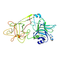
2FIM
 
 | | Structure of the C-terminal domain of Human Tubby-like protein 1 | | 分子名称: | 3-(N,N-DIMETHYLOCTYLAMMONIO)PROPANESULFONATE, SULFATE ION, Tubby related protein 1 | | 著者 | Hallberg, B.M, Ogg, D, Arrowsmith, C, Berglund, H, Edwards, A, Ehn, M, Flodin, S, Graslund, S, Hammarstrom, M, Hogbom, M, Holmberg-Schiavone, L, Kotenyova, T, Kursula, P, Nilsson-Ehle, P, Nordlund, P, Nyman, T, Sagemark, J, Stenmark, P, Sundstrom, M, Thorsell, A.G, Van Den Berg, S, Weigelt, J, Persson, C. | | 登録日 | 2005-12-29 | | 公開日 | 2006-02-07 | | 最終更新日 | 2024-03-13 | | 実験手法 | X-RAY DIFFRACTION (1.9 Å) | | 主引用文献 | Structure of the C-terminal domain of Human Tubby-like protein 1
To be published
|
|
2JE2
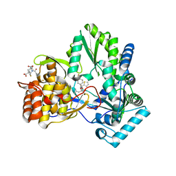
 | | Cytochrome P460 from Nitrosomonas europaea - probable nonphysiological oxidized form | | 分子名称: | CYTOCHROME P460, HEME C, PHOSPHATE ION | | 著者 | Pearson, A.R, Elmore, B.O, Yang, C, Ferrara, J.D, Hooper, A.B, Wilmot, C.M. | | 登録日 | 2007-01-13 | | 公開日 | 2007-07-03 | | 最終更新日 | 2024-11-06 | | 実験手法 | X-RAY DIFFRACTION (1.8 Å) | | 主引用文献 | The Crystal Structure of Cytochrome P460 of Nitrosomonas Europaea Reveals a Novel Cytochrome Fold and Heme-Protein Cross-Link.
Biochemistry, 46, 2007
|
|
2JE3
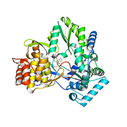
 | | Cytochrome P460 from Nitrosomonas europaea - probable physiological form | | 分子名称: | CYTOCHROME P460, HEME C, PHOSPHATE ION | | 著者 | Pearson, A.R, Elmore, B.O, Yang, C, Ferrara, J.D, Hooper, A.B, Wilmot, C.M. | | 登録日 | 2007-01-13 | | 公開日 | 2007-07-03 | | 最終更新日 | 2024-10-16 | | 実験手法 | X-RAY DIFFRACTION (1.8 Å) | | 主引用文献 | The Crystal Structure of Cytochrome P460 of Nitrosomonas Europaea Reveals a Novel Cytochrome Fold and Heme-Protein Cross-Link.
Biochemistry, 46, 2007
|
|
2JC0
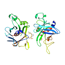
 | | CRYSTAL STRUCTURE OF HEPATITIS C VIRUS POLYMERASE IN COMPLEX WITH INHIBITOR SB655264 | | 分子名称: | (2S,4S,5R)-2-ISOBUTYL-5-(2-THIENYL)-1-[4-(TRIFLUOROMETHYL)BENZOYL]PYRROLIDINE-2,4-DICARBOXYLIC ACID, RNA-DEPENDENT RNA-POLYMERASE | | 著者 | Wonacott, A, Skarzynski, T, Singh, O.M. | | 登録日 | 2006-12-18 | | 公開日 | 2007-02-13 | | 最終更新日 | 2024-05-08 | | 実験手法 | X-RAY DIFFRACTION (2.2 Å) | | 主引用文献 | Optimization of Novel Acyl Pyrrolidine Inhibitors of Hepatitis C Virus RNA-Dependent RNA Polymerase Leading to a Development Candidate.
J.Med.Chem., 50, 2007
|
|
2JC1
 | | CRYSTAL STRUCTURE OF HEPATITIS C VIRUS POLYMERASE IN COMPLEX WITH INHIBITOR SB698223 | | 分子名称: | (2S,4S,5R)-1-(4-TERT-BUTYLBENZOYL)-2-ISOBUTYL-5-(1,3-THIAZOL-2-YL)PYRROLIDINE-2,4-DICARBOXYLIC ACID, RNA-DEPENDENT RNA-POLYMERASE | | 著者 | Wonacott, A, Skarzynski, T, Singh, O.M. | | 登録日 | 2006-12-18 | | 公開日 | 2007-02-13 | | 最終更新日 | 2024-05-08 | | 実験手法 | X-RAY DIFFRACTION (2 Å) | | 主引用文献 | Optimization of Novel Acyl Pyrrolidine Inhibitors of Hepatitis C Virus RNA-Dependent RNA Polymerase Leading to a Development Candidate.
J.Med.Chem., 50, 2007
|
|
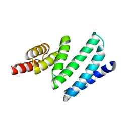
3G2S
 | | VHS Domain of human GGA1 complexed with SorLA C-terminal Peptide | | 分子名称: | ADP-ribosylation factor-binding protein GGA1, C-terminal fragment of Sortilin-related receptor, HEXANE-1,6-DIOL | | 著者 | Cramer, J.F, Behrens, M.A, Gustafsen, C, Oliveira, C.L.P, Pedersen, J.S, Madsen, P, Petersen, C.M, Thirup, S.S. | | 登録日 | 2009-02-01 | | 公開日 | 2009-12-15 | | 最終更新日 | 2024-11-06 | | 実験手法 | X-RAY DIFFRACTION (1.7 Å) | | 主引用文献 | GGA autoinhibition revisited
Traffic, 11, 2010
|
|
5CIH
 | |
3G2U
 | | VHS Domain of human GGA1 complexed with Sotilin C-terminal Peptide | | 分子名称: | ADP-ribosylation factor-binding protein GGA1, C-terminal fragment of Sortilin, IODIDE ION | | 著者 | Cramer, J.F, Behrens, M.A, Gustafsen, C, Oliveira, C.L.P, Pedersen, J.S, Madsen, P, Petersen, C.M, Thirup, S.S. | | 登録日 | 2009-02-01 | | 公開日 | 2009-12-15 | | 最終更新日 | 2024-10-30 | | 実験手法 | X-RAY DIFFRACTION (2.301 Å) | | 主引用文献 | GGA autoinhibition revisited
Traffic, 11, 2010
|
|
1D4E
 | | CRYSTAL STRUCTURE OF THE FLAVOCYTOCHROME C FUMARATE REDUCTASE OF SHEWANELLA PUTREFACIENS STRAIN MR-1 COMPLEXED WITH FUMARATE | | 分子名称: | FLAVIN-ADENINE DINUCLEOTIDE, FLAVOCYTOCHROME C FUMARATE REDUCTASE, FUMARIC ACID, ... | | 著者 | Leys, D, Tsapin, A.S, Meyer, T.E, Cusanovich, M.A, Van Beeumen, J.J. | | 登録日 | 1999-10-03 | | 公開日 | 1999-12-01 | | 最終更新日 | 2024-10-30 | | 実験手法 | X-RAY DIFFRACTION (2.8 Å) | | 主引用文献 | Structure and mechanism of the flavocytochrome c fumarate reductase of Shewanella putrefaciens MR-1.
Nat.Struct.Biol., 6, 1999
|
|
1OB1
 | | Crystal structure of a Fab complex whith Plasmodium falciparum MSP1-19 | | 分子名称: | ANTIBODY, HEAVY CHAIN, LIGHT CHAIN, ... | | 著者 | Pizarro, J.C, Chitarra, V, Verger, D, Holm, I, Petres, S, Dartville, S, Nato, F, Longacre, S, Bentley, G.A. | | 登録日 | 2003-01-22 | | 公開日 | 2003-05-01 | | 最終更新日 | 2024-10-23 | | 実験手法 | X-RAY DIFFRACTION (2.9 Å) | | 主引用文献 | Crystal Structure of a Fab Complex Formed with Pfmsp1-19, the C-Terminal Fragment of Merozoite Surface Protein 1 from Plasmodium Falciparum: A Malaria Vaccine Candidate
J.Mol.Biol., 328, 2003
|
|
2WRM
 | |
1CSJ
 | | CRYSTAL STRUCTURE OF THE RNA-DEPENDENT RNA POLYMERASE OF HEPATITIS C VIRUS | | 分子名称: | HEPATITIS C VIRUS RNA POLYMERASE (NS5B) | | 著者 | Bressanelli, S, Tomei, L, Roussel, A, Incitti, I, Vitale, R.L, Mathieu, M, De Francesco, R, Rey, F.A. | | 登録日 | 1999-08-18 | | 公開日 | 1999-11-08 | | 最終更新日 | 2024-11-20 | | 実験手法 | X-RAY DIFFRACTION (2.8 Å) | | 主引用文献 | Crystal structure of the RNA-dependent RNA polymerase of hepatitis C virus.
Proc.Natl.Acad.Sci.USA, 96, 1999
|
|
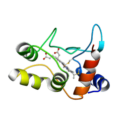
1YWU
 | | Solution NMR structure of Pseudomonas Aeruginosa protein PA4608. Northeast Structural Genomics target PaT7 | | 分子名称: | hypothetical protein PA4608 | | 著者 | Ramelot, T.A, Yee, A.A, Cort, J.R, Semesi, A, Arrowsmith, C.H, Kennedy, M.A, Northeast Structural Genomics Consortium (NESG) | | 登録日 | 2005-02-18 | | 公開日 | 2005-03-29 | | 最終更新日 | 2024-05-01 | | 実験手法 | SOLUTION NMR | | 主引用文献 | NMR structure and binding studies confirm that PA4608 from Pseudomonas aeruginosa is a PilZ domain and a c-di-GMP binding protein.
Proteins, 66, 2007
|
|
5CIC
 | |
315D
 | |
2JJL
 | | Structure of avian reovirus sigmaC117-326, P321 crystal form | | 分子名称: | CHLORIDE ION, SIGMA-C CAPSID PROTEIN, SULFATE ION, ... | | 著者 | Guardado-Calvo, P, Fox, G.C, Llamas-Saiz, A.L, Benavente, J, van Raaij, M.J. | | 登録日 | 2008-04-14 | | 公開日 | 2009-01-13 | | 最終更新日 | 2023-12-13 | | 実験手法 | X-RAY DIFFRACTION (2.3 Å) | | 主引用文献 | Crystallographic structure of the alpha-helical triple coiled-coil domain of avian reovirus S1133 fibre.
J. Gen. Virol., 90, 2009
|
|
2YKZ
 | |
2YL0
 | |
1GIW
 | | SOLUTION STRUCTURE OF REDUCED HORSE HEART CYTOCHROME C, NMR, MINIMIZED AVERAGE STRUCTURE | | 分子名称: | CYTOCHROME C, HEME C | | 著者 | Banci, L, Bertini, I, Huber, J.G, Spyroulias, G.A, Turano, P. | | 登録日 | 1998-06-17 | | 公開日 | 1998-12-09 | | 最終更新日 | 2024-10-30 | | 実験手法 | SOLUTION NMR | | 主引用文献 | Solution structure of reduced horse heart cytochrome c.
J.Biol.Inorg.Chem., 4, 1999
|
|
5CID
 | |
2C1D
 | | Crystal structure of SoxXA from P. pantotrophus | | 分子名称: | HEME C, SOXA, SOXX, ... | | 著者 | Dambe, T, Quentmeier, A, Rother, D, Friedrich, C, Scheidig, A.J. | | 登録日 | 2005-09-13 | | 公開日 | 2005-10-06 | | 最終更新日 | 2024-10-16 | | 実験手法 | X-RAY DIFFRACTION (1.92 Å) | | 主引用文献 | Structure of the Cytochrome Complex Soxxa of Paracoccus Pantotrophus, a Heme Enzyme Initiating Chemotrophic Sulfur Oxidation.
J.Struct.Biol., 152, 2005
|
|
5CIG
 | |
1DLL
 | | The HC fragement of tetanus toxin complexed with lactose | | 分子名称: | GLYCEROL, TETANUS TOXIN, beta-D-galactopyranose-(1-4)-beta-D-glucopyranose | | 著者 | Emsley, P, Fotinou, C, Black, I, Fairweather, N.F, Charles, I.G, Watts, C, Hewitt, E, Isaacs, N.W. | | 登録日 | 1999-12-10 | | 公開日 | 2000-03-24 | | 最終更新日 | 2024-02-07 | | 実験手法 | X-RAY DIFFRACTION (1.8 Å) | | 主引用文献 | The structures of the H(C) fragment of tetanus toxin with carbohydrate subunit complexes provide insight into ganglioside binding.
J.Biol.Chem., 275, 2000
|
|
4APO
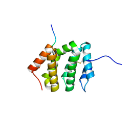
 | | AIP TPR domain in complex with human Tomm20 peptide | | 分子名称: | AH RECEPTOR-INTERACTING PROTEIN, DODECAETHYLENE GLYCOL, MITOCHONDRIAL IMPORT RECEPTOR SUBUNIT TOM20 HOMOLOG, ... | | 著者 | Morgan, R.M.L, Roe, S.M, Pearl, L.H, Prodromou, C. | | 登録日 | 2012-04-04 | | 公開日 | 2013-01-23 | | 最終更新日 | 2023-12-20 | | 実験手法 | X-RAY DIFFRACTION (1.895 Å) | | 主引用文献 | Structure of the Tpr Domain of Aip: Lack of Client Protein Interaction with the C-Terminal Alpha-7 Helix of the Tpr Domain of Aip is Sufficient for Pituitary Adenoma Predisposition.
Plos One, 7, 2012
|
|
2BGV
 | | X-ray structure of ferric cytochrome c-550 from Paracoccus versutus | | 分子名称: | CYTOCHROME C-550, HEME C | | 著者 | Worrall, J.A.R, Van Roon, A.-M.M, Ubbink, M, Canters, G.W. | | 登録日 | 2005-01-05 | | 公開日 | 2005-05-11 | | 最終更新日 | 2024-10-23 | | 実験手法 | X-RAY DIFFRACTION (1.9 Å) | | 主引用文献 | The Effect of Replacing the Axial Methionine Ligand with a Lysine Residue in Cytochrome C-550 from Paracoccus Versutus Assessed by X-Ray Crystallography and Unfolding.
FEBS J., 272, 2005
|
|


