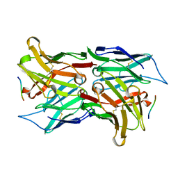
1SVR
 
 | |
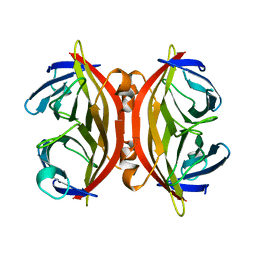
1SVS
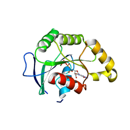
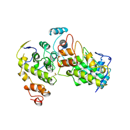
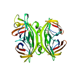
 | | Structure of the K180P mutant of Gi alpha subunit bound to GppNHp. | | 分子名称: | Guanine nucleotide-binding protein G(i), alpha-1 subunit, MAGNESIUM ION, ... | | 著者 | Thomas, C.J, Du, X, Li, P, Wang, Y, Ross, E.M, Sprang, S.R. | | 登録日 | 2004-03-29 | | 公開日 | 2004-06-01 | | 最終更新日 | 2024-02-14 | | 実験手法 | X-RAY DIFFRACTION (1.5 Å) | | 主引用文献 | Uncoupling conformational change from GTP hydrolysis in a heterotrimeric G protein {alpha}-subunit.
Proc.Natl.Acad.Sci.USA, 101, 2004
|
|
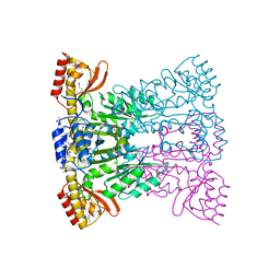
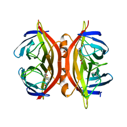
1SVT
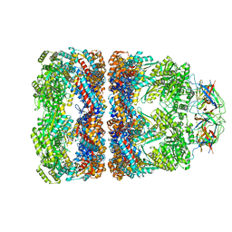 | | Crystal structure of GroEL14-GroES7-(ADP-AlFx)7 | | 分子名称: | ADENOSINE-5'-DIPHOSPHATE, ALUMINUM FLUORIDE, MAGNESIUM ION, ... | | 著者 | Chaudhry, C, Horwich, A.L, Brunger, A.T, Adams, P.D. | | 登録日 | 2004-03-29 | | 公開日 | 2005-03-01 | | 最終更新日 | 2024-02-14 | | 実験手法 | X-RAY DIFFRACTION (2.808 Å) | | 主引用文献 | Exploring the structural dynamics of the E.coli chaperonin GroEL using translation-libration-screw crystallographic refinement of intermediate states.
J.Mol.Biol., 342, 2004
|
|
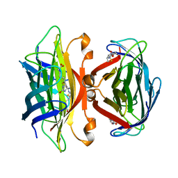
1SVU
 | | Structure of the Q237W mutant of HhaI DNA methyltransferase: an insight into protein-protein interactions | | 分子名称: | Modification methylase HhaI, S-ADENOSYL-L-HOMOCYSTEINE, SULFATE ION, ... | | 著者 | Dong, A, Zhou, L, Zhang, X, Stickel, S, Roberts, R.J, Cheng, X. | | 登録日 | 2004-03-30 | | 公開日 | 2004-06-29 | | 最終更新日 | 2023-08-23 | | 実験手法 | X-RAY DIFFRACTION (2.66 Å) | | 主引用文献 | Structure of the Q237W mutant of HhaI DNA methyltransferase: an insight into protein-protein interactions
Biol.Chem., 385, 2004
|
|
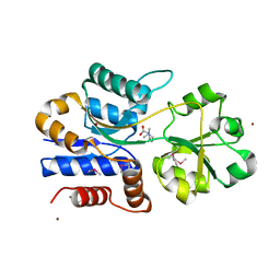
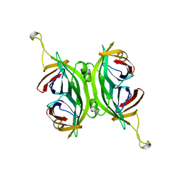
1SVV
 | |
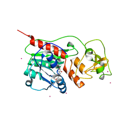
1SVW
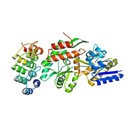
 | | Crystal Structure of YsxC complexed with GMPPNP | | 分子名称: | GTP-binding protein YsxC, GUANOSINE-5'-TRIPHOSPHATE, MAGNESIUM ION | | 著者 | Ruzheinikov, S.N, Das, S.K, Sedelnikova, S.E, Baker, P.J, Artymiuk, P.J, Garcia-Lara, J, Foster, S.J, Rice, D.W. | | 登録日 | 2004-03-30 | | 公開日 | 2004-05-25 | | 最終更新日 | 2024-02-14 | | 実験手法 | X-RAY DIFFRACTION (2.8 Å) | | 主引用文献 | Analysis of the Open and Closed Conformations of the GTP-binding Protein YsxC from Bacillus subtilis.
J.Mol.Biol., 339, 2004
|
|
1SVX
 | | Crystal structure of a designed selected Ankyrin Repeat protein in complex with the Maltose Binding Protein | | 分子名称: | Ankyrin Repeat Protein off7, Maltose-binding periplasmic protein | | 著者 | Binz, H.K, Amstutz, P, Kohl, A, Stumpp, M.T, Briand, C, Forrer, P, Gruetter, M.G, Plueckthun, A. | | 登録日 | 2004-03-30 | | 公開日 | 2004-05-25 | | 最終更新日 | 2024-02-14 | | 実験手法 | X-RAY DIFFRACTION (2.24 Å) | | 主引用文献 | High-affinity binders selected from designed ankyrin repeat protein libraries
NAT.BIOTECHNOL., 22, 2004
|
|
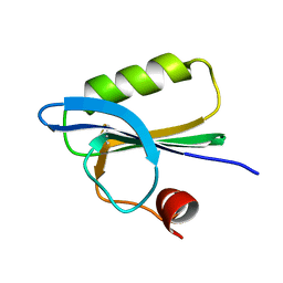
1SVY
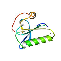 | | SEVERIN DOMAIN 2, 1.75 ANGSTROM CRYSTAL STRUCTURE | | 分子名称: | CALCIUM ION, SEVERIN, SODIUM ION | | 著者 | Puius, Y.A, Fedorov, E.V, Eichinger, L, Sullivan, M, Schleicher, M, Almo, S.C. | | 登録日 | 1998-08-10 | | 公開日 | 1999-08-10 | | 最終更新日 | 2024-06-05 | | 実験手法 | X-RAY DIFFRACTION (1.75 Å) | | 主引用文献 | Mapping the functional surface of domain 2 in the gelsolin superfamily.
Biochemistry, 39, 2000
|
|
1SVZ
 | | Crystal structure of the single-chain Fv fragment 1696 in complex with the epitope peptide corresponding to N-terminus of HIV-2 protease | | 分子名称: | epitope peptide corresponding to N-terminus of HIV-2 protease, single-chain Fv fragment 1696 | | 著者 | Rezacova, P, Brynda, J, Lescar, J, Bentley, G.A, Fabry, M, Horejsi, M, Sedlacek, J. | | 登録日 | 2004-03-30 | | 公開日 | 2005-03-01 | | 最終更新日 | 2023-08-23 | | 実験手法 | X-RAY DIFFRACTION (1.89 Å) | | 主引用文献 | Crystal structure of a cross-reaction complex between an anti-HIV-1 protease antibody and an HIV-2 protease peptide
J.Struct.Biol., 149, 2005
|
|
1SW0
 | | Triosephosphate isomerase from Gallus gallus, loop 6 hinge mutant K174L, T175W | | 分子名称: | 2-PHOSPHOGLYCOLIC ACID, Triosephosphate isomerase | | 著者 | Kursula, I, Salin, M, Sun, J, Norledge, B.V, Haapalainen, A.M, Sampson, N.S, Wierenga, R.K. | | 登録日 | 2004-03-30 | | 公開日 | 2004-08-24 | | 最終更新日 | 2023-10-25 | | 実験手法 | X-RAY DIFFRACTION (1.71 Å) | | 主引用文献 | Understanding protein lids: structural analysis of active hinge mutants in triosephosphate isomerase
Protein Eng.Des.Sel., 17, 2004
|
|
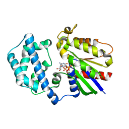
1SW1
 | | Crystal structure of ProX from Archeoglobus fulgidus in complex with proline betaine | | 分子名称: | 1,1-DIMETHYL-PROLINIUM, ZINC ION, osmoprotection protein (proX) | | 著者 | Schiefner, A, Holtmann, G, Diederichs, K, Welte, W, Bremer, E. | | 登録日 | 2004-03-30 | | 公開日 | 2004-09-14 | | 最終更新日 | 2021-10-27 | | 実験手法 | X-RAY DIFFRACTION (1.9 Å) | | 主引用文献 | Structural basis for the binding of compatible solutes by ProX from the hyperthermophilic archaeon Archaeoglobus fulgidus.
J.Biol.Chem., 279, 2004
|
|
1SW2
 | | Crystal structure of ProX from Archeoglobus fulgidus in complex with glycine betaine | | 分子名称: | TRIMETHYL GLYCINE, osmoprotection protein (proX) | | 著者 | Schiefner, A, Holtmann, G, Diederichs, K, Welte, W, Bremer, E. | | 登録日 | 2004-03-30 | | 公開日 | 2004-09-14 | | 最終更新日 | 2023-11-15 | | 実験手法 | X-RAY DIFFRACTION (2.1 Å) | | 主引用文献 | Structural basis for the binding of compatible solutes by ProX from the hyperthermophilic archaeon Archaeoglobus fulgidus.
J.Biol.Chem., 279, 2004
|
|
1SW3
 | | Triosephosphate isomerase from Gallus gallus, loop 6 mutant T175V | | 分子名称: | 2-PHOSPHOGLYCOLIC ACID, Triosephosphate isomerase | | 著者 | Kursula, I, Salin, M, Sun, J, Norledge, B.V, Haapalainen, A.M, Sampson, N.S, Wierenga, R.K. | | 登録日 | 2004-03-30 | | 公開日 | 2004-08-24 | | 最終更新日 | 2023-10-25 | | 実験手法 | X-RAY DIFFRACTION (2.03 Å) | | 主引用文献 | Understanding protein lids: structural analysis of active hinge mutants in triosephosphate isomerase
Protein Eng.Des.Sel., 17, 2004
|
|
1SW4
 | | Crystal structure of ProX from Archeoglobus fulgidus in complex with trimethyl ammonium | | 分子名称: | CHLORIDE ION, TETRAMETHYLAMMONIUM ION, ZINC ION, ... | | 著者 | Schiefner, A, Holtmann, G, Diederichs, K, Welte, W, Bremer, E. | | 登録日 | 2004-03-30 | | 公開日 | 2004-09-14 | | 最終更新日 | 2023-08-23 | | 実験手法 | X-RAY DIFFRACTION (1.9 Å) | | 主引用文献 | Structural basis for the binding of compatible solutes by ProX from the hyperthermophilic archaeon Archaeoglobus fulgidus.
J.Biol.Chem., 279, 2004
|
|
1SW5
 | | Crystal structure of ProX from Archeoglobus fulgidus in the ligand free form | | 分子名称: | CHLORIDE ION, MAGNESIUM ION, osmoprotection protein (proX) | | 著者 | Schiefner, A, Holtmann, G, Diederichs, K, Welte, W, Bremer, E. | | 登録日 | 2004-03-30 | | 公開日 | 2004-09-14 | | 最終更新日 | 2023-08-23 | | 実験手法 | X-RAY DIFFRACTION (1.8 Å) | | 主引用文献 | Structural basis for the binding of compatible solutes by ProX from the hyperthermophilic archaeon Archaeoglobus fulgidus.
J.Biol.Chem., 279, 2004
|
|
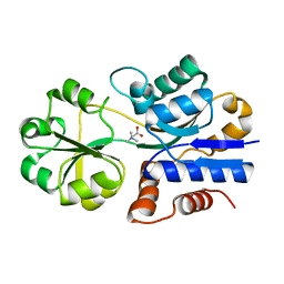
1SW6
 | |
1SW7
 | | Triosephosphate isomerase from Gallus gallus, loop 6 mutant K174N, T175S, A176S | | 分子名称: | 2-PHOSPHOGLYCOLIC ACID, Triosephosphate isomerase | | 著者 | Kursula, I, Salin, M, Sun, J, Norledge, B.V, Haapalainen, A.M, Sampson, N.S, Wierenga, R.K. | | 登録日 | 2004-03-30 | | 公開日 | 2004-08-24 | | 最終更新日 | 2023-10-25 | | 実験手法 | X-RAY DIFFRACTION (2.22 Å) | | 主引用文献 | Understanding protein lids: structural analysis of active hinge mutants in triosephosphate isomerase
Protein Eng.Des.Sel., 17, 2004
|
|
1SW8
 | | Solution structure of the N-terminal domain of Human N60D calmodulin refined with paramagnetism based strategy | | 分子名称: | CALCIUM ION, Calmodulin | | 著者 | Bertini, I, Del Bianco, C, Gelis, I, Katsaros, N, Luchinat, C, Parigi, G, Peana, M, Provenzani, A, Zoroddu, M.A, Structural Proteomics in Europe (SPINE) | | 登録日 | 2004-03-30 | | 公開日 | 2004-04-06 | | 最終更新日 | 2024-05-22 | | 実験手法 | SOLUTION NMR | | 主引用文献 | Experimentally exploring the conformational space sampled by domain reorientation in calmodulin
Proc.Natl.Acad.Sci.USA, 101, 2004
|
|
1SWA
 | | APO-CORE-STREPTAVIDIN AT PH 4.5 | | 分子名称: | STREPTAVIDIN | | 著者 | Freitag, S, Le Trong, I, Klumb, L, Stayton, P.S, Stenkamp, R.E. | | 登録日 | 1997-03-02 | | 公開日 | 1998-03-04 | | 最終更新日 | 2024-04-03 | | 実験手法 | X-RAY DIFFRACTION (1.9 Å) | | 主引用文献 | Structural studies of the streptavidin binding loop.
Protein Sci., 6, 1997
|
|
1SWB
 | | APO-CORE-STREPTAVIDIN AT PH 7.5 | | 分子名称: | STREPTAVIDIN | | 著者 | Freitag, S, Le Trong, I, Klumb, L, Stayton, P.S, Stenkamp, R.E. | | 登録日 | 1997-03-04 | | 公開日 | 1998-03-04 | | 最終更新日 | 2024-05-22 | | 実験手法 | X-RAY DIFFRACTION (1.85 Å) | | 主引用文献 | Structural studies of the streptavidin binding loop.
Protein Sci., 6, 1997
|
|
1SWC
 | | APO-CORE-STREPTAVIDIN AT PH 4.5 | | 分子名称: | STREPTAVIDIN | | 著者 | Freitag, S, Le Trong, I, Klumb, L, Stayton, P.S, Stenkamp, R.E. | | 登録日 | 1997-03-04 | | 公開日 | 1998-03-04 | | 最終更新日 | 2024-05-22 | | 実験手法 | X-RAY DIFFRACTION (1.8 Å) | | 主引用文献 | Structural studies of the streptavidin binding loop.
Protein Sci., 6, 1997
|
|
1SWD
 | | APO-CORE-STREPTAVIDIN IN COMPLEX WITH BIOTIN (TWO UNOCCUPIED BINDING SITES) AT PH 4.5 | | 分子名称: | BIOTIN, STREPTAVIDIN | | 著者 | Freitag, S, Le Trong, I, Klumb, L, Stayton, P.S, Stenkamp, R.E. | | 登録日 | 1997-03-04 | | 公開日 | 1998-03-04 | | 最終更新日 | 2024-05-22 | | 実験手法 | X-RAY DIFFRACTION (1.9 Å) | | 主引用文献 | Structural studies of the streptavidin binding loop.
Protein Sci., 6, 1997
|
|
1SWE
 | | APO-CORE-STREPTAVIDIN IN COMPLEX WITH BIOTIN AT PH 4.5 | | 分子名称: | BIOTIN, STREPTAVIDIN | | 著者 | Freitag, S, Le Trong, I, Klumb, L, Stayton, P.S, Stenkamp, R.E. | | 登録日 | 1997-03-04 | | 公開日 | 1998-03-04 | | 最終更新日 | 2024-05-22 | | 実験手法 | X-RAY DIFFRACTION (2.06 Å) | | 主引用文献 | Structural studies of the streptavidin binding loop.
Protein Sci., 6, 1997
|
|
1SWF
 | | CIRCULAR PERMUTED STREPTAVIDIN E51/A46 | | 分子名称: | CIRCULARLY PERMUTED CORE-STREPTAVIDIN E51/A46 | | 著者 | Freitag, S, Chu, V, Le Trong, I, Stayton, P.S, Stenkamp, R.E. | | 登録日 | 1997-04-23 | | 公開日 | 1998-04-29 | | 最終更新日 | 2024-05-22 | | 実験手法 | X-RAY DIFFRACTION (2 Å) | | 主引用文献 | Thermodynamic and structural consequences of flexible loop deletion by circular permutation in the streptavidin-biotin system.
Protein Sci., 7, 1998
|
|
1SWG
 | | CIRCULAR PERMUTED STREPTAVIDIN E51/A46 IN COMPLEX WITH BIOTIN | | 分子名称: | BIOTIN, CIRCULARLY PERMUTED CORE-STREPTAVIDIN E51/A46 | | 著者 | Freitag, S, Chu, V, Le Trong, I, Stayton, P.S, Stenkamp, R.E. | | 登録日 | 1997-07-12 | | 公開日 | 1998-07-15 | | 最終更新日 | 2024-05-22 | | 実験手法 | X-RAY DIFFRACTION (1.8 Å) | | 主引用文献 | Thermodynamic and structural consequences of flexible loop deletion by circular permutation in the streptavidin-biotin system.
Protein Sci., 7, 1998
|
|


