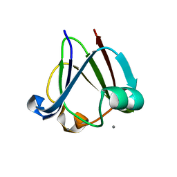
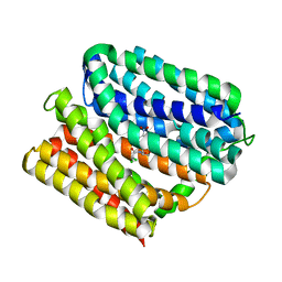
5LJA
 
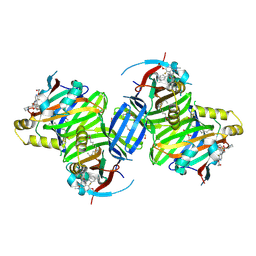
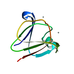
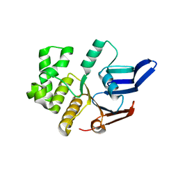 | | Structure of the E. coli MacB ABC domain (P6122) | | 分子名称: | Macrolide export ATP-binding/permease protein MacB | | 著者 | Crow, A. | | 登録日 | 2016-07-18 | | 公開日 | 2017-11-15 | | 最終更新日 | 2024-01-10 | | 実験手法 | X-RAY DIFFRACTION (2.4 Å) | | 主引用文献 | Structure and mechanotransmission mechanism of the MacB ABC transporter superfamily.
Proc. Natl. Acad. Sci. U.S.A., 114, 2017
|
|
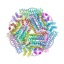
7DY9
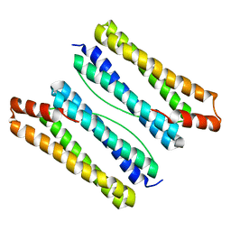 | | Thermotoga maritima ferritin mutant-FLAL | | 分子名称: | Ferritin | | 著者 | Zhao, G, Zhang, X. | | 登録日 | 2021-01-20 | | 公開日 | 2021-09-01 | | 最終更新日 | 2023-11-29 | | 実験手法 | X-RAY DIFFRACTION (2.303 Å) | | 主引用文献 | Protein interface redesign facilitates the transformation of nanocage building blocks to 1D and 2D nanomaterials.
Nat Commun, 12, 2021
|
|
7DY8
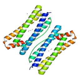 | | Thermotoga maritima ferritin mutant-FLAL | | 分子名称: | CALCIUM ION, FE (III) ION, Ferritin | | 著者 | Zhang, X, Zhao, G. | | 登録日 | 2021-01-20 | | 公開日 | 2021-09-01 | | 最終更新日 | 2023-11-29 | | 実験手法 | X-RAY DIFFRACTION (2.103 Å) | | 主引用文献 | Protein interface redesign facilitates the transformation of nanocage building blocks to 1D and 2D nanomaterials.
Nat Commun, 12, 2021
|
|
7DYB
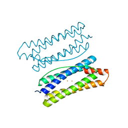 | | Thermotoga maritima ferritin mutant-FLAL-L | | 分子名称: | FE (III) ION, Ferritin | | 著者 | Zhao, G, Zhang, X. | | 登録日 | 2021-01-20 | | 公開日 | 2021-09-01 | | 最終更新日 | 2023-11-29 | | 実験手法 | X-RAY DIFFRACTION (1.779 Å) | | 主引用文献 | Protein interface redesign facilitates the transformation of nanocage building blocks to 1D and 2D nanomaterials.
Nat Commun, 12, 2021
|
|
3I9H
 | |
7DYA
 | | Crystal structure of TmFtn with calcium ions | | 分子名称: | CALCIUM ION, FE (III) ION, Ferritin | | 著者 | Zhang, X, Zhao, G. | | 登録日 | 2021-01-20 | | 公開日 | 2021-09-01 | | 最終更新日 | 2023-11-29 | | 実験手法 | X-RAY DIFFRACTION (2.197 Å) | | 主引用文献 | Protein interface redesign facilitates the transformation of nanocage building blocks to 1D and 2D nanomaterials.
Nat Commun, 12, 2021
|
|
6AW2
 | | Crystal structure of the HopQ-CEACAM1 complex | | 分子名称: | Carcinoembryonic antigen-related cell adhesion molecule 1, HopQ | | 著者 | Bonsor, D.A, Sundberg, E.J. | | 登録日 | 2017-09-05 | | 公開日 | 2018-05-16 | | 最終更新日 | 2023-10-04 | | 実験手法 | X-RAY DIFFRACTION (2.68 Å) | | 主引用文献 | TheHelicobacter pyloriadhesin protein HopQ exploits the dimer interface of human CEACAMs to facilitate translocation of the oncoprotein CagA.
EMBO J., 37, 2018
|
|
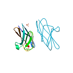
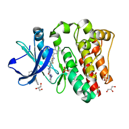
2VMH
 | | The structure of CBM51 from Clostridium perfringens GH95 | | 分子名称: | CALCIUM ION, FIBRONECTIN TYPE III DOMAIN PROTEIN | | 著者 | Gregg, K, Finn, R, Abbott, D.W, Boraston, A.B. | | 登録日 | 2008-01-25 | | 公開日 | 2008-02-19 | | 最終更新日 | 2024-05-08 | | 実験手法 | X-RAY DIFFRACTION (1.5 Å) | | 主引用文献 | Divergent Modes of Glycan Recognition by a New Family of Carbohydrate-Binding Modules
J.Biol.Chem., 283, 2008
|
|
8P0M
 | | Crystal structure of TEAD3 in complex with IAG933 | | 分子名称: | 2-(2-{2-[2-(2-METHOXY-ETHOXY)-ETHOXY]-ETHOXY}-ETHOXY)-ETHANOL, 4-[(2~{S})-5-chloranyl-6-fluoranyl-2-phenyl-2-[(2~{S})-pyrrolidin-2-yl]-3~{H}-1-benzofuran-4-yl]-5-fluoranyl-6-(2-hydroxyethyloxy)-~{N}-methyl-pyridine-3-carboxamide, DIMETHYL SULFOXIDE, ... | | 著者 | Scheufler, C, Villard, F, Chau, S. | | 登録日 | 2023-05-10 | | 公開日 | 2024-04-10 | | 最終更新日 | 2024-04-17 | | 実験手法 | X-RAY DIFFRACTION (1.961 Å) | | 主引用文献 | Direct and selective pharmacological disruption of the YAP-TEAD interface by IAG933 inhibits Hippo-dependent and RAS-MAPK-altered cancers.
Nat Cancer, 2024
|
|
6AVZ
 | | Crystal structure of the HopQ-CEACAM3 WT complex | | 分子名称: | CALCIUM ION, Carcinoembryonic antigen-related cell adhesion molecule 3, HopQ, ... | | 著者 | Bonsor, D.A, Sundberg, E.J. | | 登録日 | 2017-09-05 | | 公開日 | 2018-05-16 | | 最終更新日 | 2018-07-11 | | 実験手法 | X-RAY DIFFRACTION (2.05 Å) | | 主引用文献 | TheHelicobacter pyloriadhesin protein HopQ exploits the dimer interface of human CEACAMs to facilitate translocation of the oncoprotein CagA.
EMBO J., 37, 2018
|
|
5TP1
 | |
6AW3
 | | Crystal structure of the HopQ-CEACAM3 L44Q complex | | 分子名称: | Carcinoembryonic antigen-related cell adhesion molecule 3, HopQ | | 著者 | Bonsor, D.A, Sundberg, E.J. | | 登録日 | 2017-09-05 | | 公開日 | 2018-05-16 | | 最終更新日 | 2018-07-11 | | 実験手法 | X-RAY DIFFRACTION (2.66 Å) | | 主引用文献 | TheHelicobacter pyloriadhesin protein HopQ exploits the dimer interface of human CEACAMs to facilitate translocation of the oncoprotein CagA.
EMBO J., 37, 2018
|
|
3IAJ
 | |
6AW0
 | | Crystal structure of CEACAM3 L44Q | | 分子名称: | CHLORIDE ION, Carcinoembryonic antigen-related cell adhesion molecule 3, GLYCEROL, ... | | 著者 | Bonsor, D.A, Sundberg, E.J. | | 登録日 | 2017-09-05 | | 公開日 | 2018-05-16 | | 最終更新日 | 2023-10-04 | | 実験手法 | X-RAY DIFFRACTION (2.43 Å) | | 主引用文献 | TheHelicobacter pyloriadhesin protein HopQ exploits the dimer interface of human CEACAMs to facilitate translocation of the oncoprotein CagA.
EMBO J., 37, 2018
|
|
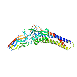
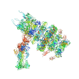
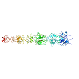
7ZHJ
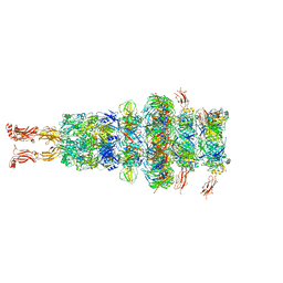 | | Tail tip of siphophage T5 : tip proteins | | 分子名称: | Distal tail protein, L-shaped tail fiber protein p132, Minor tail protein, ... | | 著者 | Linares, R, Arnaud, C.A, Effantin, G, Darnault, C, Epalle, N, Boeri Erba, E, Schoehn, G, Breyton, C. | | 登録日 | 2022-04-06 | | 公開日 | 2023-02-08 | | 最終更新日 | 2023-07-05 | | 実験手法 | ELECTRON MICROSCOPY (3.53 Å) | | 主引用文献 | Structural basis of bacteriophage T5 infection trigger and E. coli cell wall perforation.
Sci Adv, 9, 2023
|
|
7ZQP
 | | Tail tip of siphophage T5 : open cone after interaction with bacterial receptor FhuA | | 分子名称: | Probable baseplate hub protein, Probable tape measure protein | | 著者 | Linares, R, Arnaud, C.A, Effantin, G, Darnault, C, Epalle, N, Boeri Erba, E, Schoehn, G, Breyton, C. | | 登録日 | 2022-05-02 | | 公開日 | 2023-02-08 | | 最終更新日 | 2023-07-05 | | 実験手法 | ELECTRON MICROSCOPY (3.6 Å) | | 主引用文献 | Structural basis of bacteriophage T5 infection trigger and E. coli cell wall perforation.
Sci Adv, 9, 2023
|
|
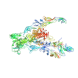
7ZN2
 | | Tail tip of siphophage T5 : full complex after interaction with its bacterial receptor FhuA | | 分子名称: | Distal tail protein, L-shaped tail fiber protein p132, Minor tail protein, ... | | 著者 | Linares, R, Arnaud, C.A, Effantin, G, Darnault, C, Epalle, N, Boeri Erba, E, Schoehn, G, Breyton, C. | | 登録日 | 2022-04-20 | | 公開日 | 2023-02-08 | | 最終更新日 | 2023-07-05 | | 実験手法 | ELECTRON MICROSCOPY (4.29 Å) | | 主引用文献 | Structural basis of bacteriophage T5 infection trigger and E. coli cell wall perforation.
Sci Adv, 9, 2023
|
|
7ZN4
 | | Tail tip of siphophage T5 : bent fibre after interaction with its bacterial receptor FhuA | | 分子名称: | Probable baseplate hub protein, Probable central straight fiber | | 著者 | Linares, R, Arnaud, C.A, Effantin, G, Darnault, C, Epalle, N, Boeri Erba, E, Schoehn, G, Breyton, C. | | 登録日 | 2022-04-20 | | 公開日 | 2023-02-08 | | 最終更新日 | 2023-07-05 | | 実験手法 | ELECTRON MICROSCOPY (4.32 Å) | | 主引用文献 | Structural basis of bacteriophage T5 infection trigger and E. coli cell wall perforation.
Sci Adv, 9, 2023
|
|
7ZLV
 | | Tail tip of siphophage T5 : central fibre protein pb4 | | 分子名称: | Probable central straight fiber | | 著者 | Linares, R, Arnaud, C.A, Effantin, G, Epalle, N, Boeri Erba, E, Schoehn, G, Breyton, C. | | 登録日 | 2022-04-15 | | 公開日 | 2023-02-08 | | 最終更新日 | 2023-07-05 | | 実験手法 | ELECTRON MICROSCOPY (4.22 Å) | | 主引用文献 | Structural basis of bacteriophage T5 infection trigger and E. coli cell wall perforation.
Sci Adv, 9, 2023
|
|
6FI9
 | |
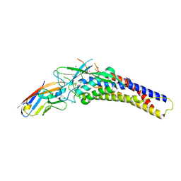
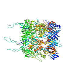
6VRZ
 | | protein A | | 分子名称: | CHLORAMPHENICOL, Multidrug transporter MdfA, PRASEODYMIUM ION | | 著者 | Lu, M, Lu, M.M. | | 登録日 | 2020-02-10 | | 公開日 | 2020-06-03 | | 最終更新日 | 2024-03-06 | | 実験手法 | X-RAY DIFFRACTION (2 Å) | | 主引用文献 | Structure and mechanism of a redesigned multidrug transporter from the Major Facilitator Superfamily.
Sci Rep, 10, 2020
|
|
6BKW
 | | BTK complex with compound 12 | | 分子名称: | GLYCEROL, N-(3-{5-[(1,5-dimethyl-1H-pyrazol-3-yl)amino]-1-methyl-6-oxo-1,6-dihydropyridazin-3-yl}-2,6-difluorophenyl)-4,5,6,7-tetrahydro-1-benzothiophene-2-carboxamide, SULFATE ION, ... | | 著者 | Kiefer, J.R, Eigenbrot, C, Yu, C.L. | | 登録日 | 2017-11-09 | | 公開日 | 2018-11-07 | | 最終更新日 | 2019-03-27 | | 実験手法 | X-RAY DIFFRACTION (1.499 Å) | | 主引用文献 | Water molecules in protein-ligand interfaces. Evaluation of software tools and SAR comparison.
J. Comput. Aided Mol. Des., 33, 2019
|
|
6BLN
 | | BTK complex with compound 13 | | 分子名称: | GLYCEROL, N-(3-{5-[(1,5-dimethyl-1H-pyrazol-3-yl)amino]-6-oxo-1,6-dihydropyridazin-3-yl}-2,6-difluorophenyl)-4,5,6,7-tetrahydro-1-benzothiophene-2-carboxamide, SULFATE ION, ... | | 著者 | Kiefer, J.R, Eigenbrot, C, Yu, C.L. | | 登録日 | 2017-11-10 | | 公開日 | 2018-11-07 | | 最終更新日 | 2019-03-27 | | 実験手法 | X-RAY DIFFRACTION (1.3 Å) | | 主引用文献 | Water molecules in protein-ligand interfaces. Evaluation of software tools and SAR comparison.
J. Comput. Aided Mol. Des., 33, 2019
|
|
6BQD
 | | TAF1-BD2 bromodomain in complex with (E)-3-(6-(but-2-en-1-yl)-7-oxo-6,7-dihydro-1H-pyrrolo[2,3-c]pyridin-4-yl)-N,N-dimethylbenzamide | | 分子名称: | 3-{6-[(2E)-but-2-en-1-yl]-7-oxo-6,7-dihydro-1H-pyrrolo[2,3-c]pyridin-4-yl}-N,N-dimethylbenzamide, Transcription initiation factor TFIID subunit 1 | | 著者 | Murray, J.M, Tang, Y. | | 登録日 | 2017-11-27 | | 公開日 | 2019-02-20 | | 最終更新日 | 2023-10-04 | | 実験手法 | X-RAY DIFFRACTION (2.136 Å) | | 主引用文献 | Water molecules in protein-ligand interfaces. Evaluation of software tools and SAR comparison.
J. Comput. Aided Mol. Des., 33, 2019
|
|
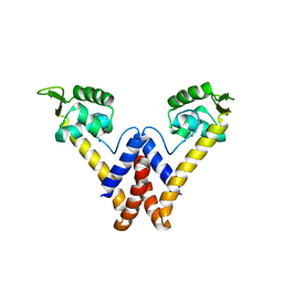
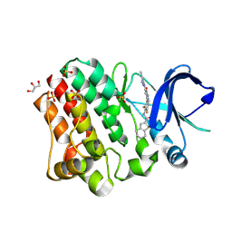
1BOB
 | | HISTONE ACETYLTRANSFERASE HAT1 FROM SACCHAROMYCES CEREVISIAE IN COMPLEX WITH ACETYL COENZYME A | | 分子名称: | ACETYL COENZYME *A, CALCIUM ION, HISTONE ACETYLTRANSFERASE | | 著者 | Dutnall, R.N, Tafrov, S.T, Sternglanz, R, Ramakrishnan, V. | | 登録日 | 1998-07-02 | | 公開日 | 1999-04-20 | | 最終更新日 | 2024-02-07 | | 実験手法 | X-RAY DIFFRACTION (2.3 Å) | | 主引用文献 | Structure of the histone acetyltransferase Hat1: a paradigm for the GCN5-related N-acetyltransferase superfamily.
Cell(Cambridge,Mass.), 94, 1998
|
|


