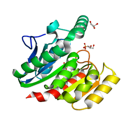
1ZBF
 
 | | Crystal structure of B. halodurans RNase H catalytic domain mutant D132N | | 分子名称: | SULFATE ION, ribonuclease H-related protein | | 著者 | Nowotny, M, Gaidamakov, S.A, Crouch, R.J, Yang, W. | | 登録日 | 2005-04-08 | | 公開日 | 2005-07-12 | | 最終更新日 | 2024-02-14 | | 実験手法 | X-RAY DIFFRACTION (1.5 Å) | | 主引用文献 | Crystal Structures of RNase H Bound to an RNA/DNA Hybrid: Substrate Specificity and Metal-Dependent Catalysis.
Cell(Cambridge,Mass.), 121, 2005
|
|
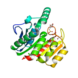
1ZHW
 | | Structure of yeast oxysterol binding protein Osh4 in complex with 20-hydroxycholesterol | | 分子名称: | 20-HYDROXYCHOLESTEROL, KES1 protein, LEAD (II) ION | | 著者 | Im, Y.J, Raychaudhuri, S, Prinz, W.A, Hurley, J.H. | | 登録日 | 2005-04-26 | | 公開日 | 2005-09-06 | | 最終更新日 | 2023-08-23 | | 実験手法 | X-RAY DIFFRACTION (1.7 Å) | | 主引用文献 | Structural mechanism for sterol sensing and transport by OSBP-related proteins
Nature, 437, 2005
|
|
1ZI7
 | | Structure of truncated yeast oxysterol binding protein Osh4 | | 分子名称: | KES1 protein, SULFATE ION | | 著者 | Im, Y.J, Raychaudhuri, S, Prinz, W.A, Hurley, J.H. | | 登録日 | 2005-04-27 | | 公開日 | 2005-09-06 | | 最終更新日 | 2023-08-23 | | 実験手法 | X-RAY DIFFRACTION (2.5 Å) | | 主引用文献 | Structural mechanism for sterol sensing and transport by OSBP-related proteins
Nature, 437, 2005
|
|
2F95
 | | M intermediate structure of sensory rhodopsin II/transducer complex in combination with the ground state structure | | 分子名称: | RETINAL, Sensory rhodopsin II, Sensory rhodopsin II transducer, ... | | 著者 | Moukhametzianov, R.I, Klare, J.P, Efremov, R.G, Baecken, C, Goeppner, A, Labahn, J, Engelhard, M, Bueldt, G, Gordeliy, V.I. | | 登録日 | 2005-12-05 | | 公開日 | 2006-03-07 | | 最終更新日 | 2023-08-30 | | 実験手法 | X-RAY DIFFRACTION (2.2 Å) | | 主引用文献 | Development of the signal in sensory rhodopsin and its transfer to the cognate transducer.
Nature, 440, 2006
|
|
1ZHX
 | | Structure of yeast oxysterol binding protein Osh4 in complex with 25-hydroxycholesterol | | 分子名称: | 25-HYDROXYCHOLESTEROL, KES1 protein | | 著者 | Im, Y.J, Raychaudhuri, S, Prinz, W.A, Hurley, J.H. | | 登録日 | 2005-04-26 | | 公開日 | 2005-09-06 | | 最終更新日 | 2024-02-14 | | 実験手法 | X-RAY DIFFRACTION (1.5 Å) | | 主引用文献 | Structural mechanism for sterol sensing and transport by OSBP-related proteins
Nature, 437, 2005
|
|
1ZI9
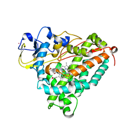
 | | Crystal Structure Analysis of the dienelactone hydrolase (E36D, C123S) mutant- 1.5 A | | 分子名称: | Carboxymethylenebutenolidase, GLYCEROL, SULFATE ION | | 著者 | Kim, H.-K, Liu, J.-W, Carr, P.D, Ollis, D.L. | | 登録日 | 2005-04-27 | | 公開日 | 2005-07-05 | | 最終更新日 | 2023-10-25 | | 実験手法 | X-RAY DIFFRACTION (1.5 Å) | | 主引用文献 | Following directed evolution with crystallography: structural changes observed in changing the substrate specificity of dienelactone hydrolase.
Acta Crystallogr.,Sect.D, 61, 2005
|
|
1ZJ4
 | | Crystal Structure Analysis of the dienelactone hydrolase mutant (E36D, C123S) bound with the PMS moiety of the protease inhibitor, Phenylmethylsulfonyl fluoride (PMSF)- 1.7 A | | 分子名称: | Carboxymethylenebutenolidase, GLYCEROL, SULFATE ION | | 著者 | Kim, H.-K, Liu, J.-W, Carr, P.D, Ollis, D.L. | | 登録日 | 2005-04-28 | | 公開日 | 2005-07-05 | | 最終更新日 | 2023-10-25 | | 実験手法 | X-RAY DIFFRACTION (1.7 Å) | | 主引用文献 | Following directed evolution with crystallography: structural changes observed in changing the substrate specificity of dienelactone hydrolase.
Acta Crystallogr.,Sect.D, 61, 2005
|
|
2FCY
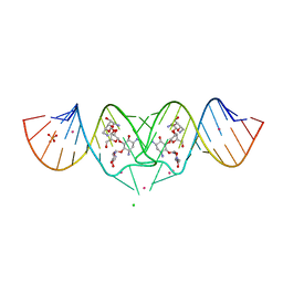 | | HIV-1 DIS kissing-loop in complex with Neomycin | | 分子名称: | CHLORIDE ION, HIV-1 DIS RNA, NEOMYCIN, ... | | 著者 | Ennifar, E, Paillart, J.C, Marquet, R, Dumas, P. | | 登録日 | 2005-12-13 | | 公開日 | 2006-05-16 | | 最終更新日 | 2023-08-30 | | 実験手法 | X-RAY DIFFRACTION (2.2 Å) | | 主引用文献 | Targeting the dimerization initiation site of HIV-1 RNA with aminoglycosides: from crystal to cell.
Nucleic Acids Res., 34, 2006
|
|
4K2J
 | |
1ZML
 | | Crystal Structure of the Catalytic Domain of Factor XI in complex with (R)-1-(4-(4-(hydroxymethyl)-1,3,2-dioxaborolan-2-yl)phenethyl)guanidine | | 分子名称: | (R)-1-(4-(4-(HYDROXYMETHYL)-1,3,2-DIOXABOROLAN-2-YL)PHENETHYL)GUANIDINE, BICARBONATE ION, Coagulation factor XI, ... | | 著者 | Lazarova, T.I, Jin, L, Rynkiewicz, M.J, Gorga, J.C, Bibbins, F, Meyers, H.V, Babine, R.E, Strickler, J.E. | | 登録日 | 2005-05-10 | | 公開日 | 2006-05-09 | | 最終更新日 | 2023-08-23 | | 実験手法 | X-RAY DIFFRACTION (2.25 Å) | | 主引用文献 | Synthesis and in vitro biological evaluation of aryl boronic acids as potential inhibitors of factor XIa.
Bioorg.Med.Chem.Lett., 16, 2006
|
|
1Z38
 | | Crystal structure of Trichomonas vaginalis purine nucleoside phosphorylase complexed with inosine | | 分子名称: | INOSINE, purine nucleoside phosphorylase | | 著者 | Zhang, Y, Wang, W.H, Wu, S.W, Wang, C.C, Ealick, S.E. | | 登録日 | 2005-03-10 | | 公開日 | 2005-03-29 | | 最終更新日 | 2023-08-23 | | 実験手法 | X-RAY DIFFRACTION (2.5 Å) | | 主引用文献 | Identification of a subversive substrate of Trichomonas vaginalis purine nucleoside phosphorylase and the crystal structure of the enzyme-substrate complex.
J.Biol.Chem., 280, 2005
|
|
1ZP6
 | | Crystal Structure of Atu3015, a Putative Cytidylate Kinase from Agrobacterium tumefaciens, Northeast Structural Genomics Target AtR62 | | 分子名称: | SULFATE ION, hypothetical protein Atu3015 | | 著者 | Forouhar, F, Abashidze, M, Vorobiev, S.M, Kuzin, A, Conover, K, Acton, T.B, Montelione, G.T, Hunt, J.F, Tong, L, Northeast Structural Genomics Consortium (NESG) | | 登録日 | 2005-05-16 | | 公開日 | 2005-05-24 | | 最終更新日 | 2017-10-11 | | 実験手法 | X-RAY DIFFRACTION (3.2 Å) | | 主引用文献 | Crystal Structure of Atu3015, a Putative Cytidylate Kinase from Agrobacterium tumefaciens, Northeast Structural Genomics Target AtR62
To be Published
|
|
1ZPB
 | | Crystal Structure of the Catalytic Domain of Coagulation Factor XI in Complex with 4-Methyl-pentanoic acid {1-[4-guanidino-1-(thiazole-2-carbonyl)-butylcarbamoyl]-2-methyl-propyl}-amide | | 分子名称: | 4-METHYL-PENTANOIC ACID {1-[4-GUANIDINO-1-(THIAZOLE-2-CARBONYL)-BUTYLCARBAMOYL]-2-METHYL-PROPYL}-AMIDE, Coagulation factor XI, SULFATE ION | | 著者 | Deng, H, Bannister, T.D, Jin, L, Nagafuji, P, Celatka, C.A, Lin, J, Lazarova, T.I, Rynkiewicz, M.J, Quinn, J, Bibbins, F, Pandey, P, Gorga, J, Babine, R.E, Meyers, H.V, Abdel-Meguid, S.S, Strickler, J.E. | | 登録日 | 2005-05-16 | | 公開日 | 2006-04-18 | | 最終更新日 | 2023-08-23 | | 実験手法 | X-RAY DIFFRACTION (2.1 Å) | | 主引用文献 | Synthesis, SAR exploration, and X-ray crystal structures of factor XIa inhibitors containing an alpha-ketothiazole arginine
Bioorg.Med.Chem.Lett., 16, 2006
|
|
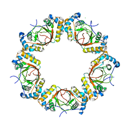
1ZA7
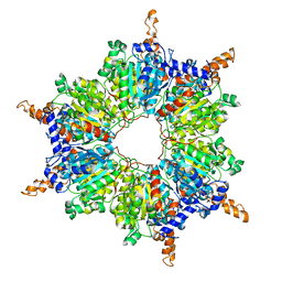
 | | The crystal structure of salt stable cowpea cholorotic mottle virus at 2.7 angstroms resolution. | | 分子名称: | Coat protein | | 著者 | Bothner, B, Speir, J.A, Qu, C, Willits, D.A, Young, M.J, Johnson, J.E. | | 登録日 | 2005-04-05 | | 公開日 | 2006-03-21 | | 最終更新日 | 2023-08-23 | | 実験手法 | X-RAY DIFFRACTION (2.7 Å) | | 主引用文献 | Enhanced local symmetry interactions globally stabilize a mutant virus capsid that maintains infectivity and capsid dynamics.
J.Virol., 80, 2006
|
|
1ZBP
 | | X-Ray Crystal Structure of Protein VPA1032 from Vibrio parahaemolyticus. Northeast Structural Genomics Consortium Target VpR44 | | 分子名称: | hypothetical protein VPA1032 | | 著者 | Forouhar, F, Yong, W, Vorobiev, S.M, Ciao, M, Acton, T.B, Montelione, G.T, Hunt, J.F, Tong, L, Northeast Structural Genomics Consortium (NESG) | | 登録日 | 2005-04-08 | | 公開日 | 2005-04-19 | | 最終更新日 | 2017-10-11 | | 実験手法 | X-RAY DIFFRACTION (2.4 Å) | | 主引用文献 | Functional insights from structural genomics.
J.STRUCT.FUNCT.GENOM., 8, 2007
|
|
1ZDL
 | | Crystal Structure of Mouse Thioredoxin Reductase Type 2 | | 分子名称: | FLAVIN-ADENINE DINUCLEOTIDE, NADPH DIHYDRO-NICOTINAMIDE-ADENINE-DINUCLEOTIDE PHOSPHATE, Thioredoxin reductase 2, ... | | 著者 | Biterova, E.I, Turanov, A.A, Gladyshev, V.N, Barycki, J.J. | | 登録日 | 2005-04-14 | | 公開日 | 2005-11-01 | | 最終更新日 | 2023-08-23 | | 実験手法 | X-RAY DIFFRACTION (3 Å) | | 主引用文献 | Crystal structures of oxidized and reduced mitochondrial thioredoxin reductase provide molecular details of the reaction mechanism.
Proc.Natl.Acad.Sci.Usa, 102, 2005
|
|
1Z35
 | | Crystal structure of Trichomonas vaginalis purine nucleoside phosphorylase complexed with 2-fluoroadenosine | | 分子名称: | 2-(6-AMINO-2-FLUORO-PURIN-9-YL)-5-HYDROXYMETHYL-TETRAHYDRO-FURAN-3,4-DIOL, purine nucleoside phosphorylase | | 著者 | Zhang, Y, Wang, W.H, Wu, S.W, Wang, C.C, Ealick, S.E. | | 登録日 | 2005-03-10 | | 公開日 | 2005-03-29 | | 最終更新日 | 2023-08-23 | | 実験手法 | X-RAY DIFFRACTION (2.5 Å) | | 主引用文献 | Identification of a subversive substrate of Trichomonas vaginalis purine nucleoside phosphorylase and the crystal structure of the enzyme-substrate complex.
J.Biol.Chem., 280, 2005
|
|
1ZHR
 | | Crystal Structure of the Catalytic Domain of Coagulation Factor XI in Complex with Benzamidine (S434A-T475A-C482S-K437A Mutant) | | 分子名称: | BENZAMIDINE, coagulation factor XI | | 著者 | Jin, L, Pandey, P, Babine, R.E, Weaver, D.T, Abdel-Meguid, S.S, Strickler, J.E. | | 登録日 | 2005-04-26 | | 公開日 | 2005-09-20 | | 最終更新日 | 2023-08-23 | | 実験手法 | X-RAY DIFFRACTION (1.73 Å) | | 主引用文献 | Mutation of surface residues to promote crystallization of activated factor XI as a complex with benzamidine: an essential step for the iterative structure-based design of factor XI inhibitors.
Acta Crystallogr.,Sect.D, 61, 2005
|
|
1ZIX
 | | Crystal Structure Analysis of the dienelactone hydrolase mutant (E36D, R105H, C123S, G211D, K234N)- 1.8 A | | 分子名称: | Carboxymethylenebutenolidase, GLYCEROL | | 著者 | Kim, H.-K, Liu, J.-W, Carr, P.D, Ollis, D.L. | | 登録日 | 2005-04-27 | | 公開日 | 2005-07-05 | | 最終更新日 | 2023-10-25 | | 実験手法 | X-RAY DIFFRACTION (1.8 Å) | | 主引用文献 | Following directed evolution with crystallography: structural changes observed in changing the substrate specificity of dienelactone hydrolase.
Acta Crystallogr.,Sect.D, 61, 2005
|
|
2ACQ
 | | AN ANION BINDING SITE IN HUMAN ALDOSE REDUCTASE: MECHANISTIC IMPLICATIONS FOR THE BINDING OF CITRATE, CACODYLATE, AND GLUCOSE-6-PHOSPHATE | | 分子名称: | 6-O-phosphono-alpha-D-glucopyranose, ALDOSE REDUCTASE, NADP NICOTINAMIDE-ADENINE-DINUCLEOTIDE PHOSPHATE | | 著者 | Harrison, D.H, Bohren, K.M, Gabbay, K.H, Petsko, G.A, Ringe, D. | | 登録日 | 1994-04-15 | | 公開日 | 1994-07-31 | | 最終更新日 | 2024-02-14 | | 実験手法 | X-RAY DIFFRACTION (1.76 Å) | | 主引用文献 | An anion binding site in human aldose reductase: mechanistic implications for the binding of citrate, cacodylate, and glucose 6-phosphate.
Biochemistry, 33, 1994
|
|
2DST
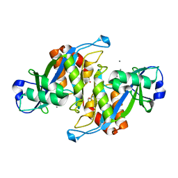
 | | Crystal Structure Analysis of TT1977 | | 分子名称: | Hypothetical protein TTHA1544 | | 著者 | Xie, Y, Kishishita, S, Murayama, K, Shirouzu, M, Chen, L, Liu, Z.J, Wang, B.C, RIKEN Structural Genomics/Proteomics Initiative (RSGI) | | 登録日 | 2006-07-06 | | 公開日 | 2007-01-06 | | 最終更新日 | 2024-03-13 | | 実験手法 | X-RAY DIFFRACTION (2 Å) | | 主引用文献 | Structure of the minimized alpha/beta-hydrolase fold protein from Thermus thermophilus HB8.
Acta Crystallogr.,Sect.F, 63, 2007
|
|
2A1N
 | |
2A6E
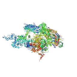 | | Crystal structure of the T. Thermophilus RNA polymerase holoenzyme | | 分子名称: | DNA-directed RNA polymerase alpha chain, DNA-directed RNA polymerase beta chain, DNA-directed RNA polymerase beta' chain, ... | | 著者 | Artsimovitch, I, Vassylyeva, M.N, Svetlov, D, Svetlov, V, Perederina, A, Igarashi, N, Matsugaki, N, Wakatsuki, S, Tahirov, T.H, Vassylyev, D.G, RIKEN Structural Genomics/Proteomics Initiative (RSGI) | | 登録日 | 2005-07-02 | | 公開日 | 2005-09-20 | | 最終更新日 | 2023-08-23 | | 実験手法 | X-RAY DIFFRACTION (2.8 Å) | | 主引用文献 | Allosteric modulation of the RNA polymerase catalytic reaction is an essential component of transcription control by rifamycins.
Cell(Cambridge,Mass.), 122, 2005
|
|
2A7S
 | | Crystal Structure of the Acyl-CoA Carboxylase, AccD5, from Mycobacterium tuberculosis | | 分子名称: | Probable propionyl-CoA carboxylase beta chain 5 | | 著者 | Lin, T, Melgar, M, Purdon, J, Tseng, T, Tsai, S.C. | | 登録日 | 2005-07-06 | | 公開日 | 2006-02-21 | | 最終更新日 | 2024-04-03 | | 実験手法 | X-RAY DIFFRACTION (2.9 Å) | | 主引用文献 | Structure-based inhibitor design of AccD5, an essential acyl-CoA carboxylase carboxyltransferase domain of Mycobacterium tuberculosis.
Proc.Natl.Acad.Sci.Usa, 103, 2006
|
|
2A8P
 | | 2.7 Angstrom Crystal Structure of the Complex Between the Nuclear SnoRNA Decapping Nudix Hydrolase X29 and Manganese | | 分子名称: | MANGANESE (II) ION, U8 snoRNA-binding protein X29 | | 著者 | Scarsdale, J.N, Peculis, B.A, Wright, H.T. | | 登録日 | 2005-07-08 | | 公開日 | 2006-03-28 | | 最終更新日 | 2023-08-23 | | 実験手法 | X-RAY DIFFRACTION (2.7 Å) | | 主引用文献 | Crystal structures of U8 snoRNA decapping nudix hydrolase, X29, and its metal and cap complexes
Structure, 14, 2006
|
|


