7O1L
 
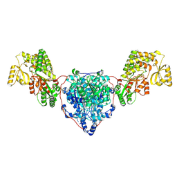
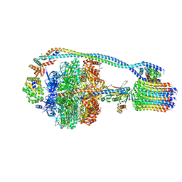
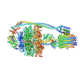
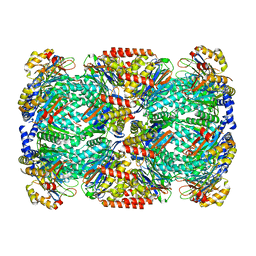
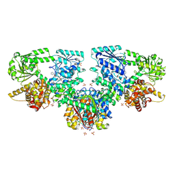
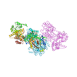 | | Structure of Mycobacterium tuberculosis beta-oxidation trifunctional enzyme alpha-H462A mutant | | 分子名称: | 3-hydroxyacyl-CoA dehydrogenase, COENZYME A, GLYCEROL, ... | | 著者 | Dalwani, S, Wierenga, R.K, Venkatesan, R. | | 登録日 | 2021-03-29 | | 公開日 | 2021-08-25 | | 最終更新日 | 2024-01-31 | | 実験手法 | X-RAY DIFFRACTION (2.38 Å) | | 主引用文献 | Substrate specificity and conformational flexibility properties of the Mycobacterium tuberculosis beta-oxidation trifunctional enzyme.
J.Struct.Biol., 213, 2021
|
|
7O1J
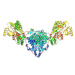
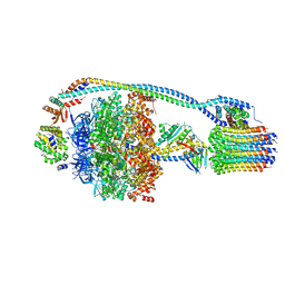
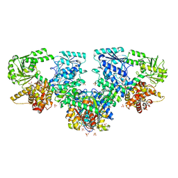
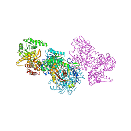 | | Structure of Mycobacterium tuberculosis beta-oxidation trifunctional enzyme beta-C92A mutant | | 分子名称: | 3-hydroxyacyl-CoA dehydrogenase, GLYCEROL, Putative acyltransferase Rv0859, ... | | 著者 | Dalwani, S, Wierenga, R.K, Venkatesan, R. | | 登録日 | 2021-03-29 | | 公開日 | 2021-08-25 | | 最終更新日 | 2024-01-31 | | 実験手法 | X-RAY DIFFRACTION (2.36 Å) | | 主引用文献 | Substrate specificity and conformational flexibility properties of the Mycobacterium tuberculosis beta-oxidation trifunctional enzyme.
J.Struct.Biol., 213, 2021
|
|
7O1M
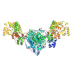
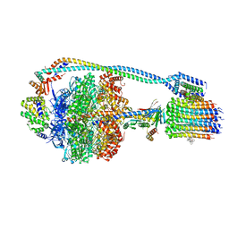
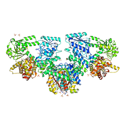
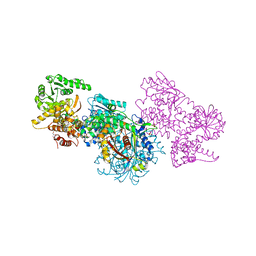 | | Structure of Mycobacterium tuberculosis beta-oxidation trifunctional enzyme alpha-H462A, beta-C92A mutant | | 分子名称: | 3-hydroxyacyl-CoA dehydrogenase, GLYCEROL, Putative acyltransferase Rv0859, ... | | 著者 | Dalwani, S, Wierenga, R.K, Venkatesan, R. | | 登録日 | 2021-03-29 | | 公開日 | 2021-08-25 | | 最終更新日 | 2024-01-31 | | 実験手法 | X-RAY DIFFRACTION (2.89 Å) | | 主引用文献 | Substrate specificity and conformational flexibility properties of the Mycobacterium tuberculosis beta-oxidation trifunctional enzyme.
J.Struct.Biol., 213, 2021
|
|
8CUY
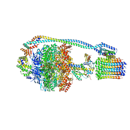
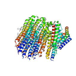
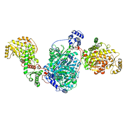 | | ACP1-KS-AT domains of mycobacterial Pks13 | | 分子名称: | 4'-PHOSPHOPANTETHEINE, Polyketide synthase PKS13, UNKNOWN LIGAND | | 著者 | Kim, S.K, Dickinson, M.S, Finer-Moore, J.S, Rosenberg, O.S, Stroud, R.M. | | 登録日 | 2022-05-17 | | 公開日 | 2023-02-15 | | 最終更新日 | 2023-03-29 | | 実験手法 | ELECTRON MICROSCOPY (2.4 Å) | | 主引用文献 | Structure and dynamics of the essential endogenous mycobacterial polyketide synthase Pks13.
Nat.Struct.Mol.Biol., 30, 2023
|
|
8CV0
 | | KS-AT domains of mycobacterial Pks13 with outward AT conformation | | 分子名称: | Polyketide synthase PKS13, UNKNOWN LIGAND | | 著者 | Kim, S.K, Dickinson, M.S, Finer-Moore, J.S, Rosenberg, O.S, Stroud, R.M. | | 登録日 | 2022-05-17 | | 公開日 | 2023-02-15 | | 最終更新日 | 2023-03-29 | | 実験手法 | ELECTRON MICROSCOPY (3.1 Å) | | 主引用文献 | Structure and dynamics of the essential endogenous mycobacterial polyketide synthase Pks13.
Nat.Struct.Mol.Biol., 30, 2023
|
|
8CUZ
 | | KS-AT domains of mycobacterial Pks13 with inward AT conformation | | 分子名称: | Polyketide synthase PKS13, UNKNOWN LIGAND | | 著者 | Kim, S.K, Dickinson, M.S, Finer-Moore, J.S, Rosenberg, O.S, Stroud, R.M. | | 登録日 | 2022-05-17 | | 公開日 | 2023-02-15 | | 最終更新日 | 2023-03-29 | | 実験手法 | ELECTRON MICROSCOPY (3 Å) | | 主引用文献 | Structure and dynamics of the essential endogenous mycobacterial polyketide synthase Pks13.
Nat.Struct.Mol.Biol., 30, 2023
|
|
8CV1
 | | ACP1-KS-AT domains of mycobacterial Pks13 | | 分子名称: | Polyketide synthase PKS13, UNKNOWN LIGAND | | 著者 | Kim, S.K, Dickinson, M.S, Finer-Moore, J.S, Rosenberg, O.S, Stroud, R.M. | | 登録日 | 2022-05-17 | | 公開日 | 2023-02-15 | | 最終更新日 | 2023-03-29 | | 実験手法 | ELECTRON MICROSCOPY (2.6 Å) | | 主引用文献 | Structure and dynamics of the essential endogenous mycobacterial polyketide synthase Pks13.
Nat.Struct.Mol.Biol., 30, 2023
|
|
6ZT4
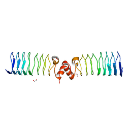 | | Pentapeptide repeat protein MfpA from Mycobacterium smegmatis | | 分子名称: | 1,2-ETHANEDIOL, Pentapeptide repeat protein MfpA | | 著者 | Feng, L, Mundy, J.E.A, Stevenson, C.E.M, Mitchenall, L.A, Lawson, D.M, Mi, K, Maxwell, A. | | 登録日 | 2020-07-17 | | 公開日 | 2021-03-03 | | 最終更新日 | 2024-01-31 | | 実験手法 | X-RAY DIFFRACTION (1.77 Å) | | 主引用文献 | The pentapeptide-repeat protein, MfpA, interacts with mycobacterial DNA gyrase as a DNA T-segment mimic.
Proc.Natl.Acad.Sci.USA, 118, 2021
|
|
7JGA
 | | Cryo-EM structure of bedaquiline-saturated Mycobacterium smegmatis ATP synthase rotational state 3 | | 分子名称: | ADENOSINE-5'-DIPHOSPHATE, ADENOSINE-5'-TRIPHOSPHATE, ATP synthase epsilon chain, ... | | 著者 | Guo, H, Courbon, G.M, Rubinstein, J.L. | | 登録日 | 2020-07-18 | | 公開日 | 2020-08-19 | | 最終更新日 | 2024-03-06 | | 実験手法 | ELECTRON MICROSCOPY (3.2 Å) | | 主引用文献 | Structure of mycobacterial ATP synthase bound to the tuberculosis drug bedaquiline.
Nature, 589, 2021
|
|
7JG6
 | | Cryo-EM structure of bedaquiline-free Mycobacterium smegmatis ATP synthase rotational state 2 (backbone model) | | 分子名称: | ATP synthase epsilon chain, ATP synthase gamma chain, ATP synthase subunit a, ... | | 著者 | Guo, H, Courbon, G.M, Rubinstein, J.L. | | 登録日 | 2020-07-18 | | 公開日 | 2020-08-19 | | 最終更新日 | 2024-03-06 | | 実験手法 | ELECTRON MICROSCOPY (3.7 Å) | | 主引用文献 | Structure of mycobacterial ATP synthase bound to the tuberculosis drug bedaquiline.
Nature, 589, 2021
|
|
7JG5
 | | Cryo-EM structure of bedaquiline-free Mycobacterium smegmatis ATP synthase rotational state 1 | | 分子名称: | ADENOSINE-5'-DIPHOSPHATE, ADENOSINE-5'-TRIPHOSPHATE, ATP synthase epsilon chain, ... | | 著者 | Guo, H, Courbon, G.M, Rubinstein, J.L. | | 登録日 | 2020-07-18 | | 公開日 | 2020-08-19 | | 最終更新日 | 2024-03-06 | | 実験手法 | ELECTRON MICROSCOPY (3.4 Å) | | 主引用文献 | Structure of mycobacterial ATP synthase bound to the tuberculosis drug bedaquiline.
Nature, 589, 2021
|
|
7JG9
 | | Cryo-EM structure of bedaquiline-saturated mycobacterium smegmatis ATP synthase rotational state 2 (backbone model) | | 分子名称: | ATP synthase epsilon chain, ATP synthase gamma chain, ATP synthase subunit a, ... | | 著者 | Guo, H, Courbon, G.M, Rubinstein, J.L. | | 登録日 | 2020-07-18 | | 公開日 | 2020-08-19 | | 最終更新日 | 2024-03-06 | | 実験手法 | ELECTRON MICROSCOPY (3.4 Å) | | 主引用文献 | Structure of mycobacterial ATP synthase bound to the tuberculosis drug bedaquiline.
Nature, 589, 2021
|
|
7JG7
 | | Cryo-EM structure of bedaquiline-free Mycobacterium smegmatis ATP synthase rotational state 3 (backbone model) | | 分子名称: | ATP synthase epsilon chain, ATP synthase gamma chain, ATP synthase subunit a, ... | | 著者 | Guo, H, Courbon, G.M, Rubinstein, J.L. | | 登録日 | 2020-07-18 | | 公開日 | 2020-08-19 | | 最終更新日 | 2024-03-06 | | 実験手法 | ELECTRON MICROSCOPY (3.5 Å) | | 主引用文献 | Structure of mycobacterial ATP synthase bound to the tuberculosis drug bedaquiline.
Nature, 589, 2021
|
|
7JG8
 | | Cryo-EM structure of bedaquiline-saturated Mycobacterium smegmatis ATP synthase rotational state 1 (backbone model) | | 分子名称: | ATP synthase epsilon chain, ATP synthase gamma chain, ATP synthase subunit a, ... | | 著者 | Guo, H, Courbon, G.M, Rubinstein, J.L. | | 登録日 | 2020-07-18 | | 公開日 | 2020-08-19 | | 最終更新日 | 2024-03-06 | | 実験手法 | ELECTRON MICROSCOPY (3.3 Å) | | 主引用文献 | Structure of mycobacterial ATP synthase bound to the tuberculosis drug bedaquiline.
Nature, 589, 2021
|
|
6DZK
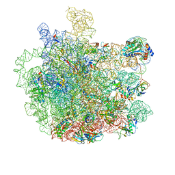
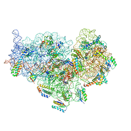 | | Cryo-EM Structure of Mycobacterium smegmatis C(minus) 30S ribosomal subunit with MPY | | 分子名称: | 16S rRNA, 30S ribosomal protein S10, 30S ribosomal protein S11, ... | | 著者 | Sharma, M.R, Li, Y, Korripella, R, Yang, Y, Kaushal, P.S, Lin, Q, Wade, J.T, Gray, A.G, Derbyshire, K.M, Agrawal, R.K, Ojha, A. | | 登録日 | 2018-07-05 | | 公開日 | 2018-09-19 | | 最終更新日 | 2024-03-13 | | 実験手法 | ELECTRON MICROSCOPY (3.6 Å) | | 主引用文献 | Zinc depletion induces ribosome hibernation in mycobacteria.
Proc. Natl. Acad. Sci. U.S.A., 115, 2018
|
|
4U5Q
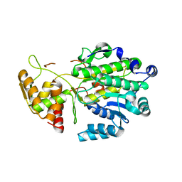 | | High resolution crystal structure of reductase (R) domain of nonribosomal peptide synthetase from Mycobacterium tuberculosis | | 分子名称: | 3,6,9,12,15,18,21,24,27-NONAOXANONACOSANE-1,29-DIOL, Peptide synthetase | | 著者 | Patel, K.D, Haque, A.S, Priyadarshan, K, Sankaranarayanan, R. | | 登録日 | 2014-07-25 | | 公開日 | 2016-01-13 | | 最終更新日 | 2023-11-08 | | 実験手法 | X-RAY DIFFRACTION (1.811 Å) | | 主引用文献 | High resolution structure of R-domain of nonribosomal peptide synthetase from Mycobacterium tuberculosis
To Be Published
|
|
8OQM
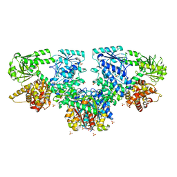
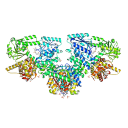 | | Structure of Mycobacterium tuberculosis beta-oxidation trifunctional enzyme in complex with Fragment-M-10 | | 分子名称: | 3-hydroxyacyl-CoA dehydrogenase, 6-[(6-azanyl-4-oxidanyl-naphthalen-2-yl)sulfonylamino]-4-oxidanyl-naphthalene-2-sulfonic acid, 6-azanyl-4-oxidanyl-naphthalene-2-sulfonic acid, ... | | 著者 | Dalwani, S, Wierenga, R.K, Venkatesan, R. | | 登録日 | 2023-04-12 | | 公開日 | 2024-01-24 | | 最終更新日 | 2024-08-14 | | 実験手法 | X-RAY DIFFRACTION (3.2 Å) | | 主引用文献 | Crystallographic fragment-binding studies of the Mycobacterium tuberculosis trifunctional enzyme suggest binding pockets for the tails of the acyl-CoA substrates at its active sites and a potential substrate-channeling path between them.
Acta Crystallogr D Struct Biol, 80, 2024
|
|
6ZT3
 | | N-terminal 47 kDa fragment of the Mycobacterium smegmatis DNA Gyrase B subunit complexed with ADPNP | | 分子名称: | 1,2-ETHANEDIOL, DNA gyrase subunit B, MAGNESIUM ION, ... | | 著者 | Feng, L, Mundy, J.E.A, Stevenson, C.E.M, Mitchenall, L.A, Lawson, D.M, Mi, K, Maxwell, A. | | 登録日 | 2020-07-17 | | 公開日 | 2021-03-03 | | 最終更新日 | 2024-01-31 | | 実験手法 | X-RAY DIFFRACTION (1.56 Å) | | 主引用文献 | The pentapeptide-repeat protein, MfpA, interacts with mycobacterial DNA gyrase as a DNA T-segment mimic.
Proc.Natl.Acad.Sci.USA, 118, 2021
|
|
7PXA
 | |
7JGC
 | | Cryo-EM structure of bedaquiline-saturated Mycobacterium smegmatis ATP synthase FO region | | 分子名称: | ATP synthase subunit a, ATP synthase subunit b, ATP synthase subunit b-delta, ... | | 著者 | Guo, H, Courbon, G.M, Rubinstein, J.L. | | 登録日 | 2020-07-18 | | 公開日 | 2020-08-19 | | 最終更新日 | 2024-03-06 | | 実験手法 | ELECTRON MICROSCOPY (3.4 Å) | | 主引用文献 | Structure of mycobacterial ATP synthase bound to the tuberculosis drug bedaquiline.
Nature, 589, 2021
|
|
5V7Q
 | | Cryo-EM structure of the large ribosomal subunit from Mycobacterium tuberculosis bound with a potent linezolid analog | | 分子名称: | 23S rRNA, 50S ribosomal protein L13, 50S ribosomal protein L14, ... | | 著者 | Yang, K, Chang, J.-Y, Cui, Z, Zhang, J. | | 登録日 | 2017-03-20 | | 公開日 | 2017-09-20 | | 最終更新日 | 2024-03-13 | | 実験手法 | ELECTRON MICROSCOPY (3.7 Å) | | 主引用文献 | Structural insights into species-specific features of the ribosome from the human pathogen Mycobacterium tuberculosis.
Nucleic Acids Res., 45, 2017
|
|
8OPX
 | | Structure of Mycobacterium tuberculosis beta-oxidation trifunctional enzyme in complex with Trehalose (Fragment-B-TRE) | | 分子名称: | 3-hydroxyacyl-CoA dehydrogenase, Putative acyltransferase Rv0859, SULFATE ION, ... | | 著者 | Dalwani, S, Wierenga, R.K, Venkatesan, R. | | 登録日 | 2023-04-10 | | 公開日 | 2024-01-24 | | 最終更新日 | 2024-08-14 | | 実験手法 | X-RAY DIFFRACTION (2.9 Å) | | 主引用文献 | Crystallographic fragment-binding studies of the Mycobacterium tuberculosis trifunctional enzyme suggest binding pockets for the tails of the acyl-CoA substrates at its active sites and a potential substrate-channeling path between them.
Acta Crystallogr D Struct Biol, 80, 2024
|
|
8OQO
 | | Structure of Mycobacterium tuberculosis beta-oxidation trifunctional enzyme in complex with Fragment-M-49 | | 分子名称: | GLYCEROL, Probable fatty oxidation protein FadB, Putative acyltransferase Rv0859, ... | | 著者 | Dalwani, S, Wierenga, R.K, Venkatesan, R. | | 登録日 | 2023-04-12 | | 公開日 | 2024-01-24 | | 最終更新日 | 2024-08-14 | | 実験手法 | X-RAY DIFFRACTION (2.6 Å) | | 主引用文献 | Crystallographic fragment-binding studies of the Mycobacterium tuberculosis trifunctional enzyme suggest binding pockets for the tails of the acyl-CoA substrates at its active sites and a potential substrate-channeling path between them.
Acta Crystallogr D Struct Biol, 80, 2024
|
|
8OQT
 | | Structure of Mycobacterium tuberculosis beta-oxidation trifunctional enzyme in complex with Fragment-M-91 | | 分子名称: | 3-hydroxyacyl-CoA dehydrogenase, 4-bromanylbenzenesulfonic acid, GLYCEROL, ... | | 著者 | Dalwani, S, Wierenga, R.K, Venkatesan, R. | | 登録日 | 2023-04-12 | | 公開日 | 2024-01-24 | | 最終更新日 | 2024-08-28 | | 実験手法 | X-RAY DIFFRACTION (2.62 Å) | | 主引用文献 | Crystallographic fragment-binding studies of the Mycobacterium tuberculosis trifunctional enzyme suggest binding pockets for the tails of the acyl-CoA substrates at its active sites and a potential substrate-channeling path between them.
Acta Crystallogr D Struct Biol, 80, 2024
|
|
8OPU
 | | Structure of Mycobacterium tuberculosis beta-oxidation trifunctional enzyme in complex with Sulfamethoxazole (Fragment-B-E1) | | 分子名称: | 3-hydroxyacyl-CoA dehydrogenase, GLYCEROL, Putative acyltransferase Rv0859, ... | | 著者 | Dalwani, S, Wierenga, R.K, Venkatesan, R. | | 登録日 | 2023-04-10 | | 公開日 | 2024-01-24 | | 最終更新日 | 2024-08-14 | | 実験手法 | X-RAY DIFFRACTION (3.04 Å) | | 主引用文献 | Crystallographic fragment-binding studies of the Mycobacterium tuberculosis trifunctional enzyme suggest binding pockets for the tails of the acyl-CoA substrates at its active sites and a potential substrate-channeling path between them.
Acta Crystallogr D Struct Biol, 80, 2024
|
|


