3TVN
 
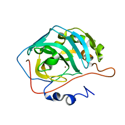 | | Human Carbonic Anhydrase II Proton Transfer Mutant | | 分子名称: | Carbonic anhydrase 2, ZINC ION | | 著者 | Mikulski, R.L, West, D.M, Sippel, K.H, Avvaru, B.S, Chingkuang, T, McKenna, R. | | 登録日 | 2011-09-20 | | 公開日 | 2012-08-08 | | 最終更新日 | 2024-02-28 | | 実験手法 | X-RAY DIFFRACTION (1.497 Å) | | 主引用文献 | Water Networks in Fast Proton Transfer during Catalysis by Human Carbonic Anhydrase II.
Biochemistry, 52, 2013
|
|
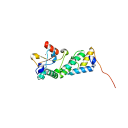
3U43
 
 | |
3W43
 
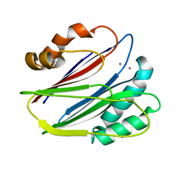 | | Crystal structure of RsbX in complex with manganese in space group P21 | | 分子名称: | MANGANESE (II) ION, Phosphoserine phosphatase RsbX | | 著者 | Teh, A.H, Makino, M, Baba, S, Shimizu, N, Yamamoto, M, Kumasaka, T. | | 登録日 | 2013-01-04 | | 公開日 | 2014-01-22 | | 最終更新日 | 2023-11-08 | | 実験手法 | X-RAY DIFFRACTION (1.22 Å) | | 主引用文献 | Structure of the RsbX phosphatase involved in the general stress response of Bacillus subtilis
Acta Crystallogr.,Sect.D, 71, 2015
|
|
3W57
 
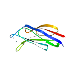 | | Structure of a C2 domain | | 分子名称: | C2 domain protein, CALCIUM ION | | 著者 | Traore, D.A.K, Whisstock, J.C. | | 登録日 | 2013-01-24 | | 公開日 | 2013-10-23 | | 最終更新日 | 2024-10-16 | | 実験手法 | X-RAY DIFFRACTION (1.662 Å) | | 主引用文献 | Defining the interaction of perforin with calcium and the phospholipid membrane.
Biochem.J., 456, 2013
|
|
3W45
 
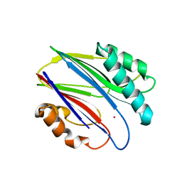 | | Crystal structure of RsbX in complex with cobalt in space group P1 | | 分子名称: | COBALT (II) ION, Phosphoserine phosphatase RsbX | | 著者 | Makino, M, Teh, A.H, Baba, S, Shimizu, N, Yamamoto, M, Kumasaka, T. | | 登録日 | 2013-01-04 | | 公開日 | 2014-01-22 | | 最終更新日 | 2024-03-20 | | 実験手法 | X-RAY DIFFRACTION (1.7 Å) | | 主引用文献 | Structure of the RsbX phosphatase involved in the general stress response of Bacillus subtilis
Acta Crystallogr.,Sect.D, 71, 2015
|
|
3OB8
 
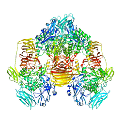 | | Structure of the beta-galactosidase from Kluyveromyces lactis in complex with galactose | | 分子名称: | Beta-galactosidase, MAGNESIUM ION, MANGANESE (II) ION, ... | | 著者 | Fernandez-Leiro, R, Pereira-Rodriguez, A, Becerra, M, Gonzalez-Siso, I, Cerdan, M.E, Sanz-Aparicio, J. | | 登録日 | 2010-08-06 | | 公開日 | 2011-08-17 | | 最終更新日 | 2024-02-21 | | 実験手法 | X-RAY DIFFRACTION (2.8 Å) | | 主引用文献 | Structural basis of specificity in tetrameric Kluyveromyces lactis beta-galactosidase.
J.Struct.Biol., 177, 2012
|
|
3OCZ
 
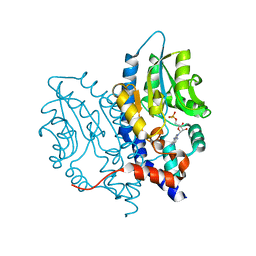 | | Structure of Recombinant Haemophilus influenzae e(P4) Acid Phosphatase Complexed with the inhibitor adenosine 5-O-thiomonophosphate | | 分子名称: | ADENOSINE -5'-THIO-MONOPHOSPHATE, Lipoprotein E, MAGNESIUM ION | | 著者 | Singh, H, Schuermann, J, Reilly, T, Calcutt, M, Tanner, J. | | 登録日 | 2010-08-10 | | 公開日 | 2011-07-20 | | 最終更新日 | 2023-09-06 | | 実験手法 | X-RAY DIFFRACTION (1.35 Å) | | 主引用文献 | Structural basis of the inhibition of class C acid phosphatases by adenosine 5'-phosphorothioate.
Febs J., 278, 2011
|
|
3OCE
 
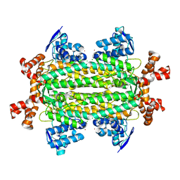 | |
3NPP
 
 | |
3NPQ
 
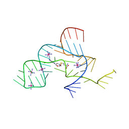 | |
3NNR
 
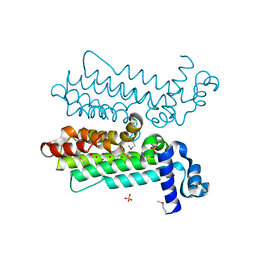 | |
3NRQ
 
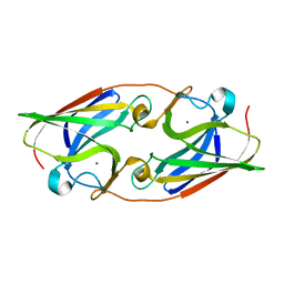 | |
3NS5
 
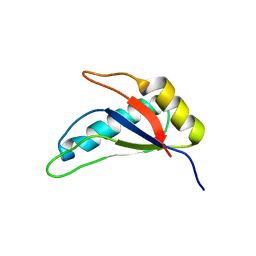 | |
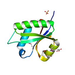
3NOH
 
 | |
3NTM
 
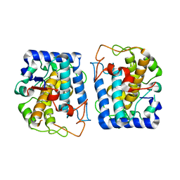 | | Crystal Structure of Tyrosinase from Bacillus megaterium crystallized in the absence of zinc, partial occupancy of CuB | | 分子名称: | COPPER (II) ION, Tyrosinase | | 著者 | Sendovski, M, Kanteev, M, Adir, N, Fishman, A. | | 登録日 | 2010-07-05 | | 公開日 | 2010-11-17 | | 最終更新日 | 2023-11-01 | | 実験手法 | X-RAY DIFFRACTION (2.3 Å) | | 主引用文献 | First structures of an active bacterial tyrosinase reveal copper plasticity
J.Mol.Biol., 405, 2011
|
|
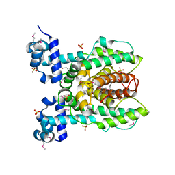
3NPI
 
 | |
3NU1
 
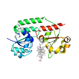 | | Structure of holo form of a periplasmic heme binding protein | | 分子名称: | Hemin-binding periplasmic protein, PROTOPORPHYRIN IX CONTAINING FE | | 著者 | Mattle, D, Goetz, B.A, Woo, J.S, Locher, K.P. | | 登録日 | 2010-07-06 | | 公開日 | 2010-10-13 | | 最終更新日 | 2024-03-20 | | 実験手法 | X-RAY DIFFRACTION (2.5 Å) | | 主引用文献 | Two stacked heme molecules in the binding pocket of the periplasmic heme-binding protein HmuT from Yersinia pestis.
J.Mol.Biol., 404, 2010
|
|
3NQO
 
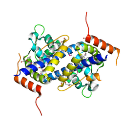 | |
3NRF
 
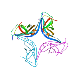 | |
3NVE
 
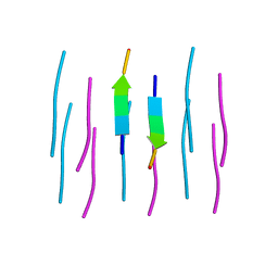 | |
3NT7
 
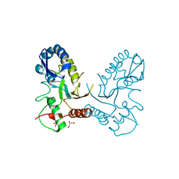 | |
3NW4
 
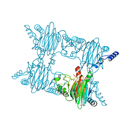 | | Crystal Structure of Salicylate 1,2-dioxygenase G106A mutant from Pseudoaminobacter salicylatoxidans in complex with gentisate | | 分子名称: | 2,5-dihydroxybenzoic acid, FE (II) ION, GLYCEROL, ... | | 著者 | Ferraroni, M, Briganti, F, Matera, I. | | 登録日 | 2010-07-09 | | 公開日 | 2011-07-13 | | 最終更新日 | 2023-09-06 | | 実験手法 | X-RAY DIFFRACTION (2 Å) | | 主引用文献 | The salicylate 1,2-dioxygenase as a model for a conventional gentisate 1,2-dioxygenase: crystal structures of the G106A mutant and its adducts with gentisate and salicylate.
FEBS J., 280, 2013
|
|
3NTY
 
 | |
3NUN
 
 | |
3NVC
 
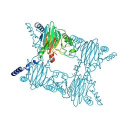 | | Crystal Structure of Salicylate 1,2-dioxygenase G106A mutant from Pseudoaminobacter salicylatoxidans in complex with salicylate | | 分子名称: | 2-HYDROXYBENZOIC ACID, FE (II) ION, GLYCEROL, ... | | 著者 | Ferraroni, M, Briganti, F, Matera, I. | | 登録日 | 2010-07-08 | | 公開日 | 2011-07-13 | | 最終更新日 | 2023-09-06 | | 実験手法 | X-RAY DIFFRACTION (2.45 Å) | | 主引用文献 | The salicylate 1,2-dioxygenase as a model for a conventional gentisate 1,2-dioxygenase: crystal structures of the G106A mutant and its adducts with gentisate and salicylate.
FEBS J., 280, 2013
|
|
