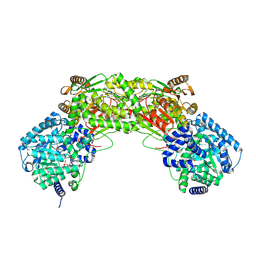
6G1N
 
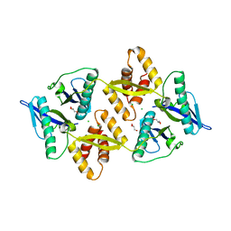 | |
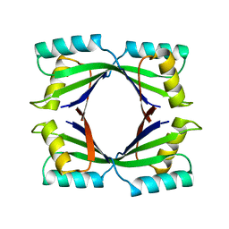
1KGJ
 | | Rat transthyretin (also called prealbumin) complex with 3',5'-dibromoflavone (EMD21388) | | 分子名称: | 6,4'-DIHYDROXY-3-METHYL-3',5'-DIBROMOFLAVONE, TRANSTHYRETIN | | 著者 | Wojtczak, A, Neumann, P, Muziol, T, Cody, V, Luft, J.R, Pangborn, W. | | 登録日 | 2001-11-27 | | 公開日 | 2002-11-27 | | 最終更新日 | 2023-08-16 | | 実験手法 | X-RAY DIFFRACTION (2.3 Å) | | 主引用文献 | Comparison of binding interactions of dibromoflavonoids with transthyretin.
Acta Biochim.Pol., 48, 2001
|
|
4WIL
 | | Crystal structure of DCoH2 S51T | | 分子名称: | Pterin-4-alpha-carbinolamine dehydratase 2 | | 著者 | Wang, D, Rose, R.B. | | 登録日 | 2014-09-26 | | 公開日 | 2014-12-31 | | 最終更新日 | 2023-09-27 | | 実験手法 | X-RAY DIFFRACTION (1.36 Å) | | 主引用文献 | Interactions with the Bifunctional Interface of the Transcriptional Coactivator DCoH1 Are Kinetically Regulated.
J.Biol.Chem., 290, 2015
|
|
6GG5
 | | Crystal structure of M2 PYK in complex with Tryptophan. | | 分子名称: | PHOSPHATE ION, POTASSIUM ION, Pyruvate kinase PKM, ... | | 著者 | McNae, I.W, Yuan, M, Walkinshaw, M.D. | | 登録日 | 2018-05-02 | | 公開日 | 2018-05-23 | | 最終更新日 | 2024-01-17 | | 実験手法 | X-RAY DIFFRACTION (3.2 Å) | | 主引用文献 | An allostatic mechanism for M2 pyruvate kinase as an amino-acid sensor.
Biochem. J., 475, 2018
|
|
1NBA
 | | CRYSTAL STRUCTURE ANALYSIS, REFINEMENT AND ENZYMATIC REACTION MECHANISM OF N-CARBAMOYLSARCOSINE AMIDOHYDROLASE FROM ARTHROBACTER SP. AT 2.0 ANGSTROMS RESOLUTION | | 分子名称: | N-CARBAMOYLSARCOSINE AMIDOHYDROLASE, SULFATE ION | | 著者 | Romao, M.J, Turk, D, Gomis-Ruth, F.-Z, Huber, R, Schumacher, G, Mollering, H, Russmann, L. | | 登録日 | 1992-05-18 | | 公開日 | 1994-06-22 | | 最終更新日 | 2024-02-14 | | 実験手法 | X-RAY DIFFRACTION (2 Å) | | 主引用文献 | Crystal structure analysis, refinement and enzymatic reaction mechanism of N-carbamoylsarcosine amidohydrolase from Arthrobacter sp. at 2.0 A resolution.
J.Mol.Biol., 226, 1992
|
|
6GG6
 | | Crystal structure of M2 PYK in complex with Serine. | | 分子名称: | MAGNESIUM ION, PHOSPHATE ION, POTASSIUM ION, ... | | 著者 | McNae, I.W, Yuan, M, Walkinshaw, M.D. | | 登録日 | 2018-05-02 | | 公開日 | 2018-05-23 | | 最終更新日 | 2024-01-17 | | 実験手法 | X-RAY DIFFRACTION (2.96 Å) | | 主引用文献 | An allostatic mechanism for M2 pyruvate kinase as an amino-acid sensor.
Biochem. J., 475, 2018
|
|
1UHE
 | | Crystal structure of aspartate decarboxylase, isoaspargine complex | | 分子名称: | Aspartate 1-decarboxylase alpha chain, Aspartate 1-decarboxylase beta chain, N~2~-(2-AMINO-1-METHYL-2-OXOETHYLIDENE)ASPARAGINATE | | 著者 | Lee, B.I, Kwon, A.-R, Han, B.W, Suh, S.W. | | 登録日 | 2003-07-01 | | 公開日 | 2004-07-13 | | 最終更新日 | 2023-12-27 | | 実験手法 | X-RAY DIFFRACTION (1.55 Å) | | 主引用文献 | Crystal structure of the schiff base intermediate prior to decarboxylation in the catalytic cycle of aspartate alpha-decarboxylase
J.Mol.Biol., 340, 2004
|
|
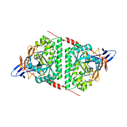
6GG3
 | |
1XY3
 | | Urate oxidase from aspergillus flavus complexed with guanine | | 分子名称: | GUANINE, Uricase | | 著者 | Retailleau, P, Colloc'h, N, Vivares, D, Bonnete, F, Castro, B, El Hajji, M, Prange, T. | | 登録日 | 2004-11-09 | | 公開日 | 2005-03-22 | | 最終更新日 | 2023-10-25 | | 実験手法 | X-RAY DIFFRACTION (3.2 Å) | | 主引用文献 | Urate oxidase from Aspergillus flavus: new crystal-packing contacts in relation to the content of the active site.
Acta Crystallogr.,Sect.D, 61, 2005
|
|
1UHD
 | | Crystal structure of aspartate decarboxylase, pyruvoly group bound form | | 分子名称: | Aspartate 1-decarboxylase alpha chain, Aspartate 1-decarboxylase beta chain | | 著者 | Lee, B.I, Kwon, A.-R, Han, B.W, Suh, S.W. | | 登録日 | 2003-07-01 | | 公開日 | 2004-07-13 | | 最終更新日 | 2023-11-15 | | 実験手法 | X-RAY DIFFRACTION (2 Å) | | 主引用文献 | Crystal structure of the schiff base intermediate prior to decarboxylation in the catalytic cycle of aspartate alpha-decarboxylase
J.Mol.Biol., 340, 2004
|
|
6GG4
 | | Crystal structure of M2 PYK in complex with Phenyalanine. | | 分子名称: | PHENYLALANINE, PHOSPHATE ION, POTASSIUM ION, ... | | 著者 | McNae, I.W, Yuan, M, Walkinshaw, M.D. | | 登録日 | 2018-05-02 | | 公開日 | 2018-05-23 | | 最終更新日 | 2024-01-17 | | 実験手法 | X-RAY DIFFRACTION (2.46 Å) | | 主引用文献 | An allostatic mechanism for M2 pyruvate kinase as an amino-acid sensor.
Biochem. J., 475, 2018
|
|
1XXJ
 | | Urate oxidase from aspergillus flavus complexed with 5-amino 6-nitro uracil | | 分子名称: | 5-AMINO-6-NITROPYRIMIDINE-2,4(1H,3H)-DIONE, BENZENE, Uricase | | 著者 | Retailleau, P, Colloc'h, N, Vivares, D, Bonnete, F, Castro, B, El Hajji, M, Prange, T. | | 登録日 | 2004-11-05 | | 公開日 | 2005-03-22 | | 最終更新日 | 2023-10-25 | | 実験手法 | X-RAY DIFFRACTION (2.8 Å) | | 主引用文献 | Urate oxidase from Aspergillus flavus: new crystal-packing contacts in relation to the content of the active site.
Acta Crystallogr.,Sect.D, 61, 2005
|
|
1NVG
 | | N249Y MUTANT OF THE ALCOHOL DEHYDROGENASE FROM THE ARCHAEON SULFOLOBUS SOLFATARICUS-TETRAGONAL CRYSTAL FORM | | 分子名称: | NAD-dependent alcohol dehydrogenase, ZINC ION | | 著者 | Esposito, L, Bruno, I, Sica, F, Raia, C.A, Giordano, A, Rossi, M, Mazzarella, L, Zagari, A. | | 登録日 | 2003-02-03 | | 公開日 | 2003-08-26 | | 最終更新日 | 2023-08-16 | | 実験手法 | X-RAY DIFFRACTION (2.5 Å) | | 主引用文献 | Structural study of a single-point mutant of Sulfolobus solfataricus alcohol dehydrogenase with enhanced activity
Febs Lett., 539, 2003
|
|
2B15
 | | The crystal structure of 2,4-dinitrophenol in complex with human transthyretin | | 分子名称: | 2,4-DINITROPHENOL, SULFATE ION, Transthyretin | | 著者 | Morais-de-Sa, E, Neto-Silva, R.M, Pereira, P.J, Saraiva, M.J, Damas, A.M. | | 登録日 | 2005-09-15 | | 公開日 | 2006-07-18 | | 最終更新日 | 2023-10-25 | | 実験手法 | X-RAY DIFFRACTION (1.7 Å) | | 主引用文献 | The binding of 2,4-dinitrophenol to wild-type and amyloidogenic transthyretin
ACTA CRYSTALLOGR.,SECT.D, 62, 2006
|
|
2B14
 | | The crystal structure of 2,4-dinitrophenol in complex with the amyloidogenic variant Transthyretin Leu 55 Pro | | 分子名称: | 2,4-DINITROPHENOL, Transthyretin | | 著者 | Morais-de-Sa, E, Neto-Silva, R.M, Pereira, P.J, Saraiva, M.J, Damas, A.M. | | 登録日 | 2005-09-15 | | 公開日 | 2006-07-18 | | 最終更新日 | 2023-10-25 | | 実験手法 | X-RAY DIFFRACTION (2 Å) | | 主引用文献 | The binding of 2,4-dinitrophenol to wild-type and amyloidogenic transthyretin
ACTA CRYSTALLOGR.,SECT.D, 62, 2006
|
|
1Y9I
 | | Crystal structure of low temperature requirement C protein from Listeria monocytogenes | | 分子名称: | CALCIUM ION, GLYCEROL, MAGNESIUM ION, ... | | 著者 | Kumaran, D, Swaminathan, S, Burley, S.K, New York SGX Research Center for Structural Genomics (NYSGXRC) | | 登録日 | 2004-12-15 | | 公開日 | 2004-12-28 | | 最終更新日 | 2021-02-03 | | 実験手法 | X-RAY DIFFRACTION (1.8 Å) | | 主引用文献 | Crystal structure of phosphatidylglycerophosphatase (PGPase), a putative membrane-bound lipid phosphatase, reveals a novel binuclear metal binding site and two "proton wires".
Proteins, 64, 2006
|
|
2B16
 | | The crystal structure of 2,4-dinitrophenol in complex with the amyloidogenic variant Transthyretin Tyr78Phe | | 分子名称: | 2,4-DINITROPHENOL, Transthyretin | | 著者 | Morais-de-Sa, E, Neto-Silva, R.M, Pereira, P.J, Saraiva, M.J, Damas, A.M. | | 登録日 | 2005-09-15 | | 公開日 | 2006-07-18 | | 最終更新日 | 2023-10-25 | | 実験手法 | X-RAY DIFFRACTION (1.75 Å) | | 主引用文献 | The binding of 2,4-dinitrophenol to wild-type and amyloidogenic transthyretin
ACTA CRYSTALLOGR.,SECT.D, 62, 2006
|
|
1YL7
 | | the crystal structure of Mycobacterium tuberculosis dihydrodipicolinate reductase (Rv2773c) in complex with NADH (crystal form C) | | 分子名称: | 1,4-DIHYDRONICOTINAMIDE ADENINE DINUCLEOTIDE, Dihydrodipicolinate reductase, MAGNESIUM ION | | 著者 | Janowski, R, Kefala, G, Weiss, M.S, TB Structural Genomics Consortium (TBSGC) | | 登録日 | 2005-01-19 | | 公開日 | 2006-01-17 | | 最終更新日 | 2023-08-23 | | 実験手法 | X-RAY DIFFRACTION (2.34 Å) | | 主引用文献 | The structure of dihydrodipicolinate reductase (DapB) from Mycobacterium tuberculosis in three crystal forms.
Acta Crystallogr.,Sect.D, 66, 2010
|
|
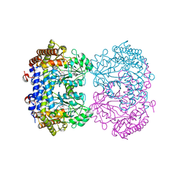
5UR2
 | |
1TWJ
 | |
5E5C
 | | Crystal structure of dihydropyrimidinase from Pseudomonas aeruginosa PAO1 | | 分子名称: | D-hydantoinase/dihydropyrimidinase, ZINC ION | | 著者 | Huang, C.C, Huang, Y.H, Hsieh, Y.C, Tzeng, C.T, Chen, C.J, Huang, C.Y. | | 登録日 | 2015-10-08 | | 公開日 | 2016-09-21 | | 最終更新日 | 2023-11-15 | | 実験手法 | X-RAY DIFFRACTION (2.1 Å) | | 主引用文献 | Crystal structure of dihydropyrimidinase from Pseudomonas aeruginosa PAO1: Insights into the molecular basis of formation of a dimer
Biochem.Biophys.Res.Commun., 478, 2016
|
|
6UXI
 | | Structure of serine hydroxymethyltransferase 8 from Glycine max cultivar Essex complexed with PLP-Glycine | | 分子名称: | 1,2-ETHANEDIOL, N-GLYCINE-[3-HYDROXY-2-METHYL-5-PHOSPHONOOXYMETHYL-PYRIDIN-4-YL-METHANE], Serine hydroxymethyltransferase | | 著者 | Korasick, D.A, Tanner, J.J, Beamer, L.J. | | 登録日 | 2019-11-07 | | 公開日 | 2020-02-12 | | 最終更新日 | 2023-10-11 | | 実験手法 | X-RAY DIFFRACTION (2.1 Å) | | 主引用文献 | Impaired folate binding of serine hydroxymethyltransferase 8 from soybean underlies resistance to the soybean cyst nematode.
J.Biol.Chem., 295, 2020
|
|
3R9Z
 | |
6UXK
 | |
4XCB
 | | Crystal Structure of HygX from Streptomyces hygroscopicus with nickel, 2-oxoglutarate, and hygromycin B bound | | 分子名称: | 2-OXOGLUTARIC ACID, HYGROMYCIN B VARIANT, NICKEL (II) ION, ... | | 著者 | McCulloch, K.M, McCranie, E.K, Sarwar, M, Mathieu, J.L, Gitschlag, B.L, Du, Y, Bachmann, B.O, Iverson, T.M. | | 登録日 | 2014-12-17 | | 公開日 | 2015-08-05 | | 最終更新日 | 2023-09-27 | | 実験手法 | X-RAY DIFFRACTION (1.6 Å) | | 主引用文献 | Oxidative cyclizations in orthosomycin biosynthesis expand the known chemistry of an oxygenase superfamily.
Proc.Natl.Acad.Sci.USA, 112, 2015
|
|


