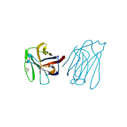
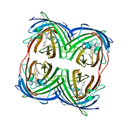
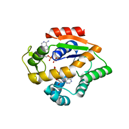
Loading
PDBj
メニューPDBj@FacebookPDBj@TwitterPDBj@YouTubewwPDB FoundationwwPDB
RCSB PDBPDBeBMRBAdv. SearchSearch help

4DXM
DownloadVisualize
BU of 4dxm by Molmil
Crystal Structure of an ancestral GFP-like protein
分子名称: GREEN FLUORESCENT PROTEIN, SULFATE ION
著者Kim, H, Fromme, R, Wachter, R.M.
登録日2012-02-27
公開日2013-02-27
最終更新日2023-11-15
実験手法X-RAY DIFFRACTION (1.4 Å)
主引用文献A hinge migration mechanism unlocks the evolution of green-to-red photoconversion in GFP-like proteins.
Structure, 23, 2015
6A66
DownloadVisualize
BU of 6a66 by Molmil
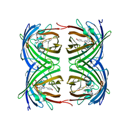
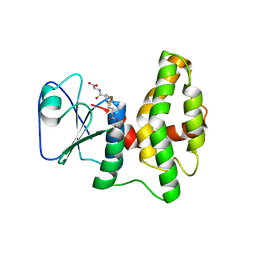
Placental protein 13/galectin-13 variant R53H with Tris
分子名称: 2-AMINO-2-HYDROXYMETHYL-PROPANE-1,3-DIOL, Galactoside-binding soluble lectin 13
著者Su, J.Y.
登録日2018-06-26
公開日2018-12-26
実験手法X-RAY DIFFRACTION (1.4 Å)
主引用文献Resetting the ligand binding site of placental protein 13/galectin-13 recovers its ability to bind lactose
Biosci. Rep., 38, 2018
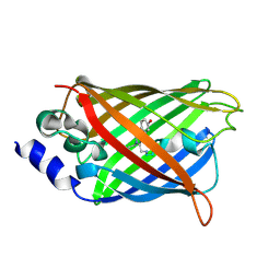
3DQ9
DownloadVisualize
BU of 3dq9 by Molmil
Structure of the Yellow Fluorescent Protein Citrine Frozen at 1500 Atmospheres Number 1: Structure 15 in a Series of 26 High Pressure Structures
分子名称: Green fluorescent protein
著者Barstow, B, Kim, C.U.
登録日2008-07-09
公開日2008-09-23
最終更新日2023-11-15
実験手法X-RAY DIFFRACTION (1.4 Å)
主引用文献Alteration of citrine structure by hydrostatic pressure explains the accompanying spectral shift.
Proc.Natl.Acad.Sci.Usa, 105, 2008
3DQD
DownloadVisualize
BU of 3dqd by Molmil
Structure of the Yellow Fluorescent Protein Citrine Frozen at 1250 Atmospheres Number 2: Structure 12 in a Series of 26 High Pressure Structures
分子名称: Green fluorescent protein
著者Barstow, B, Kim, C.U.
登録日2008-07-09
公開日2008-09-23
最終更新日2023-11-15
実験手法X-RAY DIFFRACTION (1.4 Å)
主引用文献Alteration of citrine structure by hydrostatic pressure explains the accompanying spectral shift.
Proc.Natl.Acad.Sci.Usa, 105, 2008
2DUG
DownloadVisualize
BU of 2dug by Molmil
crystal structure of a green fluorescent protein S65T/H148N at pH 5
分子名称: Green fluorescent protein
著者Shu, X, Remington, S.J.
登録日2006-07-23
公開日2007-07-10
最終更新日2024-07-10
実験手法X-RAY DIFFRACTION (1.4 Å)
主引用文献Ultrafast excited-state dynamics in the green fluorescent protein variant S65T/H148D. 1. Mutagenesis and structural studies.
Biochemistry, 46, 2007
1ZGO
DownloadVisualize
BU of 1zgo by Molmil
High Resolution Crystal Structure of the Discosoma Red Fluorescent Protein (DsRed)
分子名称: Red fluorescent protein drFP583
著者Tubbs, J.L, Tainer, J.A, Getzoff, E.D.
登録日2005-04-21
公開日2005-08-02
最終更新日2023-11-15
実験手法X-RAY DIFFRACTION (1.4 Å)
主引用文献Crystallographic structures of discosoma red fluorescent protein with immature and mature chromophores: linking Peptide bond trans-cis isomerization and acylimine formation in chromophore maturation.
Biochemistry, 44, 2005
4PQH
DownloadVisualize
BU of 4pqh by Molmil
Crystal structure of glutathione transferase lambda1 from Populus trichocarpa
分子名称: GLUTATHIONE, SODIUM ION, glutathione transferase lambda1
著者Lallement, P.A, Meux, E, Gualberto, J.M, Prosper, P, Didierjean, C, Haouz, A, Saul, F, Rouhier, N, Hecker, A.
登録日2014-03-03
公開日2014-06-25
最終更新日2023-09-20
実験手法X-RAY DIFFRACTION (1.4 Å)
主引用文献Structural and enzymatic insights into Lambda glutathione transferases from Populus trichocarpa, monomeric enzymes constituting an early divergent class specific to terrestrial plants.
Biochem.J., 462, 2014
1KS8
DownloadVisualize
BU of 1ks8 by Molmil
The structure of Endoglucanase from termite, Nasutitermes takasagoensis, at pH 2.5.
分子名称: Endo-b-1,4-glucanase, SULFATE ION
著者Khademi, S, Guarino, L.A, Watanabe, H, Tokuda, G, Meyer, E.F.
登録日2002-01-11
公開日2003-01-21
最終更新日2023-08-16
実験手法X-RAY DIFFRACTION (1.4 Å)
主引用文献Structure of an endoglucanase from termite, Nasutitermes takasagoensis.
Acta Crystallogr.,Sect.D, 58, 2002
3AI5
DownloadVisualize
BU of 3ai5 by Molmil
Crystal structure of yeast enhanced green fluorescent protein-ubiquitin fusion protein
分子名称: 1,2-ETHANEDIOL, yeast enhanced green fluorescent protein,Ubiquitin
著者Suzuki, N, Wakatsuki, S, Kawasaki, M.
登録日2010-05-10
公開日2010-09-29
最終更新日2023-11-15
実験手法X-RAY DIFFRACTION (1.4 Å)
主引用文献Crystallization of small proteins assisted by green fluorescent protein
Acta Crystallogr.,Sect.D, 66, 2010
6F7U
DownloadVisualize
BU of 6f7u by Molmil
Molecular Mechanism of ATP versus GTP Selectivity of Adenylate Kinase
分子名称: Adenylate kinase, MAGNESIUM ION, PHOSPHOMETHYLPHOSPHONIC ACID GUANYLATE ESTER
著者Rogne, P, Rosselin, M, Grundstrom, C, Hedberg, C, Wolf-Watz, M, Sauer, U.H.
登録日2017-12-12
公開日2018-03-14
最終更新日2024-05-01
実験手法X-RAY DIFFRACTION (1.4 Å)
主引用文献Molecular mechanism of ATP versus GTP selectivity of adenylate kinase.
Proc. Natl. Acad. Sci. U.S.A., 115, 2018
2B3P
DownloadVisualize
BU of 2b3p by Molmil
Crystal structure of a superfolder green fluorescent protein
分子名称: ACETIC ACID, CADMIUM ION, green fluorescent protein
著者Pedelacq, J.D, Cabantous, S, Tran, T.H, Terwilliger, T.C, Waldo, G.S.
登録日2005-09-20
公開日2005-11-08
最終更新日2023-11-15
実験手法X-RAY DIFFRACTION (1.4 Å)
主引用文献Engineering and characterization of a superfolder green fluorescent protein.
Nat.Biotechnol., 24, 2006
2ZO6
DownloadVisualize
BU of 2zo6 by Molmil
Crystal Structure of Kusabira-Cyan (KCY), a Cyan-Emitting GFP-Like Protein
分子名称: CYAN-EMITTING GFP-LIKE PROTEIN, KUSABIRA-CYAN (KCY)
著者Kikuchi, A, Fukumura, E, Karasawa, S, Miyawaki, A, Shiro, Y, RIKEN Structural Genomics/Proteomics Initiative (RSGI)
登録日2008-05-06
公開日2009-05-12
最終更新日2023-11-15
実験手法X-RAY DIFFRACTION (1.4 Å)
主引用文献Crystal structure of a new cyan fluorescent protein and its hue-shifted variants
Biochemistry, 48, 2009
2PSD
DownloadVisualize
BU of 2psd by Molmil
Crystal Structures of the Luciferase and Green Fluorescent Protein from Renilla Reniformis
分子名称: IMIDAZOLE, Renilla-luciferin 2-monooxygenase
著者Loening, A.M, Fenn, T.D, Gambhir, S.S.
登録日2007-05-06
公開日2007-06-05
最終更新日2024-02-21
実験手法X-RAY DIFFRACTION (1.4 Å)
主引用文献Crystal Structures of the Luciferase and Green Fluorescent Protein from Renilla reniformis.
J.Mol.Biol., 374, 2007
2PSF
DownloadVisualize
BU of 2psf by Molmil
Crystal Structures of the Luciferase and Green Fluorescent Protein from Renilla Reniformis
分子名称: Renilla-luciferin 2-monooxygenase
著者Loening, A.M, Fenn, T.D, Gambhir, S.S.
登録日2007-05-06
公開日2007-05-22
最終更新日2024-02-21
実験手法X-RAY DIFFRACTION (1.4 Å)
主引用文献Crystal Structures of the Luciferase and Green Fluorescent Protein from Renilla reniformis.
J.Mol.Biol., 374, 2007
3OM0
DownloadVisualize
BU of 3om0 by Molmil
Crystal structure of the GluK5 (KA2) ATD crystallographic dimer at 1.4 Angstrom resolution
分子名称: 2-acetamido-2-deoxy-beta-D-glucopyranose, ACETATE ION, GLYCEROL, ...
著者Kumar, J, Mayer, M.L.
登録日2010-08-26
公開日2010-11-03
最終更新日2024-04-03
実験手法X-RAY DIFFRACTION (1.401 Å)
主引用文献Crystal Structures of the Glutamate Receptor Ion Channel GluK3 and GluK5 Amino-Terminal Domains.
J.Mol.Biol., 404, 2010
3SST
DownloadVisualize
BU of 3sst by Molmil
Engineered low-affinity halide-binding protein derived from YFP: chloride complex
分子名称: CHLORIDE ION, Green fluorescent protein
著者Wang, W, Grimley, J.S, Beese, L.S, Hellinga, H.W.
登録日2011-07-08
公開日2012-07-11
最終更新日2023-11-15
実験手法X-RAY DIFFRACTION (1.403 Å)
主引用文献Determination of engineered chloride-binding site structures in fluorescent proteins reveals principles of halide recognition
To be Published
4EEU
DownloadVisualize
BU of 4eeu by Molmil
Crystal structure of phiLOV2.1
分子名称: FLAVIN MONONUCLEOTIDE, Phototropin-2
著者Hitomi, K, Christie, J.M, Arvai, A.S, Hartfield, K.A, Pratt, A.J, Tainer, J.A, Getzoff, E.D.
登録日2012-03-28
公開日2012-05-16
最終更新日2023-09-13
実験手法X-RAY DIFFRACTION (1.4068 Å)
主引用文献Structural Tuning of the Fluorescent Protein iLOV for Improved Photostability.
J.Biol.Chem., 287, 2012
3DQI
DownloadVisualize
BU of 3dqi by Molmil
Structure of the Yellow Fluorescent Protein Citrine Frozen at 1000 Atmospheres Number 4: Structure 8 in a Series of 26 High Pressure Structures
分子名称: Green fluorescent protein
著者Barstow, B, Kim, C.U.
登録日2008-07-09
公開日2008-09-23
最終更新日2023-11-15
実験手法X-RAY DIFFRACTION (1.42 Å)
主引用文献Alteration of citrine structure by hydrostatic pressure explains the accompanying spectral shift.
Proc.Natl.Acad.Sci.Usa, 105, 2008
3DQU
DownloadVisualize
BU of 3dqu by Molmil
Structure of the Yellow Fluorescent Protein Citrine Frozen at 2000 Atmospheres Number 1: Structure 20 in a Series of 26 High Pressure Structures
分子名称: Green fluorescent protein
著者Barstow, B, Kim, C.U.
登録日2008-07-09
公開日2008-09-23
最終更新日2023-11-15
実験手法X-RAY DIFFRACTION (1.42 Å)
主引用文献Alteration of citrine structure by hydrostatic pressure explains the accompanying spectral shift.
Proc.Natl.Acad.Sci.Usa, 105, 2008
5DPH
DownloadVisualize
BU of 5dph by Molmil
sfGFP mutant - 149 p-cyano-L-phenylalanine
分子名称: CARBON DIOXIDE, Green fluorescent protein, MAGNESIUM ION
著者Dippel, A.B, Olenginski, G.M, Maurici, N, Liskov, M.T, Brewer, S.H, Phillips-Piro, C.M.
登録日2015-09-12
公開日2016-01-13
最終更新日2023-11-15
実験手法X-RAY DIFFRACTION (1.42 Å)
主引用文献Probing the effectiveness of spectroscopic reporter unnatural amino acids: a structural study.
Acta Crystallogr D Struct Biol, 72, 2016
7PNN
DownloadVisualize
BU of 7pnn by Molmil
mVenus released from fusion protein.
分子名称: CHLORIDE ION, mVenus
著者Bloch, Y, Savvides, S.N.
登録日2021-09-07
公開日2021-11-03
最終更新日2024-01-31
実験手法X-RAY DIFFRACTION (1.43 Å)
主引用文献mVenus released from fusion protein.
To Be Published
3DQE
DownloadVisualize
BU of 3dqe by Molmil
Structure of the Yellow Fluorescent Protein Citrine Frozen at 1250 Atmospheres Number 1: Structure 11 in a Series of 26 High Pressure Structures
分子名称: Green fluorescent protein
著者Barstow, B, Kim, C.U.
登録日2008-07-09
公開日2008-09-23
最終更新日2023-11-15
実験手法X-RAY DIFFRACTION (1.43 Å)
主引用文献Alteration of citrine structure by hydrostatic pressure explains the accompanying spectral shift.
Proc.Natl.Acad.Sci.Usa, 105, 2008
4JLP
DownloadVisualize
BU of 4jlp by Molmil
Crystal structure of R150K aquifex adenylate kinase mutant
分子名称: ADENOSINE-5'-DIPHOSPHATE, Adenylate kinase
著者Cho, Y.-J, Phung, L, Kern, D.
登録日2013-03-12
公開日2014-06-25
最終更新日2024-02-28
実験手法X-RAY DIFFRACTION (1.433 Å)
主引用文献The energy landscape of adenylate kinase during catalysis.
Nat.Struct.Mol.Biol., 22, 2015
3DQA
DownloadVisualize
BU of 3dqa by Molmil
Structure of the Yellow Fluorescent Protein Citrine Frozen at 1250 Atmospheres Number 4: Structure 14 in a Series of 26 High Pressure Structures
分子名称: Green fluorescent protein
著者Barstow, B, Kim, C.U.
登録日2008-07-09
公開日2008-09-23
最終更新日2023-11-15
実験手法X-RAY DIFFRACTION (1.44 Å)
主引用文献Alteration of citrine structure by hydrostatic pressure explains the accompanying spectral shift.
Proc.Natl.Acad.Sci.Usa, 105, 2008
3DQM
DownloadVisualize
BU of 3dqm by Molmil
Structure of the Yellow Fluorescent Protein Citrine Frozen at 750 Atmospheres: Structure 4 in a Series of 26 High Pressure Structures
分子名称: Green fluorescent protein
著者Barstow, B, Kim, C.U.
登録日2008-07-09
公開日2008-09-23
最終更新日2023-11-15
実験手法X-RAY DIFFRACTION (1.44 Å)
主引用文献Alteration of citrine structure by hydrostatic pressure explains the accompanying spectral shift.
Proc.Natl.Acad.Sci.Usa, 105, 2008

223532

件を2024-08-07に公開中

PDB statisticsPDBj update infoContact PDBjnumon