3V7I
 
 | |
3VO2
 | |
4U7A
 | |
4Q5O
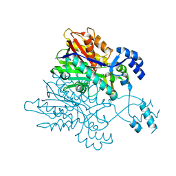
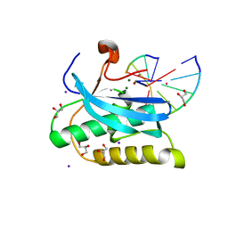
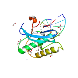
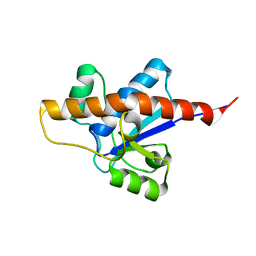
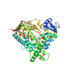
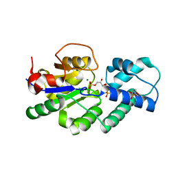
 | | Crystal structure of EctD from S. alaskensis with 2-oxoglutarate and 5-hydroxyectoine | | 分子名称: | (4S,5S)-5-HYDROXY-2-METHYL-1,4,5,6-TETRAHYDROPYRIMIDINE-4-CARBOXYLIC ACID, 2-OXOGLUTARIC ACID, Ectoine hydroxylase, ... | | 著者 | Hoeppner, A, Widderich, N, Bremer, E, Smits, S.H. | | 登録日 | 2014-04-17 | | 公開日 | 2014-09-10 | | 最終更新日 | 2023-09-20 | | 実験手法 | X-RAY DIFFRACTION (2.64 Å) | | 主引用文献 | Crystal structure of the ectoine hydroxylase, a snapshot of the active site.
J.Biol.Chem., 289, 2014
|
|
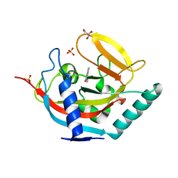
2RAW
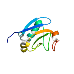
 | | Crystal structure of the Borealin-Survivin complex | | 分子名称: | Baculoviral IAP repeat-containing protein 5, Borealin, ZINC ION | | 著者 | Hymowitz, S.G. | | 登録日 | 2007-09-17 | | 公開日 | 2007-10-30 | | 最終更新日 | 2023-08-30 | | 実験手法 | X-RAY DIFFRACTION (2.4 Å) | | 主引用文献 | The mitotic regulator Survivin binds as a monomer to its functional interactor Borealin.
J.Biol.Chem., 282, 2007
|
|
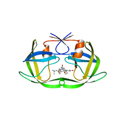
2R7F
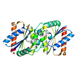
 | | Crystal structure of ribonuclease II family protein from Deinococcus radiodurans, hexagonal crystal form. NorthEast Structural Genomics target DrR63 | | 分子名称: | Ribonuclease II family protein | | 著者 | Seetharaman, J, Neely, H, Forouhar, F, Wang, D, Fang, Y, Cunningham, K, Ma, L.-C, Xia, R, Liu, J, Baran, M.C, Acton, T.B, Rost, B, Montelione, G.T, Hunt, J.F, Tong, L, Northeast Structural Genomics Consortium (NESG) | | 登録日 | 2007-09-07 | | 公開日 | 2007-10-02 | | 最終更新日 | 2018-01-24 | | 実験手法 | X-RAY DIFFRACTION (2.7 Å) | | 主引用文献 | The Structure and Enzymatic Properties of a Novel RNase II Family Enzyme from Deinococcus radiodurans.
J.Mol.Biol., 415, 2012
|
|
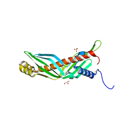
4UAV
 | | Crystal structure of CbbY (AT3G48420) from Arabidobsis thaliana | | 分子名称: | Haloacid dehalogenase-like hydrolase domain-containing protein At3g48420, MAGNESIUM ION | | 著者 | Bracher, A, Sharma, A, Starling-Windhof, A, Hartl, F.U, Hayer-Hartl, M. | | 登録日 | 2014-08-11 | | 公開日 | 2014-12-31 | | 最終更新日 | 2023-12-20 | | 実験手法 | X-RAY DIFFRACTION (1.3 Å) | | 主引用文献 | Degradation of potent Rubisco inhibitor by selective sugar phosphatase.
Nat.Plants, 1, 2015
|
|
6DOM
 | |
6DP2
 | |
6DPO
 | |
6DQI
 | |
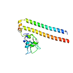
2M1M
 | | Solution structure of the PsIAA4 oligomerization domain reveals interaction modes for transcription factors in early auxin response | | 分子名称: | Auxin-induced protein IAA4 | | 著者 | Kovermann, M, Dinesh, D.C, Gopalswamy, M, Abel, S, Balbach, J. | | 登録日 | 2012-12-03 | | 公開日 | 2013-12-11 | | 最終更新日 | 2024-05-01 | | 実験手法 | SOLUTION NMR | | 主引用文献 | Solution structure of the PsIAA4 oligomerization domain reveals interaction modes for transcription factors in early auxin response.
Proc.Natl.Acad.Sci.USA, 112, 2015
|
|
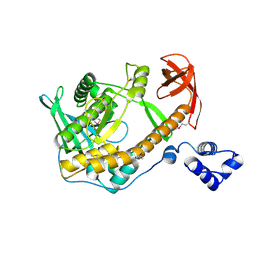
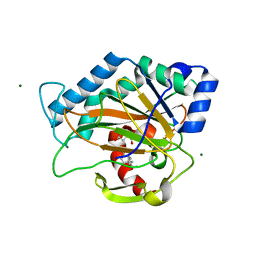
2LUO
 | | NMR solution structure of apo-MptpA | | 分子名称: | LOW MOLECULAR WEIGHT PROTEIN-TYROSINE-PHOSPHATASE A | | 著者 | Stehle, T, Sreeramulu, S, Loehr, F, Richter, C, Saxena, K, Jonker, H.R.A, Schwalbe, H. | | 登録日 | 2012-06-19 | | 公開日 | 2012-08-15 | | 最終更新日 | 2024-05-15 | | 実験手法 | SOLUTION NMR | | 主引用文献 | The Apo-structure of the Low Molecular Weight Protein-tyrosine Phosphatase A (MptpA) from Mycobacterium tuberculosis Allows for Better Target-specific Drug Development.
J.Biol.Chem., 287, 2012
|
|
4UHG
 | |
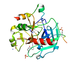
2R5Q
 | | Crystal Structure Analysis of HIV-1 Subtype C Protease Complexed with Nelfinavir | | 分子名称: | 2-[2-HYDROXY-3-(3-HYDROXY-2-METHYL-BENZOYLAMINO)-4-PHENYL SULFANYL-BUTYL]-DECAHYDRO-ISOQUINOLINE-3-CARBOXYLIC ACID TERT-BUTYLAMIDE, Protease | | 著者 | Coman, R.M, Robbins, A.H, McKenna, R, Dunn, B.M. | | 登録日 | 2007-09-04 | | 公開日 | 2007-11-20 | | 最終更新日 | 2024-02-21 | | 実験手法 | X-RAY DIFFRACTION (2.3 Å) | | 主引用文献 | The Contribution of Naturally Occurring Polymorphisms in Altering the Biochemical and Structural Characteristics of HIV-1 Subtype C Protease
Biochemistry, 47, 2008
|
|
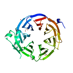
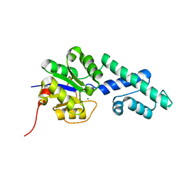
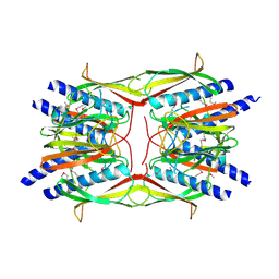
2RCK
 | | Crystal structure of juvenile hormone binding protein from Galleria mellonella hemolymph | | 分子名称: | 2-acetamido-2-deoxy-beta-D-glucopyranose, GLYCEROL, Juvenile hormone binding protein, ... | | 著者 | Kolodziejczyk, R, Bujacz, G, Jakob, M, Ozyhar, A, Jaskolski, M, Kochman, M. | | 登録日 | 2007-09-20 | | 公開日 | 2008-03-04 | | 最終更新日 | 2020-07-29 | | 実験手法 | X-RAY DIFFRACTION (2.44 Å) | | 主引用文献 | Insect Juvenile Hormone Binding Protein Shows Ancestral Fold Present in Human Lipid-Binding Proteins.
J.Mol.Biol., 377, 2008
|
|
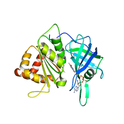
6DWN
 | | Structure of Human Cytochrome P450 1A1 with Erlotinib | | 分子名称: | 3-[(3-CHOLAMIDOPROPYL)DIMETHYLAMMONIO]-1-PROPANESULFONATE, Cytochrome P450 1A1, PROTOPORPHYRIN IX CONTAINING FE, ... | | 著者 | Bart, A.G, Scott, E.E. | | 登録日 | 2018-06-26 | | 公開日 | 2018-10-03 | | 最終更新日 | 2023-10-11 | | 実験手法 | X-RAY DIFFRACTION (3 Å) | | 主引用文献 | Structures of human cytochrome P450 1A1 with bergamottin and erlotinib reveal active-site modifications for binding of diverse ligands.
J. Biol. Chem., 293, 2018
|
|
4UOW
 | | Crystal structure of the titin M10-Obscurin Ig domain 1 complex | | 分子名称: | CHLORIDE ION, Obscurin, SODIUM ION, ... | | 著者 | Pernigo, S, Fukuzawa, A, Gautel, M, Steiner, R.A. | | 登録日 | 2014-06-10 | | 公開日 | 2014-12-17 | | 最終更新日 | 2024-01-10 | | 実験手法 | X-RAY DIFFRACTION (3.3 Å) | | 主引用文献 | The Crystal Structure of the Human Titin:Obscurin Complex Reveals a Conserved Yet Specific Muscle M-Band Zipper Module.
J.Mol.Biol., 427, 2015
|
|
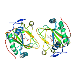
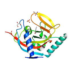
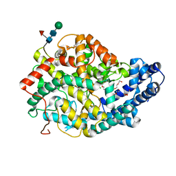
2RDN
 | | Crystal Structure of PtlH with AKG and ent-1PL bound | | 分子名称: | (1S,3aS,5aR,8aS)-1,7,7-trimethyl-1,2,3,3a,5a,6,7,8-octahydrocyclopenta[c]pentalene-4-carboxylic acid, 1-deoxypentalenic acid 11-beta hydroxylase; Fe(II)/alpha-ketoglutarate dependent hydroxylase, 2-OXOGLUTARIC ACID, ... | | 著者 | You, Z, Omura, S, Ikeda, H, Cane, D.E, Jogl, G. | | 登録日 | 2007-09-24 | | 公開日 | 2007-10-16 | | 最終更新日 | 2024-02-21 | | 実験手法 | X-RAY DIFFRACTION (1.35 Å) | | 主引用文献 | Crystal Structure of the Non-heme Iron Dioxygenase PtlH in Pentalenolactone Biosynthesis.
J.Biol.Chem., 282, 2007
|
|
4UFY
 | |
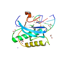
2RET
 | | The crystal structure of a binary complex of two pseudopilins: EpsI and EpsJ from the Type 2 Secretion System of Vibrio vulnificus | | 分子名称: | CHLORIDE ION, EpsJ, Pseudopilin EpsI, ... | | 著者 | Yanez, M.E, Korotkov, K.V, Abendroth, J, Hol, W.G.J. | | 登録日 | 2007-09-27 | | 公開日 | 2008-02-05 | | 最終更新日 | 2024-02-21 | | 実験手法 | X-RAY DIFFRACTION (2.21 Å) | | 主引用文献 | The crystal structure of a binary complex of two pseudopilins: EpsI and EpsJ from the type 2 secretion system of Vibrio vulnificus.
J.Mol.Biol., 375, 2008
|
|
4UAU
 | | Crystal structure of CbbY (mutant D10N) from Rhodobacter sphaeroides in complex with Xylulose-(1,5)bisphosphate, crystal form II | | 分子名称: | 2-(N-MORPHOLINO)-ETHANESULFONIC ACID, MAGNESIUM ION, Protein CbbY, ... | | 著者 | Bracher, A, Sharma, A, Starling-Windhof, A, Hartl, F.U, Hayer-Hartl, M. | | 登録日 | 2014-08-11 | | 公開日 | 2014-12-31 | | 最終更新日 | 2023-12-20 | | 実験手法 | X-RAY DIFFRACTION (1.45 Å) | | 主引用文献 | Degradation of potent Rubisco inhibitor by selective sugar phosphatase.
Nat.Plants, 1, 2015
|
|
4UDW
 | | Thrombin in complex with 1-(2R)-2-amino-3-phenyl-propanoyl-N-(2, 5dichlorophenyl)methylpyrrolidine-2-carboxamide | | 分子名称: | 2-acetamido-2-deoxy-beta-D-glucopyranose, D-phenylalanyl-N-(2,5-dichlorobenzyl)-L-prolinamide, GLYCEROL, ... | | 著者 | Ruehmann, E, Heine, A, Klebe, G. | | 登録日 | 2014-12-12 | | 公開日 | 2015-08-26 | | 最終更新日 | 2023-12-20 | | 実験手法 | X-RAY DIFFRACTION (1.16 Å) | | 主引用文献 | Fragments Can Bind Either More Enthalpy or Entropy-Driven: Crystal Structures and Residual Hydration Pattern Suggest Why.
J.Med.Chem., 58, 2015
|
|
6DEO
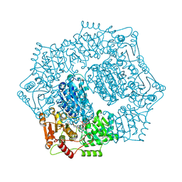 | |
4UFA
 | | Crystal structure of the Angiotensin-1 converting enzyme N-domain in complex with Ac-SD | | 分子名称: | 2-acetamido-2-deoxy-beta-D-glucopyranose, 2-acetamido-2-deoxy-beta-D-glucopyranose-(1-4)-2-acetamido-2-deoxy-beta-D-glucopyranose, ANGIOTENSIN-CONVERTING ENZYME, ... | | 著者 | Masuyer, G, Douglas, R.G, Sturrock, E.D, Acharya, K.R. | | 登録日 | 2015-03-16 | | 公開日 | 2015-10-07 | | 最終更新日 | 2023-12-20 | | 実験手法 | X-RAY DIFFRACTION (1.8 Å) | | 主引用文献 | Structural Basis of Ac-Sdkp Hydrolysis by Angiotensin-I Converting Enzyme
Sci.Rep., 5, 2015
|
|


