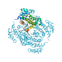
4PO9
 
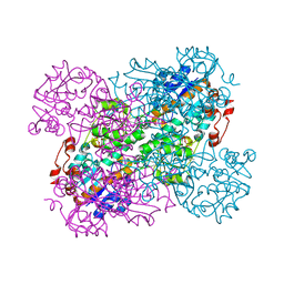
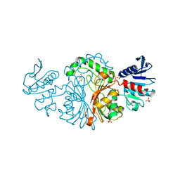
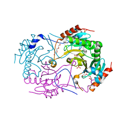
 | | Mycobacterium tuberculosis RecA glycerol bound low temperature structure IIC-BR | | 分子名称: | 1,2-ETHANEDIOL, GLYCEROL, Protein RecA, ... | | 著者 | Chandran, A.V, Prabu, J.R, Patil, N.K, Muniyappa, K, Vijayan, M. | | 登録日 | 2014-02-25 | | 公開日 | 2015-03-18 | | 最終更新日 | 2023-11-08 | | 実験手法 | X-RAY DIFFRACTION (2.75 Å) | | 主引用文献 | Structural studies on Mycobacterium tuberculosis RecA: Molecular plasticity and interspecies variability
J.Biosci., 40, 2015
|
|
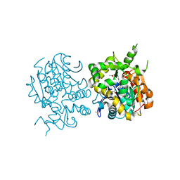
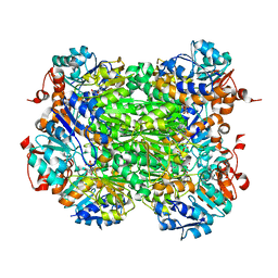
4PSV
 | | Mycobacterium tuberculosis RecA phosphate bound room temperature structure I-RT | | 分子名称: | PHOSPHATE ION, Protein RecA, 1st part, ... | | 著者 | Chandran, A.V, Prabu, J.R, Patil, N.K, Muniyappa, K, Vijayan, M. | | 登録日 | 2014-03-08 | | 公開日 | 2015-03-18 | | 最終更新日 | 2023-11-08 | | 実験手法 | X-RAY DIFFRACTION (2.6 Å) | | 主引用文献 | Structural studies on Mycobacterium tuberculosis RecA: Molecular plasticity and interspecies variability
J.Biosci., 40, 2015
|
|
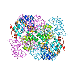
4PPN
 | | Mycobacterium tuberculosis RecA citrate bound low temperature structure IIA-BN | | 分子名称: | 1,2-ETHANEDIOL, CITRATE ANION, Protein RecA, ... | | 著者 | Chandran, A.V, Prabu, J.R, Patil, N.K, Muniyappa, K, Vijayan, M. | | 登録日 | 2014-02-27 | | 公開日 | 2015-03-18 | | 最終更新日 | 2023-11-08 | | 実験手法 | X-RAY DIFFRACTION (2.6 Å) | | 主引用文献 | Structural studies on Mycobacterium tuberculosis RecA: Molecular plasticity and interspecies variability
J.Biosci., 40, 2015
|
|
4E1R
 | |
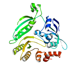
4TZK
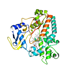
 | | Crystal structure of Mycobacterium tuberculosis enoyl reductase (INHA) complexed WITH 1-CYCLOHEXYL-N-(3,5-DICHLOROPHENYL)-5-OXOPYRROLIDINE-3-CARBOXAMIDE | | 分子名称: | (3S)-1-CYCLOHEXYL-N-(3,5-DICHLOROPHENYL)-5-OXOPYRROLIDINE-3-CARBOXAMIDE, Enoyl-[acyl-carrier-protein] reductase [NADH], NICOTINAMIDE-ADENINE-DINUCLEOTIDE | | 著者 | He, X, Alian, A, Stroud, R.M, Ortiz de Montellano, P.R. | | 登録日 | 2014-07-10 | | 公開日 | 2014-08-20 | | 最終更新日 | 2023-12-27 | | 実験手法 | X-RAY DIFFRACTION (1.62 Å) | | 主引用文献 | Pyrrolidine carboxamides as a novel class of inhibitors of enoyl acyl carrier protein reductase from Mycobacterium tuberculosis
J. Med. Chem., 2006
|
|
2ZJF
 | |
2LX5
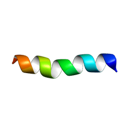 | |
2MMU
 | | Structure of CrgA, a Cell Division Structural and Regulatory Protein from Mycobacterium tuberculosis, in Lipid Bilayers | | 分子名称: | Cell division protein CrgA | | 著者 | Das, N, Dai, J, Hung, I, Rajagopalan, M, Zhou, H, Cross, T.A. | | 登録日 | 2014-03-18 | | 公開日 | 2014-12-17 | | 最終更新日 | 2024-05-15 | | 実験手法 | SOLID-STATE NMR | | 主引用文献 | Structure of CrgA, a cell division structural and regulatory protein from Mycobacterium tuberculosis, in lipid bilayers.
Proc.Natl.Acad.Sci.USA, 112, 2015
|
|
2VHZ
 | |
2VHW
 | |
4G3T
 | |
2VHV
 | |
1PFE
 | | Echinomycin-(gcgtacgc)2 complex | | 分子名称: | 2-CARBOXYQUINOXALINE, 5'-D(*GP*CP*GP*TP*AP*CP*GP*C)-3', CHLORIDE ION, ... | | 著者 | Cuesta-Seijo, J.A. | | 登録日 | 2003-05-26 | | 公開日 | 2004-06-08 | | 最終更新日 | 2024-04-03 | | 実験手法 | X-RAY DIFFRACTION (1.1 Å) | | 主引用文献 | Structures of Complexes between Echinomycin and Duplex DNA.
Acta Crystallogr.,Sect.D, 61, 2005
|
|
3VOM
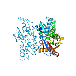
 | | Structure of a putative phosphoserine aminotransferase from mycobacterium tuberculosis | | 分子名称: | GLYCEROL, PYRIDOXAL-5'-PHOSPHATE, Putative phosphoserine aminotransferase, ... | | 著者 | Coulibaly, F, Lassalle, E, Baker, H.M, Baker, E.N. | | 登録日 | 2012-01-30 | | 公開日 | 2012-02-22 | | 最終更新日 | 2023-11-08 | | 実験手法 | X-RAY DIFFRACTION (2.1 Å) | | 主引用文献 | Structure of phosphoserine aminotransferase from Mycobacterium tuberculosis.
Acta Crystallogr.,Sect.D, 68, 2012
|
|
6R29
 | | Crystal structure of the SucA domain of Mycobacterium smegmatis KGD cocrystallized with succinylphosphonate | | 分子名称: | (4S)-2-METHYL-2,4-PENTANEDIOL, (4~{S})-4-[(2~{R})-3-[(4-azanyl-2-methyl-pyrimidin-5-yl)methyl]-4-methyl-5-[2-[oxidanyl(phosphonooxy)phosphoryl]oxyethyl]-2~{H}-1,3-thiazol-2-yl]-4-oxidanyl-4-phosphono-butanoic acid, MAGNESIUM ION, ... | | 著者 | Wagner, T, Alzari, P.M, Bellinzoni, M. | | 登録日 | 2019-03-15 | | 公開日 | 2019-09-11 | | 最終更新日 | 2024-01-24 | | 実験手法 | X-RAY DIFFRACTION (1.67 Å) | | 主引用文献 | Conformational transitions in the active site of mycobacterial 2-oxoglutarate dehydrogenase upon binding phosphonate analogues of 2-oxoglutarate: From a Michaelis-like complex to ThDP adducts.
J.Struct.Biol., 208, 2019
|
|
4G3U
 | |
2W3G
 | |
3VOS
 | | Crystal structure of Aspartate semialdehyde dehydrogenase Complexed With glycerol and sulfate From Mycobacterium tuberculosis H37Rv | | 分子名称: | Aspartate-semialdehyde dehydrogenase, GLYCEROL, SULFATE ION | | 著者 | Vyas, R, Tewari, R, Weiss, M.S, Karthikeyan, S. | | 登録日 | 2012-02-07 | | 公開日 | 2012-05-30 | | 最終更新日 | 2023-11-08 | | 実験手法 | X-RAY DIFFRACTION (2.18 Å) | | 主引用文献 | Structures of ternary complexes of aspartate-semialdehyde dehydrogenase (Rv3708c) from Mycobacterium tuberculosis H37Rv
Acta Crystallogr.,Sect.D, 68, 2012
|
|
4QA8
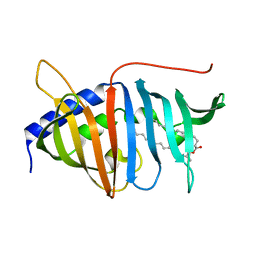 | | Crystal structure of LprF from Mycobacterium bovis | | 分子名称: | (2R)-2-(dodecanoyloxy)propyl (4E,6E,8E,10E,12E)-pentadeca-4,6,8,10,12-pentaenoate, Putative lipoprotein LprF | | 著者 | Ha, N.C, Jiao, L, Kim, J.S. | | 登録日 | 2014-05-02 | | 公開日 | 2014-10-22 | | 最終更新日 | 2024-03-20 | | 実験手法 | X-RAY DIFFRACTION (1.1 Å) | | 主引用文献 | Crystal structure and functional implications of LprF from Mycobacterium tuberculosis and M. bovis
Acta Crystallogr.,Sect.D, 70, 2014
|
|
4QII
 | | Crystal Structure of type II MenB from Mycobacteria tuberculosis | | 分子名称: | 1,4-Dihydroxy-2-naphthoyl-CoA synthase, Salicylyl CoA, TRIETHYLENE GLYCOL | | 著者 | Song, H.G, Tse, Y.S, Sung, H.P, Guo, Z.H. | | 登録日 | 2014-05-31 | | 公開日 | 2014-11-19 | | 最終更新日 | 2023-11-08 | | 実験手法 | X-RAY DIFFRACTION (1.64 Å) | | 主引用文献 | Ligand-dependent active-site closure revealed in the crystal structure of Mycobacterium tuberculosis MenB complexed with product analogues
Acta Crystallogr.,Sect.D, 70, 2014
|
|
4GO5
 | |
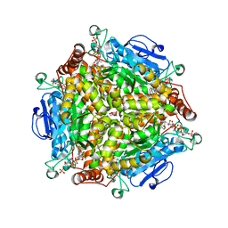
3ANO
 | | Crystal Structure of a Novel Diadenosine 5',5'''-P1,P4-Tetraphosphate Phosphorylase from Mycobacterium tuberculosis H37Rv | | 分子名称: | AP-4-A phosphorylase, PHOSPHATE ION, TETRAETHYLENE GLYCOL | | 著者 | Mori, S, Shibayama, K, Wachino, J, Arakawa, Y. | | 登録日 | 2010-09-06 | | 公開日 | 2011-05-18 | | 最終更新日 | 2024-03-13 | | 実験手法 | X-RAY DIFFRACTION (1.894 Å) | | 主引用文献 | Structural insights into the novel diadenosine 5',5-P1,P4-tetraphosphate phosphorylase from Mycobacterium tuberculosis H37Rv
J.Mol.Biol., 410, 2011
|
|
4GO7
 | |
2WH8
 | | Interaction of Mycobacterium tuberculosis CYP130 with heterocyclic arylamines | | 分子名称: | 5-AMINO-2-{4-[(4-AMINOPHENYL)SULFANYL]PHENYL}-1H-ISOINDOLE-1,3(2H)-DIONE, PROTOPORPHYRIN IX CONTAINING FE, PUTATIVE CYTOCHROME P450 130 | | 著者 | Podust, L.M, Ouellet, H, von Kries, J.P, Ortiz de Montellano, P.R. | | 登録日 | 2009-05-01 | | 公開日 | 2009-07-14 | | 最終更新日 | 2023-12-13 | | 実験手法 | X-RAY DIFFRACTION (1.7 Å) | | 主引用文献 | Interaction of Mycobacterium tuberculosis CYP130 with heterocyclic arylamines.
J. Biol. Chem., 284, 2009
|
|
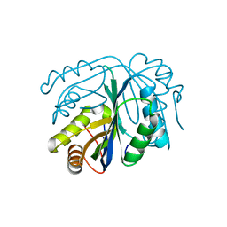
3ZEI
 | | Structure of the Mycobacterium tuberculosis O-Acetylserine Sulfhydrylase (OASS) CysK1 in complex with a small molecule inhibitor | | 分子名称: | (4S)-2-METHYL-2,4-PENTANEDIOL, 3-[(Z)-[(5Z)-5-[[2-(2-hydroxy-2-oxoethyloxy)phenyl]methylidene]-3-methyl-4-oxidanylidene-1,3-thiazolidin-2-ylidene]amino]benzoic acid, O-ACETYLSERINE SULFHYDRYLASE, ... | | 著者 | Poyraz, O, Schnell, R, Schneider, G. | | 登録日 | 2012-12-05 | | 公開日 | 2013-08-07 | | 最終更新日 | 2023-12-20 | | 実験手法 | X-RAY DIFFRACTION (2 Å) | | 主引用文献 | Structure-Guided Design of Novel Thiazolidine Inhibitors of O-Acetyl Serine Sulfhydrylase from Mycobacterium Tuberculosis.
J.Med.Chem., 56, 2013
|
|


