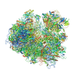
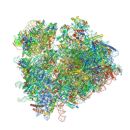
6SWC
 
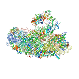 | | IC2B model of cryo-EM structure of a full archaeal ribosomal translation initiation complex devoid of aIF1 in P. abyssi | | 分子名称: | 16S ribosomal rRNA, 30S ribosomal protein S10, 30S ribosomal protein S11, ... | | 著者 | Coureux, P.-D, Mechulam, Y, Schmitt, E. | | 登録日 | 2019-09-20 | | 公開日 | 2020-02-19 | | 最終更新日 | 2024-04-24 | | 実験手法 | ELECTRON MICROSCOPY (3.3 Å) | | 主引用文献 | Cryo-EM study of an archaeal 30S initiation complex gives insights into evolution of translation initiation.
Commun Biol, 3, 2020
|
|
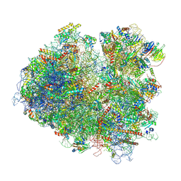
6SWD
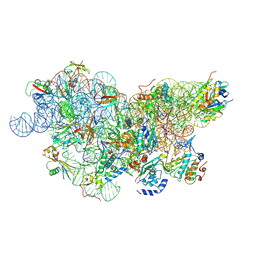 | | IC2 body model of cryo-EM structure of a full archaeal ribosomal translation initiation complex devoid of aIF1 in P. abyssi | | 分子名称: | 16S ribosomal RNA, 30S ribosomal protein S11, 30S ribosomal protein S12, ... | | 著者 | Coureux, P.-D, Mechulam, Y, Schmitt, E. | | 登録日 | 2019-09-20 | | 公開日 | 2020-02-19 | | 最終更新日 | 2024-04-24 | | 実験手法 | ELECTRON MICROSCOPY (3.2 Å) | | 主引用文献 | Cryo-EM study of an archaeal 30S initiation complex gives insights into evolution of translation initiation.
Commun Biol, 3, 2020
|
|
6SWZ
 | |
6SX4
 | |
6SXL
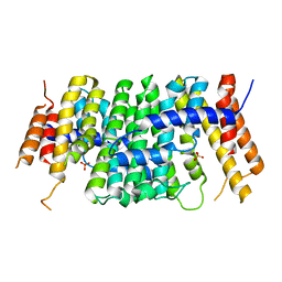 | | Crystal structure of CrtE | | 分子名称: | Geranylgeranyl pyrophosphate synthase, PHOSPHATE ION | | 著者 | Feng, Y, Morgan, R.M.L, Nixon, P.J. | | 登録日 | 2019-09-26 | | 公開日 | 2020-06-24 | | 最終更新日 | 2024-01-24 | | 実験手法 | X-RAY DIFFRACTION (2.5 Å) | | 主引用文献 | Crystal Structure of Geranylgeranyl Pyrophosphate Synthase (CrtE) Involved in Cyanobacterial Terpenoid Biosynthesis.
Front Plant Sci, 11, 2020
|
|
6SXN
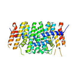 | |
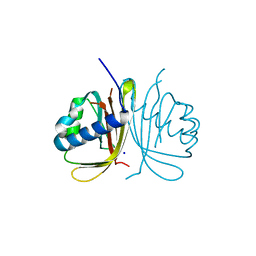
6T36
 | | Crystal structure of the PTPN3 PDZ domain bound to the HBV core protein C-terminal peptide | | 分子名称: | BROMIDE ION, Capsid protein, Tyrosine-protein phosphatase non-receptor type 3 | | 著者 | Genera, M, Mechaly, A, Haouz, A, Caillet-Saguy, C. | | 登録日 | 2019-10-10 | | 公開日 | 2021-01-20 | | 最終更新日 | 2024-01-24 | | 実験手法 | X-RAY DIFFRACTION (1.86 Å) | | 主引用文献 | Molecular basis of the interaction of the human tyrosine phosphatase PTPN3 with the hepatitis B virus core protein.
Sci Rep, 11, 2021
|
|
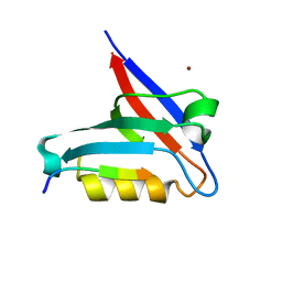
6T39
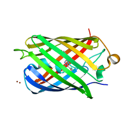
 | | Crystal structure of rsEGFP2 in its off-state determined by SFX | | 分子名称: | Green fluorescent protein | | 著者 | Woodhouse, J, Coquelle, N, Adam, V, Barends, T.R.M, De La Mora, E, Bourgeois, D, Colletier, J.P, Schlichting, I, Weik, M. | | 登録日 | 2019-10-10 | | 公開日 | 2020-02-19 | | 最終更新日 | 2024-01-24 | | 実験手法 | X-RAY DIFFRACTION (1.6 Å) | | 主引用文献 | Photoswitching mechanism of a fluorescent protein revealed by time-resolved crystallography and transient absorption spectroscopy.
Nat Commun, 11, 2020
|
|
6T3A
 | | Difference-refined structure of rsEGFP2 10 ns following 400-nm laser irradiation of the off-state determined by SFX | | 分子名称: | Green fluorescent protein | | 著者 | Woodhouse, J, Coquelle, N, Adam, V, Barends, T.R.M, De La Mora, E, Bourgeois, D, Colletier, J.P, Schlichting, I, Weik, M. | | 登録日 | 2019-10-10 | | 公開日 | 2020-02-19 | | 最終更新日 | 2024-01-24 | | 実験手法 | X-RAY DIFFRACTION (1.85 Å) | | 主引用文献 | Photoswitching mechanism of a fluorescent protein revealed by time-resolved crystallography and transient absorption spectroscopy.
Nat Commun, 11, 2020
|
|
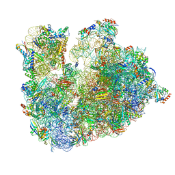
6T4Q
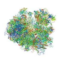 | | Structure of yeast 80S ribosome stalled on the CGA-CCG inhibitory codon combination. | | 分子名称: | 18S rRNA, 25S rRNA, 40S ribosomal protein S0-A, ... | | 著者 | Tesina, P, Buschauer, R, Cheng, J, Berninghausen, O, Becker, R, Beckmann, R. | | 登録日 | 2019-10-14 | | 公開日 | 2019-12-25 | | 最終更新日 | 2024-05-22 | | 実験手法 | ELECTRON MICROSCOPY (2.6 Å) | | 主引用文献 | Molecular mechanism of translational stalling by inhibitory codon combinations and poly(A) tracts.
Embo J., 39, 2020
|
|
6T7I
 | | Structure of yeast 80S ribosome stalled on the CGA-CGA inhibitory codon combination. | | 分子名称: | 18S ribosomal RNA, 25S ribosomal RNA, 40S ribosomal protein S0-A, ... | | 著者 | Tesina, P, Buschauer, R, Cheng, J, Berninghausen, O, Becker, R, Beckmann, R. | | 登録日 | 2019-10-22 | | 公開日 | 2019-12-25 | | 最終更新日 | 2024-05-22 | | 実験手法 | ELECTRON MICROSCOPY (3.2 Å) | | 主引用文献 | Molecular mechanism of translational stalling by inhibitory codon combinations and poly(A) tracts.
Embo J., 39, 2020
|
|
6T7T
 | | Structure of yeast 80S ribosome stalled on poly(A) tract. | | 分子名称: | 18S rRNA, 25S rRNA, 40S ribosomal protein S0-A, ... | | 著者 | Tesina, P, Buschauer, R, Cheng, J, Berninghausen, O, Becker, R, Beckmann, R. | | 登録日 | 2019-10-23 | | 公開日 | 2019-12-25 | | 最終更新日 | 2020-02-19 | | 実験手法 | ELECTRON MICROSCOPY (3.1 Å) | | 主引用文献 | Molecular mechanism of translational stalling by inhibitory codon combinations and poly(A) tracts.
Embo J., 39, 2020
|
|
6T83
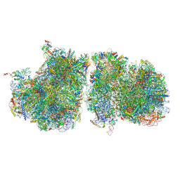 | | Structure of yeast disome (di-ribosome) stalled on poly(A) tract. | | 分子名称: | 18S rRNA, 25S rRNA, 40S ribosomal protein S0-A, ... | | 著者 | Tesina, P, Buschauer, R, Cheng, J, Berninghausen, O, Becker, R, Beckmann, R. | | 登録日 | 2019-10-24 | | 公開日 | 2019-12-25 | | 最終更新日 | 2020-02-12 | | 実験手法 | ELECTRON MICROSCOPY (4 Å) | | 主引用文献 | Molecular mechanism of translational stalling by inhibitory codon combinations and poly(A) tracts.
Embo J., 39, 2020
|
|
6T90
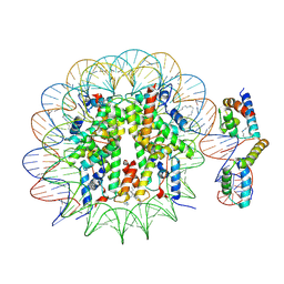 | | OCT4-SOX2-bound nucleosome - SHL-6 | | 分子名称: | DNA (146-MER), Green fluorescent protein,POU domain, class 5, ... | | 著者 | Michael, A.K, Kempf, G, Cavadini, S, Bunker, R.D, Thoma, N.H. | | 登録日 | 2019-10-25 | | 公開日 | 2020-05-06 | | 最終更新日 | 2020-07-08 | | 実験手法 | ELECTRON MICROSCOPY (3.05 Å) | | 主引用文献 | Mechanisms of OCT4-SOX2 motif readout on nucleosomes.
Science, 368, 2020
|
|
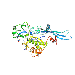
6TA7
 | | CRYSTAL STRUCTURE OF HUMAN G3BP1-NTF2 IN COMPLEX WITH HUMAN CAPRIN1-DERIVED SOLOMON MOTIF | | 分子名称: | CHLORIDE ION, Caprin-1, Ras GTPase-activating protein-binding protein 1, ... | | 著者 | Schulte, T, Achour, A, Panas, M.D, McInerney, G.M. | | 登録日 | 2019-10-29 | | 公開日 | 2021-05-12 | | 最終更新日 | 2024-01-24 | | 実験手法 | X-RAY DIFFRACTION (1.93 Å) | | 主引用文献 | Caprin-1 binding to the critical stress granule protein G3BP1 is regulated by pH
Biorxiv, 2021
|
|
6TB3
 | | yeast 80S ribosome in complex with the Not5 subunit of the CCR4-NOT complex | | 分子名称: | 25S rRNA, 40S ribosomal protein S0-A, 40S ribosomal protein S1-A, ... | | 著者 | Buschauer, R, Cheng, J, Berninghausen, O, Tesina, P, Becker, T, Beckmann, R. | | 登録日 | 2019-10-31 | | 公開日 | 2020-04-22 | | 最終更新日 | 2024-05-22 | | 実験手法 | ELECTRON MICROSCOPY (2.8 Å) | | 主引用文献 | The Ccr4-Not complex monitors the translating ribosome for codon optimality.
Science, 368, 2020
|
|
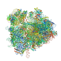
6TH6
 | | Cryo-EM Structure of T. kodakarensis 70S ribosome | | 分子名称: | 16S ribosomal RNA, 23S ribosomal RNA, 30S ribosomal protein S10, ... | | 著者 | Matzov, D, Sas-Chen, A, Thomas, J.M, Santangelo, T, Meier, J.L, Schwartz, S, Shalev-Benami, M. | | 登録日 | 2019-11-18 | | 公開日 | 2020-07-29 | | 最終更新日 | 2024-04-24 | | 実験手法 | ELECTRON MICROSCOPY (2.55 Å) | | 主引用文献 | Dynamic RNA acetylation revealed by quantitative cross-evolutionary mapping.
Nature, 583, 2020
|
|
6TIT
 | | VSV G_440 | | 分子名称: | 2-acetamido-2-deoxy-beta-D-glucopyranose, ACETATE ION, CALCIUM ION, ... | | 著者 | Albertini, A.A, Belot, L, Abouhamdan, A, Gaudin, Y. | | 登録日 | 2019-11-22 | | 公開日 | 2020-09-02 | | 最終更新日 | 2024-01-24 | | 実験手法 | X-RAY DIFFRACTION (2.07 Å) | | 主引用文献 | Identification of a pH-Sensitive Switch in VSV-G and a Crystal Structure of the G Pre-fusion State Highlight the VSV-G Structural Transition Pathway.
Cell Rep, 32, 2020
|
|
6TNU
 | | Yeast 80S ribosome in complex with eIF5A and decoding A-site and P-site tRNAs. | | 分子名称: | 18S rRNA, 25S rRNA, 4-{(2R)-2-[(1S,3S,5S)-3,5-dimethyl-2-oxocyclohexyl]-2-hydroxyethyl}piperidine-2,6-dione, ... | | 著者 | Buschauer, R, Cheng, J, Berninghausen, O, Tesina, P, Becker, T, Beckmann, R. | | 登録日 | 2019-12-10 | | 公開日 | 2020-04-22 | | 最終更新日 | 2020-04-29 | | 実験手法 | ELECTRON MICROSCOPY (3.1 Å) | | 主引用文献 | The Ccr4-Not complex monitors the translating ribosome for codon optimality.
Science, 368, 2020
|
|
6U1A
 | | Crystal Structure of Fluorescent Protein FusionRed | | 分子名称: | CALCIUM ION, NICKEL (II) ION, Red fluorescent protein | | 著者 | Pletnev, S, Muslinkina, L, Pletneva, N, Pletnev, V.Z. | | 登録日 | 2019-08-15 | | 公開日 | 2020-04-22 | | 最終更新日 | 2023-11-15 | | 実験手法 | X-RAY DIFFRACTION (1.09 Å) | | 主引用文献 | Two independent routes of post-translational chemistry in fluorescent protein FusionRed.
Int.J.Biol.Macromol., 155, 2020
|
|
6U95
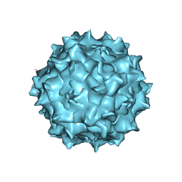 | | Adeno-associated virus strain AAVhu.37 capsid icosahedral structure | | 分子名称: | Capsid protein VP1 | | 著者 | Kaelber, J.T, Yost, S.A, Firlar, E, Mercer, A.C. | | 登録日 | 2019-09-06 | | 公開日 | 2019-09-18 | | 最終更新日 | 2024-03-20 | | 実験手法 | ELECTRON MICROSCOPY (2.56 Å) | | 主引用文献 | Structure of the AAVhu.37 capsid by cryoelectron microscopy.
Acta Crystallogr.,Sect.F, 76, 2020
|
|
6UHJ
 | | X-ray Structure of C148 mGFP | | 分子名称: | C148 mGFP | | 著者 | Winegar, P.W, Hayes, O.G, McMillan, J.R, Figg, C.A, Focia, P.J, Mirkin, C.A. | | 登録日 | 2019-09-27 | | 公開日 | 2020-03-18 | | 最終更新日 | 2023-11-15 | | 実験手法 | X-RAY DIFFRACTION (1.5 Å) | | 主引用文献 | DNA-Directed Protein Packing within Single Crystals.
Chem, 6, 2020
|
|
6UHK
 | | Crystal Structure of C176 mGFP | | 分子名称: | C176 mGFP | | 著者 | Winegar, P.W, Hayes, O.G, McMillan, J.R, Figg, C.A, Focia, P.J, Mirkin, C.A. | | 登録日 | 2019-09-27 | | 公開日 | 2020-03-18 | | 最終更新日 | 2023-11-15 | | 実験手法 | X-RAY DIFFRACTION (1.9 Å) | | 主引用文献 | DNA-Directed Protein Packing within Single Crystals.
Chem, 6, 2020
|
|
6UHL
 | | Crystal Structure of C148 mGFP-scDNA-1 | | 分子名称: | C148 mGFP-scDNA-1, UNKNOWN LIGAND | | 著者 | Winegar, P.W, Hayes, O.G, McMillan, J.R, Figg, C.A, Focia, P.J, Mirkin, C.A. | | 登録日 | 2019-09-27 | | 公開日 | 2020-03-18 | | 最終更新日 | 2023-11-15 | | 実験手法 | X-RAY DIFFRACTION (1.91 Å) | | 主引用文献 | DNA-Directed Protein Packing within Single Crystals.
Chem, 6, 2020
|
|
6UHM
 | | Crystal Structure of a Physical Mixture of C148 mGFP and scDNA-1 | | 分子名称: | C148 mGFP | | 著者 | Winegar, P.W, Hayes, O.G, McMillan, J.R, Figg, C.A, Focia, P.J, Mirkin, C.A. | | 登録日 | 2019-09-27 | | 公開日 | 2020-03-18 | | 最終更新日 | 2023-11-15 | | 実験手法 | X-RAY DIFFRACTION (2.1 Å) | | 主引用文献 | DNA-Directed Protein Packing within Single Crystals.
Chem, 6, 2020
|
|


