8A6R
 
 | |
8A6N
 | |
8A6Q
 | |
8A6G
 | |
7Y40
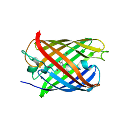
 | | Crystal structure of a bright green fluorescent protein (StayGold) in jellyfish Cytaeis uchidae from Biortus | | 分子名称: | 1,2-ETHANEDIOL, staygold | | 著者 | Wu, J, Wang, F, Gui, W, Cheng, W, Yang, Y. | | 登録日 | 2022-06-13 | | 公開日 | 2023-07-05 | | 最終更新日 | 2024-04-03 | | 実験手法 | X-RAY DIFFRACTION (1.7 Å) | | 主引用文献 | Crystal structure of a bright green fluorescent protein (StayGold) in jellyfish Cytaeis uchidae from Biortus
To Be Published
|
|
7XSU
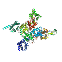 | | Cardiac sodium channel in complex with LqhIII | | 分子名称: | (3beta,14beta,17beta,25R)-3-[4-methoxy-3-(methoxymethyl)butoxy]spirost-5-en, 2-acetamido-2-deoxy-beta-D-glucopyranose, Alpha-like toxin Lqh3, ... | | 著者 | Jiang, D, Catterall, W.A. | | 登録日 | 2022-05-15 | | 公開日 | 2023-11-29 | | 実験手法 | ELECTRON MICROSCOPY (3.4 Å) | | 主引用文献 | Structural Basis for Nav1.5 Opening Modulated by a Gating Modifier Toxin
To Be Published
|
|
7ZCT
 | |
7UGS
 | |
7UGT
 | |
7UGR
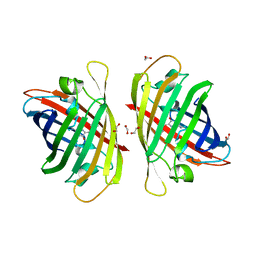
 | | Crystal structure of hyperfolder YFP | | 分子名称: | 1,2-ETHANEDIOL, DI(HYDROXYETHYL)ETHER, Hyperfolder yellow fluorescent protein, ... | | 著者 | Campbell, B.C, Liu, C.F, Petsko, G.A. | | 登録日 | 2022-03-25 | | 公開日 | 2022-10-26 | | 最終更新日 | 2023-11-15 | | 実験手法 | X-RAY DIFFRACTION (1.74 Å) | | 主引用文献 | Chemically stable fluorescent proteins for advanced microscopy.
Nat.Methods, 19, 2022
|
|
7Z7P
 | |
7Z7O
 | |
7Z7Q
 | |
7X5V
 | |
7TSR
 | |
7TSU
 | |
7TSS
 | |
7TSV
 | |
7QLL
 | |
7QLK
 | |
7QLI
 | |
7QLJ
 | |
7QLM
 | |
7QLN
 | |
7QLO
 | |


