8OTS
 
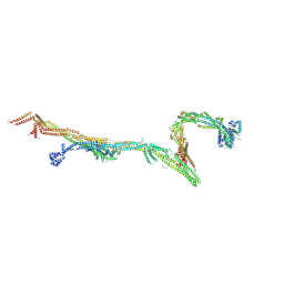
 | | OCT4 and MYC-MAX co-bound to a nucleosome | | 分子名称: | DNA (127-MER), Green fluorescent protein,POU domain, class 5, ... | | 著者 | Michael, A.K, Stoos, L, Kempf, G, Cavadini, S, Thoma, N. | | 登録日 | 2023-04-21 | | 公開日 | 2023-05-24 | | 最終更新日 | 2023-07-26 | | 実験手法 | ELECTRON MICROSCOPY (3.3 Å) | | 主引用文献 | Cooperation between bHLH transcription factors and histones for DNA access.
Nature, 619, 2023
|
|
8OVP
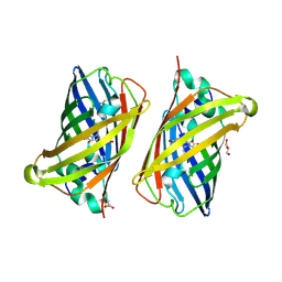
 | | X-ray structure of the iAspSnFR in complex with L-aspartate | | 分子名称: | ACETATE ION, ASPARTIC ACID, MAGNESIUM ION, ... | | 著者 | Tarnawski, M, Hellweg, L, Bergner, A, Hiblot, J, Leippe, P, Johnsson, K. | | 登録日 | 2023-04-26 | | 公開日 | 2023-05-17 | | 実験手法 | X-RAY DIFFRACTION (1.7 Å) | | 主引用文献 | X-ray structure of the SF-iAspSnFR in complex with L-aspartate
To Be Published
|
|
8OVN
 | | X-ray structure of the SF-iGluSnFR-S72A | | 分子名称: | CITRIC ACID, Putative periplasmic binding transport protein,Green fluorescent protein | | 著者 | Tarnawski, M, Hellweg, L, Bergner, A, Hiblot, J, Leippe, P, Johnsson, K. | | 登録日 | 2023-04-26 | | 公開日 | 2023-05-17 | | 実験手法 | X-RAY DIFFRACTION (2.6 Å) | | 主引用文献 | X-ray structure of the SF-iGluSnFR-S72A
To Be Published
|
|
8OVO
 | | X-ray structure of the SF-iGluSnFR-S72A in complex with L-aspartate | | 分子名称: | ASPARTIC ACID, Putative periplasmic binding transport protein,Green fluorescent protein | | 著者 | Tarnawski, M, Hellweg, L, Bergner, A, Hiblot, J, Leippe, P, Johnsson, K. | | 登録日 | 2023-04-26 | | 公開日 | 2023-05-17 | | 実験手法 | X-RAY DIFFRACTION (1.7 Å) | | 主引用文献 | X-ray structure of the SF-iGluSnFR-S72A in complex with L-aspartate
To Be Published
|
|
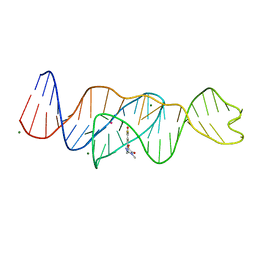
8FHX
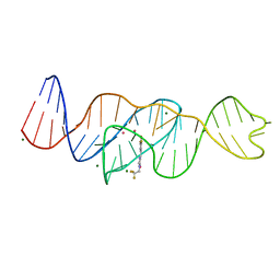 | | Structure of Lettuce aptamer bound to DFHBI-1T | | 分子名称: | (5Z)-5-(3,5-difluoro-4-hydroxybenzylidene)-2-methyl-3-(2,2,2-trifluoroethyl)-3,5-dihydro-4H-imidazol-4-one, Lettuce DNA aptamer, MAGNESIUM ION, ... | | 著者 | Passalacqua, L.F.M, Ferre-D'Amare, A.R. | | 登録日 | 2022-12-15 | | 公開日 | 2023-05-10 | | 最終更新日 | 2023-10-25 | | 実験手法 | X-RAY DIFFRACTION (2.5 Å) | | 主引用文献 | Intricate 3D architecture of a DNA mimic of GFP.
Nature, 618, 2023
|
|
8FI8
 | |
8FI0
 | |
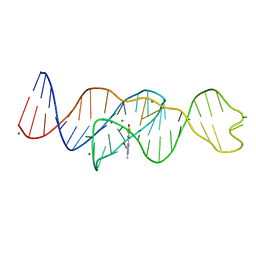
8FI2
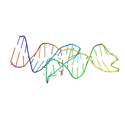
 | | Structure of Lettuce C20T bound to DFHBI-1T | | 分子名称: | (5Z)-5-(3,5-difluoro-4-hydroxybenzylidene)-2-methyl-3-(2,2,2-trifluoroethyl)-3,5-dihydro-4H-imidazol-4-one, Lettuce DNA aptamer, MAGNESIUM ION, ... | | 著者 | Passalacqua, L.F.M, Ferre-D'Amare, A.R. | | 登録日 | 2022-12-15 | | 公開日 | 2023-05-10 | | 最終更新日 | 2023-10-25 | | 実験手法 | X-RAY DIFFRACTION (3 Å) | | 主引用文献 | Intricate 3D architecture of a DNA mimic of GFP.
Nature, 618, 2023
|
|
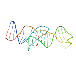
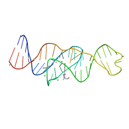
8FI7
 | | Structure of Lettuce C20T bound to DFHO | | 分子名称: | (5Z)-5-[(3,5-difluoro-4-hydroxyphenyl)methylidene]-2-[(E)-(hydroxyimino)methyl]-3-methyl-3,5-dihydro-4H-imidazol-4-one, Lettuce DNA aptamer, MAGNESIUM ION, ... | | 著者 | Passalacqua, L.F.M, Ferre-D'Amare, A.R. | | 登録日 | 2022-12-15 | | 公開日 | 2023-05-10 | | 最終更新日 | 2023-10-25 | | 実験手法 | X-RAY DIFFRACTION (2.9 Å) | | 主引用文献 | Intricate 3D architecture of a DNA mimic of GFP.
Nature, 618, 2023
|
|
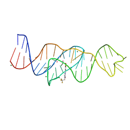
8FI1
 | | Structure of Lettuce C20G bound to DFHO | | 分子名称: | (5Z)-5-[(3,5-difluoro-4-hydroxyphenyl)methylidene]-2-[(E)-(hydroxyimino)methyl]-3-methyl-3,5-dihydro-4H-imidazol-4-one, Lettuce DNA aptamer, MAGNESIUM ION, ... | | 著者 | Passalacqua, L.F.M, Ferre-D'Amare, A.R. | | 登録日 | 2022-12-15 | | 公開日 | 2023-05-10 | | 最終更新日 | 2023-10-25 | | 実験手法 | X-RAY DIFFRACTION (2.6 Å) | | 主引用文献 | Intricate 3D architecture of a DNA mimic of GFP.
Nature, 618, 2023
|
|
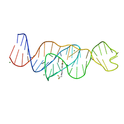
8FHZ
 | | Structure of Lettuce aptamer bound to DFHO | | 分子名称: | (5Z)-5-[(3,5-difluoro-4-hydroxyphenyl)methylidene]-2-[(E)-(hydroxyimino)methyl]-3-methyl-3,5-dihydro-4H-imidazol-4-one, Lettuce DNA aptamer, MAGNESIUM ION, ... | | 著者 | Passalacqua, L.F.M, Ferre-D'Amare, A.R. | | 登録日 | 2022-12-15 | | 公開日 | 2023-05-10 | | 最終更新日 | 2023-10-25 | | 実験手法 | X-RAY DIFFRACTION (2.6 Å) | | 主引用文献 | Intricate 3D architecture of a DNA mimic of GFP.
Nature, 618, 2023
|
|
8FHV
 | | Structure of Lettuce aptamer bound to DFHBI-1T with thallium I ions | | 分子名称: | (5Z)-5-(3,5-difluoro-4-hydroxybenzylidene)-2-methyl-3-(2,2,2-trifluoroethyl)-3,5-dihydro-4H-imidazol-4-one, Lettuce DNA aptamer, MAGNESIUM ION, ... | | 著者 | Passalacqua, L.F.M, Ferre-D'Amare, A.R. | | 登録日 | 2022-12-15 | | 公開日 | 2023-05-10 | | 最終更新日 | 2024-05-22 | | 実験手法 | X-RAY DIFFRACTION (2.5 Å) | | 主引用文献 | Intricate 3D architecture of a DNA mimic of GFP.
Nature, 618, 2023
|
|
7YDQ
 | | Structure of PfNT1(Y190A)-GFP in complex with GSK4 | | 分子名称: | 5-methyl-N-[2-(2-oxidanylideneazepan-1-yl)ethyl]-2-phenyl-1,3-oxazole-4-carboxamide, Nucleoside transporter 1,Green fluorescent protein | | 著者 | Wang, C, Yu, L.Y, Li, J.L, Ren, R.B, Deng, D. | | 登録日 | 2022-07-04 | | 公開日 | 2023-04-26 | | 実験手法 | ELECTRON MICROSCOPY (4.04 Å) | | 主引用文献 | Structural basis of the substrate recognition and inhibition mechanism of Plasmodium falciparum nucleoside transporter PfENT1.
Nat Commun, 14, 2023
|
|
8FCK
 | | Structure of the vertebrate augmin complex | | 分子名称: | HAUS augmin like complex subunit 2 L homeolog, Green fluorescent protein chimera, HAUS augmin like complex subunit 4 L homeolog, ... | | 著者 | Travis, S.M, Huang, W, Zhang, R, Petry, S. | | 登録日 | 2022-12-01 | | 公開日 | 2023-04-19 | | 最終更新日 | 2023-04-26 | | 実験手法 | ELECTRON MICROSCOPY (6.88 Å) | | 主引用文献 | Integrated model of the vertebrate augmin complex.
Nat Commun, 14, 2023
|
|
7Q6B
 | | mRubyFT/S148I, a mutant of blue-to-red fluorescent timer in its blue state | | 分子名称: | mRubyFT S148I, a mutant of blue-to-red fluorescent timer | | 著者 | Boyko, K.M, Nikolaeva, A.Y, Vlaskina, A.V, Dorovatovskii, P.V, Khrenova, M.G, Subach, O.M, Popov, V.O, Subach, F.M. | | 登録日 | 2021-11-06 | | 公開日 | 2023-04-12 | | 最終更新日 | 2024-02-07 | | 実験手法 | X-RAY DIFFRACTION (1.8 Å) | | 主引用文献 | Combined Structural and Computational Study of the mRubyFT Fluorescent Timer Locked in Its Blue Form.
Int J Mol Sci, 24, 2023
|
|
8BVG
 | |
8F6P
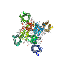 | | Rat Cardiac Sodium Channel with Ranolazine Bound | | 分子名称: | (R)-ranolazine, 2-acetamido-2-deoxy-beta-D-glucopyranose, 2-acetamido-2-deoxy-beta-D-glucopyranose-(1-4)-2-acetamido-2-deoxy-beta-D-glucopyranose, ... | | 著者 | Lenaeus, M.J, Tonggu, L. | | 登録日 | 2022-11-16 | | 公開日 | 2023-04-12 | | 実験手法 | ELECTRON MICROSCOPY (3.2 Å) | | 主引用文献 | Structural Basis for Inhibition of the Cardiac Sodium Channel by the Atypical Antiarrhythmic Drug Ranolazine
To Be Published
|
|
8C0N
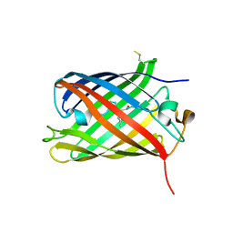 | | Crystal structure of the red form of the mTagFT fluorescent timer | | 分子名称: | Blue-to-red TagFT fluorescent timer | | 著者 | Boyko, K.M, Nikolaeva, A.Y, Vlaskina, A.V, Agapova, Y.K, Subach, O.M, Popov, V.O, Subach, F.V. | | 登録日 | 2022-12-19 | | 公開日 | 2023-03-08 | | 最終更新日 | 2023-11-15 | | 実験手法 | X-RAY DIFFRACTION (2.9 Å) | | 主引用文献 | Blue-to-Red TagFT, mTagFT, mTsFT, and Green-to-FarRed mNeptusFT2 Proteins, Genetically Encoded True and Tandem Fluorescent Timers.
Int J Mol Sci, 24, 2023
|
|
8FEE
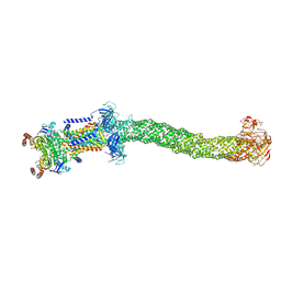 | | Structure of Mce1 transporter from Mycobacterium smegmatis in the absence of LucB (Map2) | | 分子名称: | ABC transporter, ATP-binding protein,Green fluorescent protein chimera, ABC-transporter integral membrane protein, ... | | 著者 | Chen, J, Bhabha, G, Ekiert, D.C. | | 登録日 | 2022-12-06 | | 公開日 | 2023-02-22 | | 最終更新日 | 2023-08-23 | | 実験手法 | ELECTRON MICROSCOPY (2.9 Å) | | 主引用文献 | Structure of an endogenous mycobacterial MCE lipid transporter.
Nature, 620, 2023
|
|
8FEF
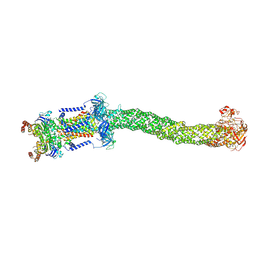 | | Structure of Mce1 transporter from Mycobacterium smegmatis (Map0) | | 分子名称: | ABC transporter, ATP-binding protein,Green fluorescent protein chimera, ABC-transporter integral membrane protein, ... | | 著者 | Chen, J, Bhabha, G, Ekiert, D.C. | | 登録日 | 2022-12-06 | | 公開日 | 2023-02-22 | | 最終更新日 | 2023-08-23 | | 実験手法 | ELECTRON MICROSCOPY (2.71 Å) | | 主引用文献 | Structure of an endogenous mycobacterial MCE lipid transporter.
Nature, 620, 2023
|
|
8FED
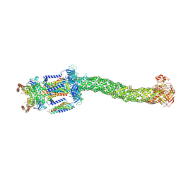 | | Structure of Mce1-LucB complex from Mycobacterium smegmatis (Map1) | | 分子名称: | ABC transporter, ATP-binding protein,Green fluorescent protein chimera, ABC-transporter integral membrane protein, ... | | 著者 | Chen, J, Bhabha, G, Ekiert, D.C. | | 登録日 | 2022-12-06 | | 公開日 | 2023-02-22 | | 最終更新日 | 2023-08-23 | | 実験手法 | ELECTRON MICROSCOPY (2.76 Å) | | 主引用文献 | Structure of an endogenous mycobacterial MCE lipid transporter.
Nature, 620, 2023
|
|
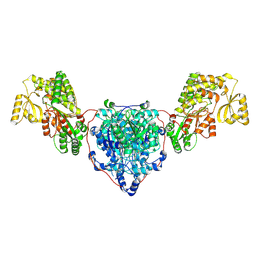
8CV1
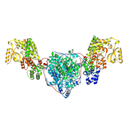 | | ACP1-KS-AT domains of mycobacterial Pks13 | | 分子名称: | Polyketide synthase PKS13, UNKNOWN LIGAND | | 著者 | Kim, S.K, Dickinson, M.S, Finer-Moore, J.S, Rosenberg, O.S, Stroud, R.M. | | 登録日 | 2022-05-17 | | 公開日 | 2023-02-15 | | 最終更新日 | 2023-03-29 | | 実験手法 | ELECTRON MICROSCOPY (2.6 Å) | | 主引用文献 | Structure and dynamics of the essential endogenous mycobacterial polyketide synthase Pks13.
Nat.Struct.Mol.Biol., 30, 2023
|
|
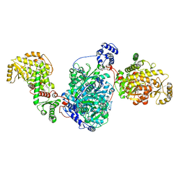
8CV0
 | | KS-AT domains of mycobacterial Pks13 with outward AT conformation | | 分子名称: | Polyketide synthase PKS13, UNKNOWN LIGAND | | 著者 | Kim, S.K, Dickinson, M.S, Finer-Moore, J.S, Rosenberg, O.S, Stroud, R.M. | | 登録日 | 2022-05-17 | | 公開日 | 2023-02-15 | | 最終更新日 | 2023-03-29 | | 実験手法 | ELECTRON MICROSCOPY (3.1 Å) | | 主引用文献 | Structure and dynamics of the essential endogenous mycobacterial polyketide synthase Pks13.
Nat.Struct.Mol.Biol., 30, 2023
|
|
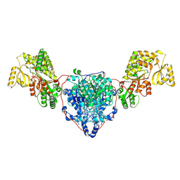
8CUY
 | | ACP1-KS-AT domains of mycobacterial Pks13 | | 分子名称: | 4'-PHOSPHOPANTETHEINE, Polyketide synthase PKS13, UNKNOWN LIGAND | | 著者 | Kim, S.K, Dickinson, M.S, Finer-Moore, J.S, Rosenberg, O.S, Stroud, R.M. | | 登録日 | 2022-05-17 | | 公開日 | 2023-02-15 | | 最終更新日 | 2023-03-29 | | 実験手法 | ELECTRON MICROSCOPY (2.4 Å) | | 主引用文献 | Structure and dynamics of the essential endogenous mycobacterial polyketide synthase Pks13.
Nat.Struct.Mol.Biol., 30, 2023
|
|
8CUZ
 | | KS-AT domains of mycobacterial Pks13 with inward AT conformation | | 分子名称: | Polyketide synthase PKS13, UNKNOWN LIGAND | | 著者 | Kim, S.K, Dickinson, M.S, Finer-Moore, J.S, Rosenberg, O.S, Stroud, R.M. | | 登録日 | 2022-05-17 | | 公開日 | 2023-02-15 | | 最終更新日 | 2023-03-29 | | 実験手法 | ELECTRON MICROSCOPY (3 Å) | | 主引用文献 | Structure and dynamics of the essential endogenous mycobacterial polyketide synthase Pks13.
Nat.Struct.Mol.Biol., 30, 2023
|
|


