1MYW
 
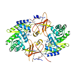
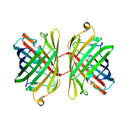
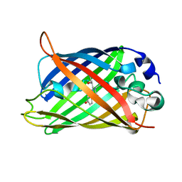
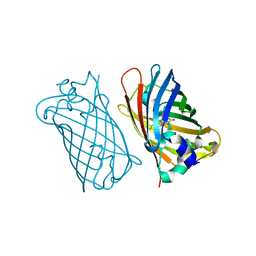 | | CRYSTAL STRUCTURE OF A YELLOW FLUORESCENT PROTEIN WITH IMPROVED MATURATION AND REDUCED ENVIRONMENTAL SENSITIVITY | | 分子名称: | Green fluorescent protein | | 著者 | Rekas, A, Alattia, J.R, Nagai, T, Miyawaki, A, Ikura, M. | | 登録日 | 2002-10-04 | | 公開日 | 2003-01-14 | | 最終更新日 | 2021-10-27 | | 実験手法 | X-RAY DIFFRACTION (2.2 Å) | | 主引用文献 | Crystal Structure of Venus, a Yellow Fluorescent
Protein with Improved Maturation and
Reduced Environmental Sensitivity
J.Biol.Chem., 277, 2002
|
|
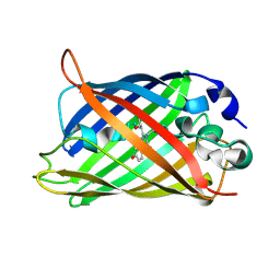
1KS8
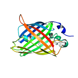
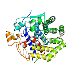 | | The structure of Endoglucanase from termite, Nasutitermes takasagoensis, at pH 2.5. | | 分子名称: | Endo-b-1,4-glucanase, SULFATE ION | | 著者 | Khademi, S, Guarino, L.A, Watanabe, H, Tokuda, G, Meyer, E.F. | | 登録日 | 2002-01-11 | | 公開日 | 2003-01-21 | | 最終更新日 | 2023-08-16 | | 実験手法 | X-RAY DIFFRACTION (1.4 Å) | | 主引用文献 | Structure of an endoglucanase from termite, Nasutitermes takasagoensis.
Acta Crystallogr.,Sect.D, 58, 2002
|
|
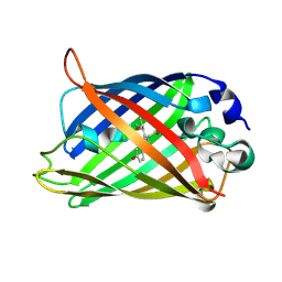
1KSD
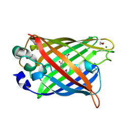
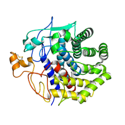 | | The structure of Endoglucanase from termite, Nasutitermes takasagoensis, at pH 6.5. | | 分子名称: | CALCIUM ION, Endo-b-1,4-glucanase | | 著者 | Khademi, S, Guarino, L.A, Watanabe, H, Tokuda, G, Meyer, E.F. | | 登録日 | 2002-01-12 | | 公開日 | 2003-01-21 | | 最終更新日 | 2023-08-16 | | 実験手法 | X-RAY DIFFRACTION (1.6 Å) | | 主引用文献 | Structure of an endoglucanase from termite, Nasutitermes takasagoensis.
Acta Crystallogr.,Sect.D, 58, 2002
|
|
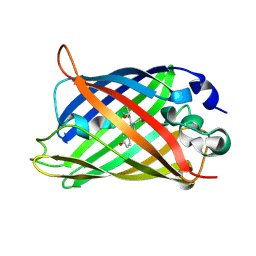
1KSC
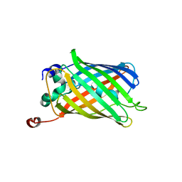
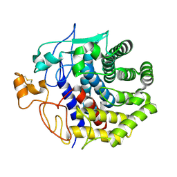 | | The structure of Endoglucanase from termite, Nasutitermes takasagoensis, at pH 5.6. | | 分子名称: | CALCIUM ION, Endo-b-1,4-glucanase | | 著者 | Khademi, S, Guarino, L.A, Watanabe, H, Tokuda, G, Meyer, E.F. | | 登録日 | 2002-01-11 | | 公開日 | 2003-01-21 | | 最終更新日 | 2023-08-16 | | 実験手法 | X-RAY DIFFRACTION (1.55 Å) | | 主引用文献 | Structure of an endoglucanase from termite, Nasutitermes takasagoensis.
Acta Crystallogr.,Sect.D, 58, 2002
|
|
1MOU
 | | Crystal structure of Coral pigment | | 分子名称: | GFP-like non-fluorescent chromoprotein, IODIDE ION | | 著者 | Prescott, M, Ling, M, Beddoe, T, Oakley, A.J, Dove, S, Hoegh-Guldberg, O, Devenish, R.J, Rossjohn, J. | | 登録日 | 2002-09-10 | | 公開日 | 2003-04-08 | | 最終更新日 | 2023-11-15 | | 実験手法 | X-RAY DIFFRACTION (2.2 Å) | | 主引用文献 | The 2.2 a crystal structure of a pocilloporin pigment reveals a nonplanar chromophore conformation.
Structure, 11, 2003
|
|
1MOV
 | | Crystal structure of Coral protein mutant | | 分子名称: | GFP-like non-fluorescent chromoprotein, IODIDE ION | | 著者 | Prescott, M, Ling, M, Beddoe, T, Oakley, A.J, Dove, S, Hoegh-Guldberg, O, Devenish, R.J, Rossjohn, J. | | 登録日 | 2002-09-10 | | 公開日 | 2003-04-08 | | 最終更新日 | 2023-11-15 | | 実験手法 | X-RAY DIFFRACTION (2.4 Å) | | 主引用文献 | The 2.2 a crystal structure of a pocilloporin pigment reveals a nonplanar chromophore conformation.
Structure, 11, 2003
|
|
1NJJ
 | |
1JC0
 | | CRYSTAL STRUCTURE ANALYSIS OF A REDOX-SENSITIVE GREEN FLUORESCENT PROTEIN VARIANT IN A REDUCED FORM | | 分子名称: | GREEN FLUORESCENT PROTEIN | | 著者 | Hanson, G.T, Aggeler, R, Oglesbee, D, Cannon, M, Capaldi, R.A, Tsien, R.Y, Remington, S.J. | | 登録日 | 2001-06-07 | | 公開日 | 2003-09-09 | | 最終更新日 | 2023-11-15 | | 実験手法 | X-RAY DIFFRACTION (2 Å) | | 主引用文献 | Investigating mitochondrial redox potential with redox-sensitive green fluorescent protein indicators.
J.Biol.Chem., 279, 2004
|
|
1JC1
 | | CRYSTAL STRUCTURE ANALYSIS OF A REDOX-SENSITIVE GREEN FLUORESCENT PROTEIN VARIANT IN A OXIDIZED FORM | | 分子名称: | GREEN FLUORESCENT PROTEIN | | 著者 | Hanson, G.T, Aggeler, R, Oglesbee, D, Cannon, M, Capaldi, R.A, Tsien, R.Y, Remington, S.J. | | 登録日 | 2001-06-07 | | 公開日 | 2003-09-09 | | 最終更新日 | 2023-11-15 | | 実験手法 | X-RAY DIFFRACTION (1.9 Å) | | 主引用文献 | Investigating mitochondrial redox potential with redox-sensitive green fluorescent protein indicators.
J.Biol.Chem., 279, 2004
|
|
1CV7
 | |
1QXT
 | | Crystal structure of precyclized intermediate for the green fluorescent protein R96A variant (A) | | 分子名称: | green-fluorescent protein | | 著者 | Barondeau, D.P, Putnam, C.D, Kassmann, C.J, Tainer, J.A, Getzoff, E.D. | | 登録日 | 2003-09-08 | | 公開日 | 2003-09-23 | | 最終更新日 | 2023-08-23 | | 実験手法 | X-RAY DIFFRACTION (2 Å) | | 主引用文献 | Mechanism and energetics of green fluorescent protein chromophore synthesis revealed by trapped intermediate structures
Proc.Natl.Acad.Sci.USA, 100
|
|
1QY3
 | | Crystal structure of precyclized intermediate for the green fluorescent protein R96A variant (B) | | 分子名称: | green-fluorescent protein | | 著者 | Barondeau, D.P, Putnam, C.D, Kassmann, C.J, Tainer, J.A, Getzoff, E.D. | | 登録日 | 2003-09-09 | | 公開日 | 2003-09-23 | | 最終更新日 | 2023-08-23 | | 実験手法 | X-RAY DIFFRACTION (2 Å) | | 主引用文献 | Mechanism and energetics of green fluorescent protein chromophore synthesis revealed by trapped intermediate structures.
Proc.Natl.Acad.Sci.Usa, 100, 2003
|
|
1QYF
 | | Crystal structure of matured green fluorescent protein R96A variant | | 分子名称: | 1,2-ETHANEDIOL, MAGNESIUM ION, green-fluorescent protein | | 著者 | Barondeau, D.P, Putnam, C.D, Kassmann, C.J, Tainer, J.A, Getzoff, E.D. | | 登録日 | 2003-09-10 | | 公開日 | 2003-09-30 | | 最終更新日 | 2023-11-15 | | 実験手法 | X-RAY DIFFRACTION (1.5 Å) | | 主引用文献 | Mechanism and energetics of green fluorescent protein chromophore synthesis revealed by trapped intermediate structures.
Proc.Natl.Acad.Sci.Usa, 100, 2003
|
|
1QYQ
 | | Crystal Structure of the cyclized S65G Y66G GFP variant | | 分子名称: | green-fluorescent protein | | 著者 | Barondeau, D.P, Putnam, C.D, Kassmann, C.J, Tainer, J.A, Getzoff, E.D. | | 登録日 | 2003-09-11 | | 公開日 | 2003-09-30 | | 最終更新日 | 2023-11-15 | | 実験手法 | X-RAY DIFFRACTION (1.8 Å) | | 主引用文献 | Mechanism and energetics of green fluorescent protein chromophore synthesis revealed by trapped intermediate structures.
Proc.Natl.Acad.Sci.Usa, 100, 2003
|
|
1QYO
 | | Anaerobic precylization intermediate crystal structure for S65G Y66G GFP variant | | 分子名称: | green-fluorescent protein | | 著者 | Barondeau, D.P, Putnam, C.D, Kassmann, C.J, Tainer, J.A, Getzoff, E.D. | | 登録日 | 2003-09-11 | | 公開日 | 2003-09-30 | | 最終更新日 | 2023-08-23 | | 実験手法 | X-RAY DIFFRACTION (1.8 Å) | | 主引用文献 | Mechanism and energetics of green fluorescent protein chromophore synthesis revealed by trapped intermediate structures.
Proc.Natl.Acad.Sci.Usa, 100, 2003
|
|
1UIS
 | | The 2.0 crystal structure of eqFP611, a far-red fluorescent protein from the sea anemone Entacmaea quadricolor | | 分子名称: | ACETIC ACID, CALCIUM ION, red fluorescent protein FP611 | | 著者 | Petersen, J, Wilmann, P.G, Beddoe, T, Oakley, A.J, Devenish, R.J, Prescott, M, Rossjohn, J. | | 登録日 | 2003-07-21 | | 公開日 | 2003-10-21 | | 最終更新日 | 2023-12-27 | | 実験手法 | X-RAY DIFFRACTION (2 Å) | | 主引用文献 | The 2.0A crystal structure of eqFP611, a far-red fluorescent protein from the sea anemone Entacmaea quadricolor
J.Biol.Chem., 278, 2003
|
|
1R9I
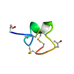 | | NMR Solution Structure of PIIIA toxin, NMR, 20 structures | | 分子名称: | Mu-conotoxin PIIIA | | 著者 | Nielsen, K.J, Watson, M, Adams, D.J, Hammarstrom, A.K, Gage, P.W, Hill, J.M, Craik, D.J, Thomas, L, Adams, D, Alewood, P.F, Lewis, R.J. | | 登録日 | 2003-10-30 | | 公開日 | 2003-11-18 | | 最終更新日 | 2019-12-25 | | 実験手法 | SOLUTION NMR | | 主引用文献 | Solution structure of mu-conotoxin PIIIA, a preferential inhibitor of persistent tetrodotoxin-sensitive sodium channels
J.Biol.Chem., 277, 2002
|
|
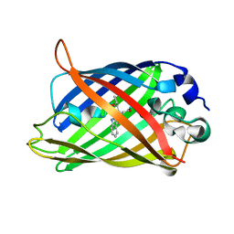
1OXF
 | | Expansion of the Genetic Code Enables Design of a Novel "Gold" Class of Green Fluorescent Proteins | | 分子名称: | cyan fluorescent protein cfp | | 著者 | Hyun Bae, J, Rubini, M, Jung, G, Wiegand, G, Seifert, M.H, Azim, M.K, Kim, J.S, Zumbusch, A, Holak, T.A, Moroder, L, Huber, R, Budisa, N. | | 登録日 | 2003-04-02 | | 公開日 | 2003-12-02 | | 最終更新日 | 2023-11-15 | | 実験手法 | X-RAY DIFFRACTION (1.69 Å) | | 主引用文献 | Expansion of the Genetic Code Enables Design of a Novel "Gold" Class of Green Fluorescent Proteins
J.Mol.Biol., 328, 2003
|
|
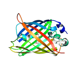
1OXE
 | | Expansion of the Genetic Code Enables Design of a Novel "Gold" Class of Green Fluorescent Proteins | | 分子名称: | cyan fluorescent protein cfp | | 著者 | Hyun Bae, J, Rubini, M, Jung, G, Wiegand, G, Seifert, M.H, Azim, M.K, Kim, J.S, Zumbusch, A, Holak, T.A, Moroder, L, Huber, R, Budisa, N. | | 登録日 | 2003-04-02 | | 公開日 | 2003-12-02 | | 最終更新日 | 2021-10-27 | | 実験手法 | X-RAY DIFFRACTION (1.15 Å) | | 主引用文献 | Expansion of the Genetic Code Enables Design of a Novel "Gold" Class of Green Fluorescent Proteins
J.Mol.Biol., 328, 2003
|
|
1OXD
 | | Expansion of the Genetic Code Enables Design of a Novel "Gold" Class of Green Fluorescent Proteins | | 分子名称: | cyan fluorescent protein cfp | | 著者 | Hyun Bae, J, Rubini, M, Jung, G, Wiegand, G, Seifert, M.H, Azim, M.K, Kim, J.S, Zumbusch, A, Holak, T.A, Moroder, L, Huber, R, Budisa, N. | | 登録日 | 2003-04-02 | | 公開日 | 2003-12-02 | | 最終更新日 | 2021-10-27 | | 実験手法 | X-RAY DIFFRACTION (1.15 Å) | | 主引用文献 | Expansion of the Genetic Code Enables Design of a Novel "Gold" Class of Green Fluorescent Proteins
J.Mol.Biol., 328, 2003
|
|
1Q73
 | |
1Q4E
 | |
1Q4A
 | |
1Q4D
 | |
1Q4B
 | |


