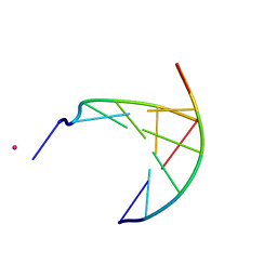
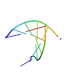
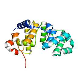
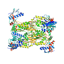
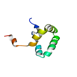
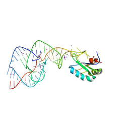
2KFP
 
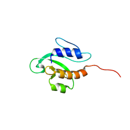 | | Solution NMR structure of PSPTO_3016 from Pseudomonas syringae. Northeast Structural Genomics Consortium target PsR293. | | 分子名称: | PSPTO_3016 protein | | 著者 | Feldmann, E.A, Ramelot, T.A, Zhao, L, Hamilton, K, Ciccosanti, C, Xiao, R, Nair, R, Everett, J.K, Swapna, G, Acton, T.B, Rost, B, Montelione, G.T, Kennedy, M.A, Northeast Structural Genomics Consortium (NESG) | | 登録日 | 2009-02-24 | | 公開日 | 2009-03-24 | | 最終更新日 | 2024-05-08 | | 実験手法 | SOLUTION NMR | | 主引用文献 | Solution NMR and X-ray crystal structures of Pseudomonas syringae Pspto_3016 from protein domain family PF04237 (DUF419) adopt a "double wing" DNA binding motif.
J.Struct.Funct.Genom., 13, 2012
|
|
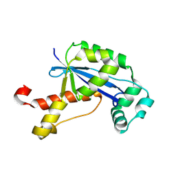
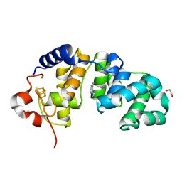
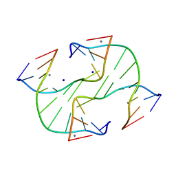
2A6M
 | | Crystal Structure of the ISHp608 Transposase | | 分子名称: | ISHp608 transposase | | 著者 | Ronning, D.R, Guynet, C, Ton-Hoang, B, Perez, Z.N, Ghirlando, R, Chandler, M, Dyda, F. | | 登録日 | 2005-07-03 | | 公開日 | 2005-10-25 | | 最終更新日 | 2024-02-14 | | 実験手法 | X-RAY DIFFRACTION (2.4 Å) | | 主引用文献 | Active site sharing and subterminal hairpin recognition in a new class of DNA transposases.
Mol.Cell, 20, 2005
|
|
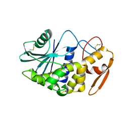
1MC8
 | | Crystal Structure of Flap Endonuclease-1 R42E mutant from Pyrococcus horikoshii | | 分子名称: | Flap Endonuclease-1 | | 著者 | Matsui, E, Musti, K.V, Abe, J, Yamazaki, K, Matsui, I, Harata, K. | | 登録日 | 2002-08-06 | | 公開日 | 2002-10-16 | | 最終更新日 | 2023-10-25 | | 実験手法 | X-RAY DIFFRACTION (3.1 Å) | | 主引用文献 | Molecular Structure and Novel DNA Binding Sites Located in Loops of Flap Endonuclease-1 from Pyrococcus horikoshii
J.BIOL.CHEM., 277, 2002
|
|
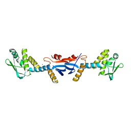
6IET
 | | The crystal structure of TRIM66 PHD-Bromo domain | | 分子名称: | Tripartite motif-containing protein 66, ZINC ION | | 著者 | Chen, J. | | 登録日 | 2018-09-17 | | 公開日 | 2019-09-18 | | 最終更新日 | 2023-11-22 | | 実験手法 | X-RAY DIFFRACTION (2.101 Å) | | 主引用文献 | TRIM66 reads unmodified H3R2K4 and H3K56ac to respond to DNA damage in embryonic stem cells.
Nat Commun, 10, 2019
|
|
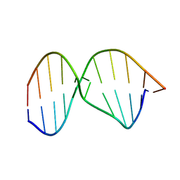
3GJK
 | | crystal structure of a DNA duplex containing 7,8-dihydropyridol[2,3-d]pyrimidin-2-one | | 分子名称: | 5'-D(*CP*GP*CP*GP*AP*A)-3', 5'-D(P*TP*TP*(B7C)P*GP*CP*G)-3', POTASSIUM ION | | 著者 | Takenaka, A, Juan, E.C.M, Shimizu, S, Haraguchi, T, Xiao, M, Kurose, T, Ohkubo, A, Sekine, M, Shibata, T, Millington, C.L, Williams, D.M. | | 登録日 | 2009-03-09 | | 公開日 | 2010-03-31 | | 最終更新日 | 2023-11-01 | | 実験手法 | X-RAY DIFFRACTION (2.2 Å) | | 主引用文献 | Insights into the stabilizing contributions of bicyclic cytosine analogues: crystal structures of DNA duplexes containing 7,8-dihydropyridol[2,3-d]pyrimidin-2-one
To be Published
|
|
2GBZ
 | | The Crystal Structure of XC847 from Xanthomonas campestris: a 3-5 Oligoribonuclease of DnaQ fold family with a Novel Opposingly-Shifted Helix | | 分子名称: | MAGNESIUM ION, Oligoribonuclease | | 著者 | Chin, K.H, Yang, C.Y, Chou, C.C, Wang, A.H.J, Chou, S.H. | | 登録日 | 2006-03-12 | | 公開日 | 2007-01-16 | | 最終更新日 | 2017-10-18 | | 実験手法 | X-RAY DIFFRACTION (2.3 Å) | | 主引用文献 | The crystal structure of XC847 from Xanthomonas campestris: a 3'-5' oligoribonuclease of DnaQ fold family with a novel opposingly shifted helix
Proteins, 65, 2006
|
|
1WEG
 | | Catalytic Domain Of Muty From Escherichia Coli K142A Mutant | | 分子名称: | 1,2-ETHANEDIOL, A/G-specific adenine glycosylase, IMIDAZOLE, ... | | 著者 | Hitomi, K, Arvai, A.S, Tainer, J.A. | | 登録日 | 2004-05-25 | | 公開日 | 2004-09-21 | | 最終更新日 | 2024-05-29 | | 実験手法 | X-RAY DIFFRACTION (1.8 Å) | | 主引用文献 | Reaction intermediates in the catalytic mechanism of Escherichia coli MutY DNA glycosylase
J.Biol.Chem., 279, 2004
|
|
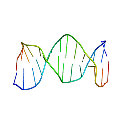
3GJL
 | | crystal structure of a DNA duplex containing 7,8-dihydropyridol[2,3-d]pyrimidin-2-one | | 分子名称: | 5'-D(*CP*GP*CP*GP*AP*A)-3', 5'-D(P*TP*TP*(B7C)P*GP*CP*G)-3', SODIUM ION | | 著者 | Takenaka, A, Juan, E.C.M, Shimizu, S, Haraguchi, T, Xiao, M, Kurose, T, Ohkubo, A, Sekine, M, Shibata, T, Millington, C.L, Williams, D.M. | | 登録日 | 2009-03-09 | | 公開日 | 2010-03-31 | | 最終更新日 | 2023-11-01 | | 実験手法 | X-RAY DIFFRACTION (1.92 Å) | | 主引用文献 | Insights into the stabilizing contributions of bicyclic cytosine analogues: crystal structures of DNA duplexes containing 7,8-dihydropyridol[2,3-d]pyrimidin-2-one
To be Published
|
|
1WEF
 | |
1WEI
 | | Catalytic Domain Of Muty From Escherichia Coli K20A Mutant Complexed To Adenine | | 分子名称: | 1,2-ETHANEDIOL, A/G-specific adenine glycosylase, ADENINE, ... | | 著者 | Hitomi, K, Arvai, A.S, Tainer, J.A. | | 登録日 | 2004-05-25 | | 公開日 | 2004-09-21 | | 最終更新日 | 2024-05-29 | | 実験手法 | X-RAY DIFFRACTION (1.45 Å) | | 主引用文献 | Reaction intermediates in the catalytic mechanism of Escherichia coli MutY DNA glycosylase
J.Biol.Chem., 279, 2004
|
|
6H09
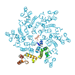 | | HIV capsid hexamer with IP6 ligand | | 分子名称: | Gag polyprotein, INOSITOL HEXAKISPHOSPHATE | | 著者 | James, L.C. | | 登録日 | 2018-07-06 | | 公開日 | 2018-08-15 | | 最終更新日 | 2020-10-07 | | 実験手法 | X-RAY DIFFRACTION (2 Å) | | 主引用文献 | IP6 is an HIV pocket factor that prevents capsid collapse and promotes DNA synthesis.
Elife, 7, 2018
|
|
1NGT
 | | The Role of Minor Groove Functional Groups in DNA Hydration | | 分子名称: | 5'-D(*CP*GP*CP*GP*AP*AP*(MTR)P*TP*CP*GP*CP*G)-3', MAGNESIUM ION | | 著者 | Woods, K.K, Lan, T, McLaughlin, L.W, Williams, L.D. | | 登録日 | 2002-12-17 | | 公開日 | 2003-03-04 | | 最終更新日 | 2024-02-14 | | 実験手法 | X-RAY DIFFRACTION (2.04 Å) | | 主引用文献 | The Role of Minor Groove Functional Groups in DNA Hydration
Nucleic Acids Res., 31, 2003
|
|
7CUX
 | | Crystal structure of human Schlafen 5 N'-terminal domain (SLFN5-N) involved in ssRNA cleaving and DNA binding | | 分子名称: | Schlafen family member 5, ZINC ION | | 著者 | Yang, J.Y, Luo, M, Ou, J.Y, Wang, Z.W, Gao, S. | | 登録日 | 2020-08-25 | | 公開日 | 2021-08-25 | | 最終更新日 | 2023-11-29 | | 実験手法 | X-RAY DIFFRACTION (3.29477072 Å) | | 主引用文献 | Crystal structure of human Schlafen 5 N'-terminal domain (SLFN5-N) involved in ssRNA cleaving and DNA binding
To Be Published
|
|
1K9L
 | |
1K5F
 | | SOLUTION STRUCTURE OF THE S-STYRENE ADDUCT IN THE RAS61 SEQUENCE | | 分子名称: | 5'-D(*CP*GP*GP*AP*CP*(ABS)P*AP*GP*AP*AP*G)-3', 5'-D(*CP*TP*TP*CP*TP*TP*GP*TP*CP*CP*G)-3' | | 著者 | Hennard, C, Finneman, J, Harris, C.M, Harris, T.M, Stone, M.P. | | 登録日 | 2001-10-10 | | 公開日 | 2002-01-23 | | 最終更新日 | 2024-05-22 | | 実験手法 | SOLUTION NMR | | 主引用文献 | The nonmutagenic (R)- and (S)-beta-(N(6)-adenyl)styrene oxide adducts are oriented in the major groove and show little perturbation to DNA
structure.
Biochemistry, 40, 2001
|
|
6IEU
 | |
1PVE
 | |
1K5E
 | | Solution Structure of R-styrene Adduct in the Ras61 Sequence | | 分子名称: | 5'-D(*CP*GP*GP*AP*CP*(ABR)P*AP*GP*AP*AP*G)-3', 5'-D(*CP*TP*TP*CP*TP*TP*GP*TP*CP*CP*G)-3' | | 著者 | Hennard, C, Finneman, J, Harris, C.M, Harris, T.M, Stone, M.P. | | 登録日 | 2001-10-10 | | 公開日 | 2002-01-23 | | 最終更新日 | 2024-05-22 | | 実験手法 | SOLUTION NMR | | 主引用文献 | The nonmutagenic (R)- and (S)-beta-(N(6)-adenyl)styrene oxide adducts are oriented in the major groove and show little perturbation to DNA
structure.
Biochemistry, 40, 2001
|
|
1K9H
 | |
2OIH
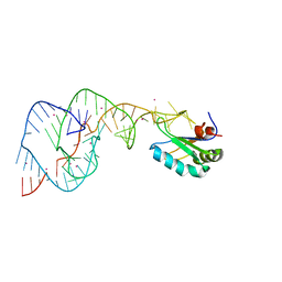 | | Hepatitis Delta Virus gemonic ribozyme precursor with C75U mutation and bound to monovalent cation Tl+ | | 分子名称: | HDV ribozyme, THALLIUM (I) ION, U1 small nuclear ribonucleoprotein A | | 著者 | Ke, A, Ding, F, Batchelor, J.D, Doudna, J.A. | | 登録日 | 2007-01-11 | | 公開日 | 2007-03-27 | | 最終更新日 | 2023-12-27 | | 実験手法 | X-RAY DIFFRACTION (2.4 Å) | | 主引用文献 | Structural roles of monovalent cations in the HDV ribozyme.
Structure, 15, 2007
|
|
2OJ3
 | | Hepatitis Delta Virus ribozyme precursor structure, with C75U mutation, bound to Tl+ and cobalt hexammine (Co(NH3)63+) | | 分子名称: | COBALT HEXAMMINE(III), HDV RIBOZYME, THALLIUM (I) ION, ... | | 著者 | Ke, A, Ding, F, Batchelor, J.D, Doudna, J.A. | | 登録日 | 2007-01-12 | | 公開日 | 2007-03-27 | | 最終更新日 | 2023-12-27 | | 実験手法 | X-RAY DIFFRACTION (2.9 Å) | | 主引用文献 | Structural roles of monovalent cations in the HDV ribozyme.
Structure, 15, 2007
|
|
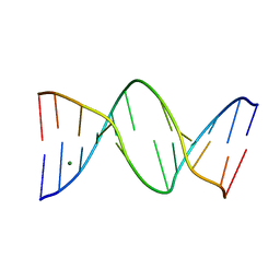
1L4J
 | | Holliday Junction TCGGTACCGA with Na and Ca Binding Sites. | | 分子名称: | 5'-D(*TP*CP*GP*GP*TP*AP*CP*CP*GP*A)-3', CALCIUM ION, SODIUM ION | | 著者 | Thorpe, J.H, Gale, B.C, Teixeira, S.C.M, Cardin, C.J. | | 登録日 | 2002-03-05 | | 公開日 | 2003-03-04 | | 最終更新日 | 2024-02-14 | | 実験手法 | X-RAY DIFFRACTION (1.85 Å) | | 主引用文献 | Conformational and Hydration Effects of Site-selective Sodium, Calcium and
Strontium Ion Binding to the DNA Holliday Junction Structure
d(TCGGTACCGA)4
J.Mol.Biol., 327, 2003
|
|
3H5K
 | | Crystal structure of the ribosome inactivating protein PDL1 | | 分子名称: | 1,2-ETHANEDIOL, 2-acetamido-2-deoxy-beta-D-glucopyranose, Ribosome-inactivating protein PD-L1/PD-L2 | | 著者 | Ruggiero, A, Di Maro, A, Severino, V, Chambery, A, Berisio, R. | | 登録日 | 2009-04-22 | | 公開日 | 2009-10-13 | | 最終更新日 | 2020-07-29 | | 実験手法 | X-RAY DIFFRACTION (1.45 Å) | | 主引用文献 | Crystal structure of PD-L1, a ribosome inactivating protein from Phytolacca dioica L. Leaves with the property to induce DNA cleavage
Biopolymers, 91, 2009
|
|
3KDK
 | |
3KDG
 | |


