7F7N
 
 | |
1DXA
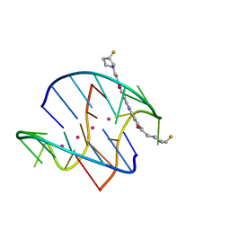
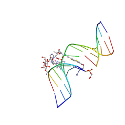
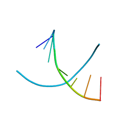
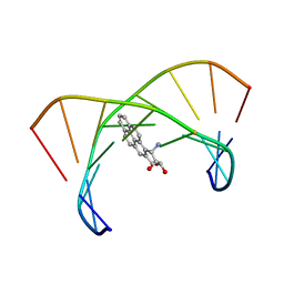 | | BENZO[A]PYRENE DIOL EPOXIDE ADDUCT OF DA IN DUPLEX DNA | | 分子名称: | 1,2,3-TRIHYDROXY-1,2,3,4-TETRAHYDROBENZO[A]PYRENE, DNA (5'-D(*CP*TP*CP*GP*GP*GP*AP*CP*C)-3'), DNA (5'-D(*GP*GP*TP*CP*AP*CP*GP*AP*G)-3') | | 著者 | Yeh, H.J.C, Sayer, J.M, Liu, X, Altieri, A.S, Byrd, R.A, Lakshman, M.K, Yagi, H, Schurter, E.J, Gorenstein, D.G, Jerina, D.M. | | 登録日 | 1995-09-01 | | 公開日 | 1995-12-07 | | 最終更新日 | 2024-03-13 | | 実験手法 | SOLUTION NMR | | 主引用文献 | NMR solution structure of a nonanucleotide duplex with a dG mismatch opposite a 10S adduct derived from trans addition of a deoxyadenosine N6-amino group to (+)-(7R,8S,9S,10R)-7,8-dihydroxy-9,10-epoxy-7,8,9,10- tetrahydrobenzo[a]pyrene: an unusual syn glycosidic torsion angle at the modified dA
Biochemistry, 34, 1995
|
|
3M7G
 | |
3M6K
 | |
3M7D
 | |
8A5D
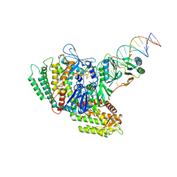
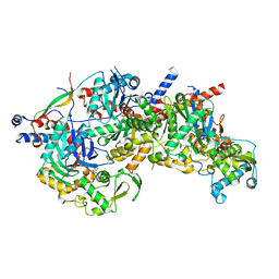 | | Structure of Arp4-Ies4-N-actin-Arp8-Ino80HSA subcomplex (A-module) of Chaetomium thermophilum INO80 | | 分子名称: | Actin, Actin related protein 4 (Arp4), Actin-related protein 8, ... | | 著者 | Kunert, F, Metzner, F.J, Eustermann, S, Jung, J, Woike, S, Schall, K, Kostrewa, D, Hopfner, K.P. | | 登録日 | 2022-06-14 | | 公開日 | 2022-12-28 | | 最終更新日 | 2024-07-24 | | 実験手法 | ELECTRON MICROSCOPY (2.9 Å) | | 主引用文献 | Structural mechanism of extranucleosomal DNA readout by the INO80 complex.
Sci Adv, 8, 2022
|
|
3NZ7
 | |
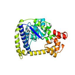
2YGK
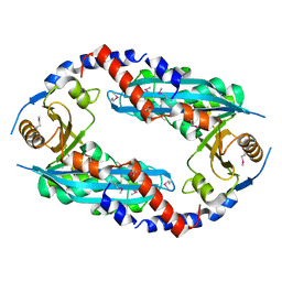 | | Crystal structure of the NurA nuclease from Sulfolobus solfataricus | | 分子名称: | MANGANESE (II) ION, NURA | | 著者 | Rzechorzek, N.J, Blackwood, J.K, Abrams, A.S, Maman, J.D, Robinson, N.P, Pellegrini, L. | | 登録日 | 2011-04-18 | | 公開日 | 2011-12-14 | | 最終更新日 | 2012-04-25 | | 実験手法 | X-RAY DIFFRACTION (2.5 Å) | | 主引用文献 | Structural and Functional Insights Into DNA-End Processing by the Archaeal Hera Helicase-Nura Nuclease Complex.
Nucleic Acids Res., 40, 2012
|
|
3M6Z
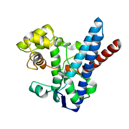
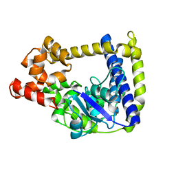
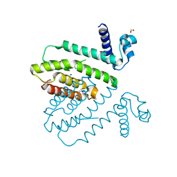
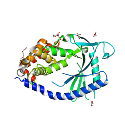
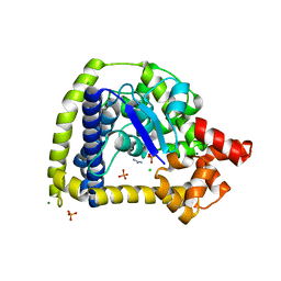 | | Crystal structure of an N-terminal 44 kDa fragment of topoisomerase V in the presence of guanidium hydrochloride | | 分子名称: | CHLORIDE ION, GUANIDINE, MAGNESIUM ION, ... | | 著者 | Rajan, R, Taneja, B, Mondragon, A. | | 登録日 | 2010-03-16 | | 公開日 | 2010-08-04 | | 最終更新日 | 2011-07-13 | | 実験手法 | X-RAY DIFFRACTION (1.4 Å) | | 主引用文献 | Structures of minimal catalytic fragments of topoisomerase v reveals conformational changes relevant for DNA binding.
Structure, 18, 2010
|
|
1GJ2
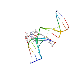
 | | CO(III)-BLEOMYCIN-OOH BOUND TO AN OLIGONUCLEOTIDE CONTAINING A PHOSPHOGLYCOLATE LESION | | 分子名称: | 2-PHOSPHOGLYCOLIC ACID, 5'-D(*CP*CP*AP*AP*AP*G)-3', 5'-D(*CP*CP*CP*AP*GP*TP*AP*CP*TP*TP*TP*GP*G)-3', ... | | 著者 | Hoehn, S.T, Junker, H.-D, Bunt, R.C, Turner, C.J, Stubbe, J. | | 登録日 | 2000-11-01 | | 公開日 | 2001-06-06 | | 最終更新日 | 2024-07-10 | | 実験手法 | SOLUTION NMR | | 主引用文献 | Solution structure of Co(III)-bleomycin-OOH bound to a phosphoglycolate lesion containing oligonucleotide: implications for bleomycin-induced double-strand DNA cleavage.
Biochemistry, 40, 2001
|
|
1G5L
 | | CO(III)-BLEOMYCIN-OOH BOUND TO AN OLIGONUCLEOTIDE CONTAINING A PHOSPHOGLYCOLATE LESION | | 分子名称: | 2-PHOSPHOGLYCOLIC ACID, 5'-D(*CP*CP*AP*AP*AP*G)-3', 5'-D(*CP*CP*CP*AP*GP*TP*AP*CP*TP*TP*TP*GP*G)-3', ... | | 著者 | Hoehn, S.T, Junker, H.-D, Bunt, R.C, Turner, C.J, Stubbe, J. | | 登録日 | 2000-11-01 | | 公開日 | 2001-06-06 | | 最終更新日 | 2024-05-22 | | 実験手法 | SOLUTION NMR | | 主引用文献 | Solution structure of Co(III)-bleomycin-OOH bound to a phosphoglycolate lesion containing oligonucleotide: implications for bleomycin-induced double-strand DNA cleavage.
Biochemistry, 40, 2001
|
|
6WH2
 | | Structure of the C-terminal BRCT domain of human XRCC1 | | 分子名称: | X-ray repair cross complementing protein 1 variant | | 著者 | Pourfarjam, Y, Ellenberger, T, Tainer, J.A, Tomkinson, A.E, Kim, I.K. | | 登録日 | 2020-04-07 | | 公開日 | 2020-12-02 | | 最終更新日 | 2023-10-18 | | 実験手法 | X-RAY DIFFRACTION (2.414 Å) | | 主引用文献 | An atypical BRCT-BRCT interaction with the XRCC1 scaffold protein compacts human DNA Ligase III alpha within a flexible DNA repair complex.
Nucleic Acids Res., 49, 2021
|
|
3NKE
 | | High resolution structure of the C-terminal domain CRISP-associated protein Cas1 from Escherichia coli str. K-12 | | 分子名称: | 1,2-ETHANEDIOL, SULFATE ION, SULFITE ION, ... | | 著者 | Nocek, B, Skarina, T, Beloglazova, N, Savchenko, A, Joachimiak, A, Yakunin, A. | | 登録日 | 2010-06-18 | | 公開日 | 2010-08-25 | | 最終更新日 | 2014-12-17 | | 実験手法 | X-RAY DIFFRACTION (1.4 Å) | | 主引用文献 | A dual function of the CRISPR-Cas system in bacterial antivirus immunity and DNA repair.
Mol.Microbiol., 79, 2011
|
|
2NSK
 | |
5NR6
 | |
3B81
 | |
3EBE
 | |
3BD3
 | |
7CSV
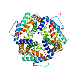
 | | Pseudomonas aeruginosa antitoxin HigA | | 分子名称: | HTH cro/C1-type domain-containing protein | | 著者 | Song, Y.J, Luo, G.H, Bao, R. | | 登録日 | 2020-08-17 | | 公開日 | 2021-01-13 | | 最終更新日 | 2023-11-29 | | 実験手法 | X-RAY DIFFRACTION (1.71 Å) | | 主引用文献 | Pseudomonas aeruginosa antitoxin HigA functions as a diverse regulatory factor by recognizing specific pseudopalindromic DNA motifs.
Environ.Microbiol., 23, 2021
|
|
5KGF
 | | Structural model of 53BP1 bound to a ubiquitylated and methylated nucleosome, at 4.5 A resolution | | 分子名称: | DNA (145-MER), Histone H2A type 1, Histone H2B type 1-C/E/F/G/I, ... | | 著者 | Wilson, M.D, Benlekbir, S, Sicheri, F, Rubinstein, J.L, Durocher, D. | | 登録日 | 2016-06-13 | | 公開日 | 2016-07-27 | | 最終更新日 | 2020-01-15 | | 実験手法 | ELECTRON MICROSCOPY (4.54 Å) | | 主引用文献 | The structural basis of modified nucleosome recognition by 53BP1.
Nature, 536, 2016
|
|
6OIT
 | | CryoEM structure of Arabidopsis DDR' complex (DRD1 peptide-DMS3-RDM1) | | 分子名称: | Protein CHROMATIN REMODELING 35, Protein DEFECTIVE IN MERISTEM SILENCING 3, Protein RDM1 | | 著者 | Wongpalee, S.P, Liu, S, Zhou, Z.H, Jacobsen, S.E. | | 登録日 | 2019-04-09 | | 公開日 | 2019-07-24 | | 最終更新日 | 2024-03-20 | | 実験手法 | ELECTRON MICROSCOPY (3.5 Å) | | 主引用文献 | CryoEM structures of Arabidopsis DDR complexes involved in RNA-directed DNA methylation.
Nat Commun, 10, 2019
|
|
7MID
 | | Sub-complex of Cas4-Cas1-Cas2 bound PAM containing DNA | | 分子名称: | CRISPR-associated endoribonuclease Cas2, CRISPR-associated exonuclease Cas4/endonuclease Cas1 fusion, DNA (33-MER), ... | | 著者 | Hu, C.Y, Ke, A.K. | | 登録日 | 2021-04-16 | | 公開日 | 2021-11-17 | | 実験手法 | ELECTRON MICROSCOPY (3.56 Å) | | 主引用文献 | Mechanism for Cas4-assisted directional spacer acquisition in CRISPR-Cas.
Nature, 598, 2021
|
|
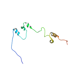
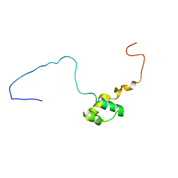
1NHN
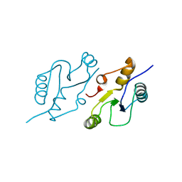
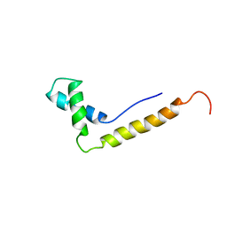 | | THE STRUCTURE OF THE HMG BOX AND ITS INTERACTION WITH DNA | | 分子名称: | HIGH MOBILITY GROUP PROTEIN 1 | | 著者 | Read, C.M, Cary, P.D, Crane-Robinson, C, Driscoll, P.C, Carillo, M.O.M, Norman, D.G. | | 登録日 | 1994-11-17 | | 公開日 | 1995-02-07 | | 最終更新日 | 2024-05-22 | | 実験手法 | SOLUTION NMR | | 主引用文献 | The Structure of the Hmg Box and its Interaction with DNA
NUCLEIC ACIDS MOL.BIOL., 9, 1995
|
|
4JLX
 | |
1NHM
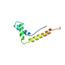 | | THE STRUCTURE OF THE HMG BOX AND ITS INTERACTION WITH DNA | | 分子名称: | HIGH MOBILITY GROUP PROTEIN 1 | | 著者 | Read, C.M, Cary, P.D, Crane-Robinson, C, Driscoll, P.C, Carillo, M.O.M, Norman, D.G. | | 登録日 | 1994-11-17 | | 公開日 | 1995-02-07 | | 最終更新日 | 2024-05-22 | | 実験手法 | SOLUTION NMR | | 主引用文献 | The Structure of the Hmg Box and its Interaction with DNA
NUCLEIC ACIDS MOL.BIOL., 9, 1995
|
|


