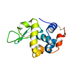
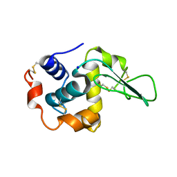
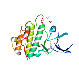
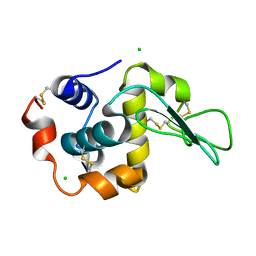
3GOK
 
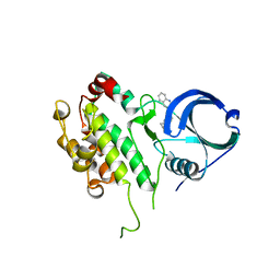 | | Binding site mapping of protein ligands | | 分子名称: | 2-(2-QUINOLIN-3-YLPYRIDIN-4-YL)-1,5,6,7-TETRAHYDRO-4H-PYRROLO[3,2-C]PYRIDIN-4-ONE, MAP kinase-activated protein kinase 2 | | 著者 | Scheich, C. | | 登録日 | 2009-03-19 | | 公開日 | 2010-03-02 | | 最終更新日 | 2023-09-06 | | 実験手法 | X-RAY DIFFRACTION (3.2 Å) | | 主引用文献 | Binding site mapping of protein ligands
To be published
|
|
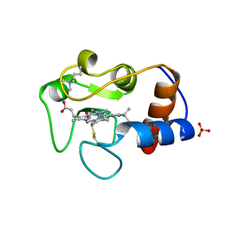
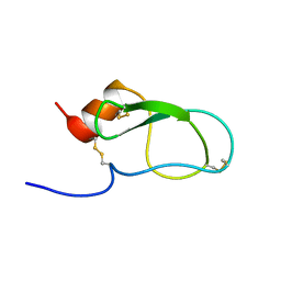
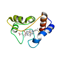
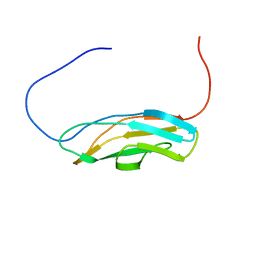
1WDX
 | | Yeast BBC1 SH3 domain, triclinic crystal form | | 分子名称: | Myosin tail region-interacting protein MTI1 | | 著者 | Wilmanns, M, Consani Textor, L, Kursula, P, Kursula, I, Lehmann, F, Song, Y.H. | | 登録日 | 2004-05-19 | | 公開日 | 2005-05-31 | | 最終更新日 | 2024-04-03 | | 実験手法 | X-RAY DIFFRACTION (2.5 Å) | | 主引用文献 | Crystal structure of Yeast BBC1 SH3 domain, triclinic crystal form
To be Published
|
|
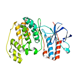
1J41
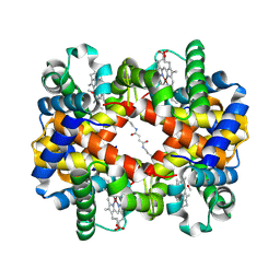 | | Direct observation of photolysis-induced tertiary structural changes in human haemoglobin; Crystal structure of alpha(Ni)-beta(Fe) hemoglobin (laser photolysed) | | 分子名称: | BUT-2-ENEDIAL, CARBON MONOXIDE, Hemoglobin alpha Chain, ... | | 著者 | Adachi, S, Park, S.-Y, Tame, J.R.H, Shiro, Y, Shibayama, N, RIKEN Structural Genomics/Proteomics Initiative (RSGI) | | 登録日 | 2003-02-21 | | 公開日 | 2003-07-22 | | 最終更新日 | 2023-12-27 | | 実験手法 | X-RAY DIFFRACTION (1.45 Å) | | 主引用文献 | Direct observation of photolysis-induced tertiary structural changes in hemoglobin
Proc.Natl.Acad.Sci.USA, 100, 2003
|
|
2QR7
 | |
1X48
 | | Solution structure of the second DSRM domain in Interferon-induced, double-stranded RNA-activated protein kinase | | 分子名称: | Interferon-induced, double-stranded RNA-activated protein kinase | | 著者 | He, F, Muto, Y, Inoue, M, Tarada, T, Shirouzu, M, Kigawa, T, Yokoyama, S, RIKEN Structural Genomics/Proteomics Initiative (RSGI) | | 登録日 | 2005-05-14 | | 公開日 | 2005-11-14 | | 最終更新日 | 2024-05-29 | | 実験手法 | SOLUTION NMR | | 主引用文献 | Solution structure of the second DSRM domain in Interferon-induced, double-stranded RNA-activated protein kinase
To be Published
|
|
1IR7
 | |
1IR9
 | |
1X5H
 | | The solution structure of the third fibronectin type III domain of human Neogenin | | 分子名称: | Neogenin | | 著者 | Tochio, N, Sasagawa, A, Koshiba, S, Inoue, M, Kigawa, T, Yokoyama, S, RIKEN Structural Genomics/Proteomics Initiative (RSGI) | | 登録日 | 2005-05-15 | | 公開日 | 2005-11-15 | | 最終更新日 | 2024-05-29 | | 実験手法 | SOLUTION NMR | | 主引用文献 | The solution structure of the third fibronectin type III domain of human Neogenin
To be Published
|
|
1X5X
 | | Solution structure of the fibronectin type-III domain of human fibronectin type III domain containing protein 3 | | 分子名称: | Fibronectin type-III domain containing protein 3a | | 著者 | Yoneyama, M, Tochio, N, Koshiba, S, Inoue, M, Kigawa, T, Yokoyama, S, RIKEN Structural Genomics/Proteomics Initiative (RSGI) | | 登録日 | 2005-05-17 | | 公開日 | 2005-11-17 | | 最終更新日 | 2024-05-29 | | 実験手法 | SOLUTION NMR | | 主引用文献 | Solution structure of the fibronectin type-III domain of human fibronectin type III domain containing protein 3
To be Published
|
|
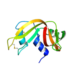
1IP4
 | | G72A HUMAN LYSOZYME | | 分子名称: | LYSOZYME C, SODIUM ION | | 著者 | Takano, K, Yamagata, Y, Yutani, K. | | 登録日 | 2001-04-20 | | 公開日 | 2001-11-14 | | 最終更新日 | 2023-12-27 | | 実験手法 | X-RAY DIFFRACTION (1.8 Å) | | 主引用文献 | Role of amino acid residues in left-handed helical conformation for the conformational stability of a protein.
Proteins, 45, 2001
|
|
1IRV
 | |
1WBO
 | | fragment based p38 inhibitors | | 分子名称: | 2-CHLOROPHENOL, MITOGEN-ACTIVATED PROTEIN KINASE 14 | | 著者 | Cleasby, A, Devine, L, Gill, A, Jhoti, H. | | 登録日 | 2004-11-04 | | 公開日 | 2005-01-27 | | 最終更新日 | 2024-05-08 | | 実験手法 | X-RAY DIFFRACTION (2.16 Å) | | 主引用文献 | Fragment-Based Lead Discovery Using X-Ray Crystallography
J.Med.Chem., 48, 2005
|
|
1WBW
 | | Identification of novel p38 alpha MAP Kinase inhibitors using fragment-based lead generation. | | 分子名称: | 3-(1-NAPHTHYLMETHOXY)PYRIDIN-2-AMINE, MITOGEN-ACTIVATED PROTEIN KINASE 14 | | 著者 | Tickle, J, Cleasby, A, Devine, L.A, Jhoti, H. | | 登録日 | 2004-11-05 | | 公開日 | 2005-11-03 | | 最終更新日 | 2024-05-08 | | 実験手法 | X-RAY DIFFRACTION (2.41 Å) | | 主引用文献 | Identification of Novel P38Alpha Map Kinase Inhibitors Using Fragment-Based Lead Generation
J.Med.Chem., 48, 2005
|
|
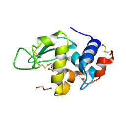
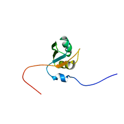
1IRH
 | | The Solution Structure of The Third Kunitz Domain of Tissue Factor Pathway Inhibitor | | 分子名称: | tissue factor pathway inhibitor | | 著者 | Mine, S, Yamazaki, T, Miyata, T, Hara, S, Kato, H. | | 登録日 | 2001-10-02 | | 公開日 | 2002-02-06 | | 最終更新日 | 2023-12-27 | | 実験手法 | SOLUTION NMR | | 主引用文献 | Structural mechanism for heparin-binding of the third Kunitz domain of human tissue factor pathway inhibitor.
Biochemistry, 41, 2002
|
|
1IS0
 | | Crystal Structure of a Complex of the Src SH2 Domain with Conformationally Constrained Peptide Inhibitor | | 分子名称: | AY0 GLU GLU ILE peptide, Tyrosine-protein kinase transforming protein SRC | | 著者 | Davidson, J.P, Lubman, O, Rose, T, Waksman, G, Martin, S.F. | | 登録日 | 2001-11-02 | | 公開日 | 2002-02-06 | | 最終更新日 | 2023-12-27 | | 実験手法 | X-RAY DIFFRACTION (1.9 Å) | | 主引用文献 | Calorimetric and structural studies of 1,2,3-trisubstituted cyclopropanes as conformationally constrained peptide inhibitors of Src SH2 domain binding.
J.Am.Chem.Soc., 124, 2002
|
|
2BLP
 | |
2BLX
 | |
1IWU
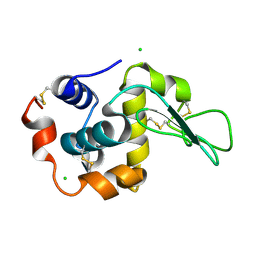 | | Crystal Structure Analysis of Human lysozyme at 127K. | | 分子名称: | CHLORIDE ION, LYSOZYME C | | 著者 | Joti, Y, Nakasako, M, Kidera, A, Go, N. | | 登録日 | 2002-06-03 | | 公開日 | 2002-09-04 | | 最終更新日 | 2023-12-27 | | 実験手法 | X-RAY DIFFRACTION (1.4 Å) | | 主引用文献 | Nonlinear temperature dependence of the crystal structure of lysozyme: correlation between coordinate shifts and thermal factors.
Acta Crystallogr.,Sect.D, 58, 2002
|
|
2XEZ
 | | Crystal structure of checkpoint kinase 1 (Chk1) in complex with inhibitors | | 分子名称: | 1,2-ETHANEDIOL, 6-(1H-PYRAZOL-3-YL)-3-(1H-PYRAZOL-4-YL)IMIDAZO[1,2-A]PYRAZINE, SERINE/THREONINE-PROTEIN KINASE CHK1 | | 著者 | Matthews, T.P, McHardy, T, Klair, S, Boxall, K, Fisher, M, Cherry, M, Allen, C.E, Addison, G.J, Ellard, J, Aherne, G.W, Westwood, I.M, vanMontfort, R, Garrett, M.D, Reader, J.C, Collins, I. | | 登録日 | 2010-05-19 | | 公開日 | 2010-07-07 | | 最終更新日 | 2023-12-20 | | 実験手法 | X-RAY DIFFRACTION (2.25 Å) | | 主引用文献 | Design and Evaluation of 3,6-Di(Hetero)Aryl Imidazo[1,2-A]Pyrazines as Inhibitors of Checkpoint and Other Kinases.
Bioorg.Med.Chem.Lett., 20, 2010
|
|
2C2C
 | |
1SGC
 | |
1IVC
 | |
1IWX
 | | Crystal Structure Analysis of Human lysozyme at 161K. | | 分子名称: | CHLORIDE ION, LYSOZYME C | | 著者 | Joti, Y, Nakasako, M, Kidera, A, Go, N. | | 登録日 | 2002-06-03 | | 公開日 | 2002-09-04 | | 最終更新日 | 2023-12-27 | | 実験手法 | X-RAY DIFFRACTION (1.4 Å) | | 主引用文献 | Nonlinear temperature dependence of the crystal structure of lysozyme: correlation between coordinate shifts and thermal factors.
Acta Crystallogr.,Sect.D, 58, 2002
|
|
1X3D
 | | Solution structure of the fibronectin type-III domain of human fibronectin type-III domain containing protein 3a | | 分子名称: | Fibronectin type-III domain containing protein 3a | | 著者 | Yoneyama, M, Tochio, N, Koshiba, S, Inoue, M, Kigawa, T, Yokoyama, S, RIKEN Structural Genomics/Proteomics Initiative (RSGI) | | 登録日 | 2005-05-02 | | 公開日 | 2005-11-02 | | 最終更新日 | 2024-05-29 | | 実験手法 | SOLUTION NMR | | 主引用文献 | Solution structure of the fibronectin type-III domain of human fibronectin type-III domain containing protein 3a
To be Published
|
|
1X4G
 | | Solution structure of RRM domain in Nucleolysin TIAR | | 分子名称: | Nucleolysin TIAR | | 著者 | He, F, Muto, Y, Inoue, M, Kigawa, T, Shirouzu, M, Terada, T, Yokoyama, S, RIKEN Structural Genomics/Proteomics Initiative (RSGI) | | 登録日 | 2005-05-14 | | 公開日 | 2005-11-14 | | 最終更新日 | 2024-05-29 | | 実験手法 | SOLUTION NMR | | 主引用文献 | Solution structure of RRM domain in Nucleolysin TIAR
To be Published
|
|


