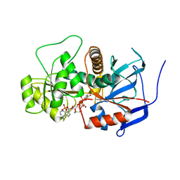
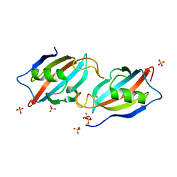
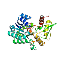
1I0V
 
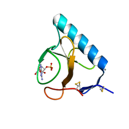 | | Ribonuclease T1 in complex with 2'GMP (form I crystal) | | 分子名称: | CALCIUM ION, GUANOSINE-2'-MONOPHOSPHATE, GUANYL-SPECIFIC RIBONUCLEASE T1 | | 著者 | De Swarte, J, De Vos, S, Langhorst, U, Steyaert, J, Loris, R. | | 登録日 | 2001-01-30 | | 公開日 | 2001-02-14 | | 最終更新日 | 2021-10-27 | | 実験手法 | X-RAY DIFFRACTION (1.234 Å) | | 主引用文献 | The contribution of metal ions to the conformational stability of ribonuclease T1: crystal versus solution.
Eur.J.Biochem., 268, 2001
|
|
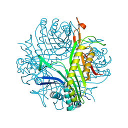
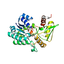
1I0X
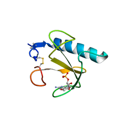 | | RIBONUCLEASE T1 IN COMPLEX WITH 2'GMP (FORM II CRYSTAL) | | 分子名称: | CALCIUM ION, GUANOSINE-2'-MONOPHOSPHATE, GUANYL-SPECIFIC RIBONUCLEASE T1 | | 著者 | De Swarte, J, De Vos, S, Langhorst, U, Steyaert, J, Loris, R. | | 登録日 | 2001-01-30 | | 公開日 | 2001-02-14 | | 最終更新日 | 2021-10-27 | | 実験手法 | X-RAY DIFFRACTION (1.65 Å) | | 主引用文献 | The contribution of metal ions to the conformational stability of ribonuclease T1: crystal versus solution.
Eur.J.Biochem., 268, 2001
|
|
5IMN
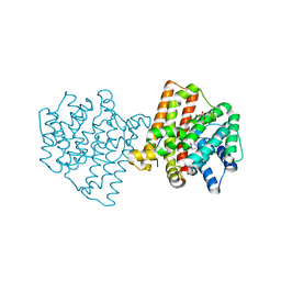 | | Crystal structure of N299A/S303A Aspergillus terreus aristolochene synthase complexed with (1S,8S,9aR)-1,9a-dimethyl-8-(prop-1-en-2-yl)decahydroquinolizin-5-ium | | 分子名称: | (1S,5S,8S,9aR)-1,9a-dimethyl-8-(prop-1-en-2-yl)octahydro-2H-quinolizinium, Aristolochene synthase, GLYCEROL, ... | | 著者 | Chen, M, Christianson, D.W. | | 登録日 | 2016-03-06 | | 公開日 | 2016-05-25 | | 最終更新日 | 2023-09-27 | | 実験手法 | X-RAY DIFFRACTION (2.527 Å) | | 主引用文献 | Probing the Role of Active Site Water in the Sesquiterpene Cyclization Reaction Catalyzed by Aristolochene Synthase.
Biochemistry, 55, 2016
|
|
5I6C
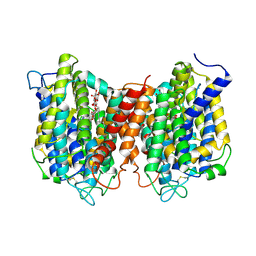 | | The structure of the eukaryotic purine/H+ symporter, UapA, in complex with Xanthine | | 分子名称: | DODECYL-BETA-D-MALTOSIDE, Uric acid-xanthine permease, XANTHINE | | 著者 | Alguel, Y, Amillis, S, Leung, J, Lambrinidis, G, Capaldi, S, Scull, N.J, Craven, G, Iwata, S, Armstrong, A, Mikros, E, Diallinas, G, Cameron, A.D, Byrne, B. | | 登録日 | 2016-02-16 | | 公開日 | 2016-04-27 | | 最終更新日 | 2017-08-30 | | 実験手法 | X-RAY DIFFRACTION (3.7 Å) | | 主引用文献 | Structure of eukaryotic purine/H(+) symporter UapA suggests a role for homodimerization in transport activity.
Nat Commun, 7, 2016
|
|
5IVG
 | |
2BVW
 | | CELLOBIOHYDROLASE II (CEL6A) FROM HUMICOLA INSOLENS IN COMPLEX WITH GLUCOSE AND CELLOTETRAOSE | | 分子名称: | 2-acetamido-2-deoxy-beta-D-glucopyranose, CELLOBIOHYDROLASE II, GLYCEROL, ... | | 著者 | Varrot, A, Davies, G.J, Schulein, M. | | 登録日 | 1999-02-18 | | 公開日 | 2000-02-25 | | 最終更新日 | 2023-08-23 | | 実験手法 | X-RAY DIFFRACTION (1.7 Å) | | 主引用文献 | Structural changes of the active site tunnel of Humicola insolens cellobiohydrolase, Cel6A, upon oligosaccharide binding.
Biochemistry, 38, 1999
|
|
1I2G
 | | Ribonuclease T1 V16T mutant | | 分子名称: | CALCIUM ION, GUANOSINE-2'-MONOPHOSPHATE, GUANYL-SPECIFIC RIBONUCLEASE T1 | | 著者 | De Vos, S, Backmann, J, Steyaert, J, Loris, R. | | 登録日 | 2001-02-09 | | 公開日 | 2001-03-07 | | 最終更新日 | 2021-10-27 | | 実験手法 | X-RAY DIFFRACTION (1.85 Å) | | 主引用文献 | Hydrophobic core manipulations in ribonuclease T1
Biochemistry, 40, 2001
|
|
1I2E
 | | Ribonuclease T1 V16A mutant, form I | | 分子名称: | CALCIUM ION, GUANOSINE-2'-MONOPHOSPHATE, GUANYL-SPECIFIC RIBONUCLEASE T1 | | 著者 | De Vos, S, Backmann, J, Steyaert, J, Loris, R. | | 登録日 | 2001-02-09 | | 公開日 | 2001-03-07 | | 最終更新日 | 2021-10-27 | | 実験手法 | X-RAY DIFFRACTION (1.8 Å) | | 主引用文献 | Hydrophobic core manipulations in ribonuclease T1
Biochemistry, 40, 2001
|
|
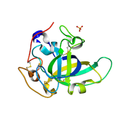
3B70
 | | Crystal structure of Aspergillus terreus trans-acting lovastatin polyketide enoyl reductase (LovC) with bound NADP | | 分子名称: | Enoyl reductase, GLYCEROL, NADP NICOTINAMIDE-ADENINE-DINUCLEOTIDE PHOSPHATE | | 著者 | Ames, B.D, Smith, P.T, Ma, S.M, Wong, E.W, Xie, X, Vederas, J.C, Tang, Y, Tsai, S.-C. | | 登録日 | 2007-10-29 | | 公開日 | 2008-09-16 | | 最終更新日 | 2023-08-30 | | 実験手法 | X-RAY DIFFRACTION (1.89 Å) | | 主引用文献 | Crystal structure and biochemical studies of the trans-acting polyketide enoyl reductase LovC from lovastatin biosynthesis.
Proc.Natl.Acad.Sci.USA, 109, 2012
|
|
1I2F
 | | Ribonuclease T1 V16A mutant, form II | | 分子名称: | CALCIUM ION, GUANOSINE-2'-MONOPHOSPHATE, GUANYL-SPECIFIC RIBONUCLEASE T1 | | 著者 | De Vos, S, Backmann, J, Steyaert, J, Loris, R. | | 登録日 | 2001-02-09 | | 公開日 | 2001-03-07 | | 最終更新日 | 2021-10-27 | | 実験手法 | X-RAY DIFFRACTION (1.95 Å) | | 主引用文献 | Hydrophobic core manipulations in ribonuclease T1
Biochemistry, 40, 2001
|
|
1I3F
 | | Ribonuclease T1 V89S mutant | | 分子名称: | CALCIUM ION, GUANOSINE-2'-MONOPHOSPHATE, GUANYL-SPECIFIC RIBONUCLEASE T1 | | 著者 | De Vos, S, Backmann, J, Steyaert, J, Loris, R. | | 登録日 | 2001-02-15 | | 公開日 | 2001-03-07 | | 最終更新日 | 2021-10-27 | | 実験手法 | X-RAY DIFFRACTION (2.35 Å) | | 主引用文献 | Hydrophobic core manipulations in ribonuclease T1
Biochemistry, 40, 2001
|
|
1I3I
 | | Ribonuclease T1 V78T mutant | | 分子名称: | CALCIUM ION, GUANOSINE-2'-MONOPHOSPHATE, GUANYL-SPECIFIC RIBONUCLEASE T1 | | 著者 | De Vos, S, Backmann, J, Steyaert, J, Loris, R. | | 登録日 | 2001-02-15 | | 公開日 | 2001-03-07 | | 最終更新日 | 2021-10-27 | | 実験手法 | X-RAY DIFFRACTION (1.76 Å) | | 主引用文献 | Hydrophobic core manipulations in ribonuclease T1
Biochemistry, 40, 2001
|
|
5KJQ
 | | X-ray structure of PcCel45A in complex with cellobiose expressed in Aspergillus nidullans | | 分子名称: | Endoglucanase V-like protein, SULFATE ION, beta-D-glucopyranose-(1-4)-beta-D-glucopyranose | | 著者 | Godoy, A.S, Ramia, M.P, Camilo, C.M, Polikarpov, I. | | 登録日 | 2016-06-20 | | 公開日 | 2017-06-21 | | 最終更新日 | 2023-09-27 | | 実験手法 | X-RAY DIFFRACTION (1.704 Å) | | 主引用文献 | Structure, computational and biochemical analysis of PcCel45A endoglucanase from Phanerochaete chrysosporium and catalytic mechanisms of GH45 subfamily C members.
Sci Rep, 8, 2018
|
|
5HWB
 | |
2D44
 | | Crystal structure of arabinofuranosidase complexed with arabinofuranosyl-alpha-1,2-xylobiose | | 分子名称: | 2-acetamido-2-deoxy-beta-D-glucopyranose-(1-4)-2-acetamido-2-deoxy-beta-D-glucopyranose, alpha-L-arabinofuranose-(1-2)-alpha-D-xylopyranose-(1-4)-alpha-D-xylopyranose, alpha-L-arabinofuranosidase B | | 著者 | Miyanaga, A, Koseki, T, Miwa, Y, Matsuzawa, H, Wakagi, T, Shoun, H, Fushinobu, S. | | 登録日 | 2005-10-07 | | 公開日 | 2006-09-19 | | 最終更新日 | 2021-11-10 | | 実験手法 | X-RAY DIFFRACTION (2.3 Å) | | 主引用文献 | The family 42 carbohydrate-binding module of family 54 alpha-L-arabinofuranosidase specifically binds the arabinofuranose side chain of hemicellulose
Biochem.J., 399, 2006
|
|
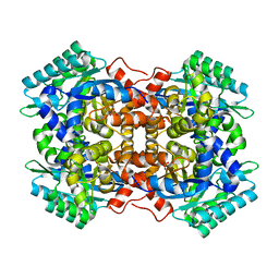
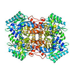
2FUB
 | | Crystal structure of urate oxidase at 140 MPa | | 分子名称: | 8-AZAXANTHINE, CYSTEINE, Uricase | | 著者 | Colloc'h, N, Girard, E, Fourme, R. | | 登録日 | 2006-01-26 | | 公開日 | 2006-02-14 | | 最終更新日 | 2023-11-15 | | 実験手法 | X-RAY DIFFRACTION (2.3 Å) | | 主引用文献 | High pressure macromolecular crystallography: The 140-MPa crystal structure at 2.3 A resolution of urate oxidase, a 135-kDa tetrameric assembly
Biochim.Biophys.Acta, 1764, 2006
|
|
1WD3
 | | Crystal structure of arabinofuranosidase | | 分子名称: | 2-acetamido-2-deoxy-beta-D-glucopyranose-(1-4)-2-acetamido-2-deoxy-beta-D-glucopyranose, alpha-L-arabinofuranosidase B | | 著者 | Miyanaga, A, Koseki, T, Matsuzawa, H, Wakagi, T, Shoun, H, Fushinobu, S. | | 登録日 | 2004-05-11 | | 公開日 | 2004-09-14 | | 最終更新日 | 2020-07-29 | | 実験手法 | X-RAY DIFFRACTION (1.75 Å) | | 主引用文献 | Crystal structure of a family 54 alpha-L-arabinofuranosidase reveals a novel carbohydrate-binding module that can bind arabinose
J.Biol.Chem., 279, 2004
|
|
1WD4
 | | Crystal structure of arabinofuranosidase complexed with arabinose | | 分子名称: | 2-acetamido-2-deoxy-beta-D-glucopyranose-(1-4)-2-acetamido-2-deoxy-beta-D-glucopyranose, alpha-L-arabinofuranose, alpha-L-arabinofuranosidase B | | 著者 | Miyanaga, A, Koseki, T, Matsuzawa, H, Wakagi, T, Shoun, H, Fushinobu, S. | | 登録日 | 2004-05-11 | | 公開日 | 2004-09-14 | | 最終更新日 | 2020-07-29 | | 実験手法 | X-RAY DIFFRACTION (2.07 Å) | | 主引用文献 | Crystal structure of a family 54 alpha-L-arabinofuranosidase reveals a novel carbohydrate-binding module that can bind arabinose
J.Biol.Chem., 279, 2004
|
|
7WGI
 | | Crystal structure of AflSQS from Aspergillus flavus | | 分子名称: | INDOLE, PHOSPHATE ION, Squalene synthase | | 著者 | Shang, N, Liu, W.D, Chen, C.C, Guo, R.T. | | 登録日 | 2021-12-28 | | 公開日 | 2022-11-09 | | 最終更新日 | 2023-11-29 | | 実験手法 | X-RAY DIFFRACTION (2.5 Å) | | 主引用文献 | A Structural and Bioinformatics Investigation of a Fungal Squalene Synthase and Comparisons with Other Membrane Proteins.
Acs Omega, 7, 2022
|
|
7WGH
 | | Crystal structure of AflSQS from Aspergillus flavus in complex with FSPP | | 分子名称: | PHOSPHATE ION, PYROPHOSPHATE 2-, S-[(2E,6E)-3,7,11-TRIMETHYLDODECA-2,6,10-TRIENYL] TRIHYDROGEN THIODIPHOSPHATE, ... | | 著者 | Shang, N, Liu, W.D, Chen, C.C, Guo, R.T. | | 登録日 | 2021-12-28 | | 公開日 | 2022-11-09 | | 最終更新日 | 2023-11-29 | | 実験手法 | X-RAY DIFFRACTION (2.36 Å) | | 主引用文献 | A Structural and Bioinformatics Investigation of a Fungal Squalene Synthase and Comparisons with Other Membrane Proteins.
Acs Omega, 7, 2022
|
|
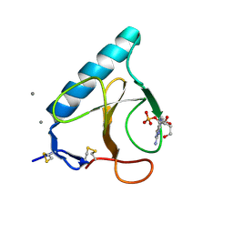
2VUU
 | | Crystal structure of NADP-bound NmrA-AreA zinc finger complex | | 分子名称: | NADP NICOTINAMIDE-ADENINE-DINUCLEOTIDE PHOSPHATE, NITROGEN METABOLITE REPRESSION REGULATOR NMRA, NITROGEN REGULATORY PROTEIN AREA, ... | | 著者 | Kotaka, M, Johnson, C, Lamb, H.K, Hawkins, A.R, Ren, J, Stammers, D.K. | | 登録日 | 2008-05-30 | | 公開日 | 2008-07-29 | | 最終更新日 | 2024-05-08 | | 実験手法 | X-RAY DIFFRACTION (2.8 Å) | | 主引用文献 | Structural Analysis of the Recognition of the Negative Regulator Nmra and DNA by the Zinc Finger from the Gata-Type Transcription Factor Area.
J.Mol.Biol., 381, 2008
|
|
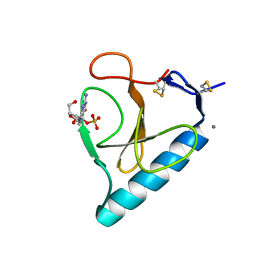
2VUT
 | | Crystal structure of NAD-bound NmrA-AreA zinc finger complex | | 分子名称: | CHLORIDE ION, GLYCEROL, NICOTINAMIDE-ADENINE-DINUCLEOTIDE, ... | | 著者 | Kotaka, M, Johnson, C, Lamb, H.K, Hawkins, A.R, Ren, J, Stammers, D.K. | | 登録日 | 2008-05-30 | | 公開日 | 2008-07-29 | | 最終更新日 | 2024-05-08 | | 実験手法 | X-RAY DIFFRACTION (2.3 Å) | | 主引用文献 | Structural Analysis of the Recognition of the Negative Regulator Nmra and DNA by the Zinc Finger from the Gata-Type Transcription Factor Area.
J.Mol.Biol., 381, 2008
|
|
7WKL
 | |
7WJR
 | |
7WMB
 | |


