5BK2
 
 | |
5BJZ
 | |
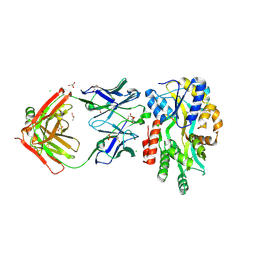
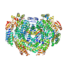
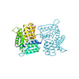
7M8R
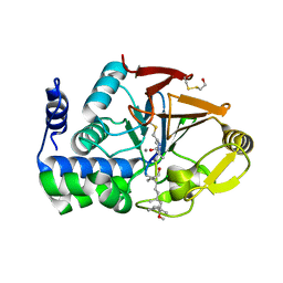
 | | Complex structure of Methane monooxygenase hydroxylase and regulatory subunit with fluorosubstituted tryptophans | | 分子名称: | 1,1,1-tris(fluoranyl)propan-2-one, 1,2-ETHANEDIOL, BENZOIC ACID, ... | | 著者 | Johns, J.C, Banerjee, R, Shi, K, Semonis, M.M, Aihara, H, Pomerantz, W.C.K, Lipscomb, J.D. | | 登録日 | 2021-03-30 | | 公開日 | 2021-07-28 | | 最終更新日 | 2023-10-18 | | 実験手法 | X-RAY DIFFRACTION (2.22 Å) | | 主引用文献 | Soluble Methane Monooxygenase Component Interactions Monitored by 19 F NMR.
Biochemistry, 60, 2021
|
|
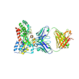
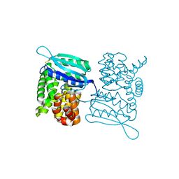
7M8Q
 | | Complex structure of Methane monooxygenase hydroxylase and regulatory subunit with fluorosubstituted tryptophans | | 分子名称: | 1,2-ETHANEDIOL, BENZOIC ACID, FE (III) ION, ... | | 著者 | Johns, J.C, Banerjee, R, Shi, K, Semonis, M.M, Aihara, H, Pomerantz, W.C.K, Lipscomb, J.D. | | 登録日 | 2021-03-30 | | 公開日 | 2021-07-28 | | 最終更新日 | 2023-10-18 | | 実験手法 | X-RAY DIFFRACTION (2.08 Å) | | 主引用文献 | Soluble Methane Monooxygenase Component Interactions Monitored by 19 F NMR.
Biochemistry, 60, 2021
|
|
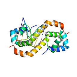
5WVN
 | | Crystal structure of MBS-BaeS fusion protein | | 分子名称: | Maltose-binding periplasmic protein,Two-component system sensor kinase, SULFATE ION | | 著者 | Wang, W, Zhang, Y, Ran, T, Xu, D. | | 登録日 | 2016-12-26 | | 公開日 | 2018-01-03 | | 最終更新日 | 2024-03-20 | | 実験手法 | X-RAY DIFFRACTION (2.8 Å) | | 主引用文献 | Crystal structure of the sensor domain of BaeS from Serratia marcescens FS14
Proteins, 85, 2017
|
|
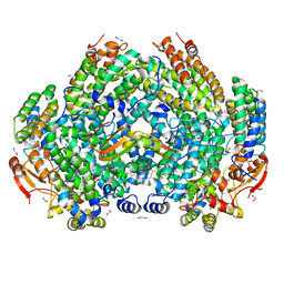
4GAM
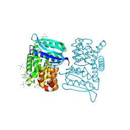
 | | Complex structure of Methane monooxygenase hydroxylase and regulatory subunit | | 分子名称: | FE (III) ION, Methane monooxygenase component A alpha chain, Methane monooxygenase component A beta chain, ... | | 著者 | Lee, S.J, Lippard, S.J, Cho, U.-S. | | 登録日 | 2012-07-25 | | 公開日 | 2013-02-06 | | 最終更新日 | 2023-09-13 | | 実験手法 | X-RAY DIFFRACTION (2.902 Å) | | 主引用文献 | Control of substrate access to the active site in methane monooxygenase.
Nature, 494, 2013
|
|
6QKQ
 | |
6QKP
 | |
3FG7
 | |
6ZZ9
 | |
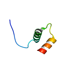
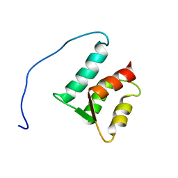
1M39
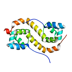
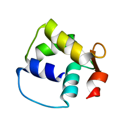 | | Solution structure of the C-terminal fragment (F86-I165) of the human centrin 2 in calcium saturated form | | 分子名称: | Caltractin, isoform 1 | | 著者 | Matei, E, Miron, S, Blouquit, Y, Duchambon, P, Durussel, P, Cox, J.A, Craescu, C.T. | | 登録日 | 2002-06-27 | | 公開日 | 2003-03-25 | | 最終更新日 | 2024-05-22 | | 実験手法 | SOLUTION NMR | | 主引用文献 | C-terminal half of human centrin 2 behaves like a regulatory EF-hand domain
Biochemistry, 42, 2003
|
|
3HPE
 | |
3LY8
 | |
3LYA
 | |
3LY9
 | |
3LY7
 | |
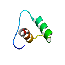
1ZMZ
 | | Solution structure of the N-terminal domain (M1-S98) of human centrin 2 | | 分子名称: | Centrin-2 | | 著者 | Yang, A, Miron, S, Duchambon, P, Assairi, L, Blouquit, Y, Craescu, C.T. | | 登録日 | 2005-05-11 | | 公開日 | 2006-04-25 | | 最終更新日 | 2024-05-22 | | 実験手法 | SOLUTION NMR | | 主引用文献 | The N-terminal domain of human centrin 2 has a closed structure, binds calcium with a very low affinity, and plays a role in the protein self-assembly
Biochemistry, 45, 2006
|
|
7CSW
 | |
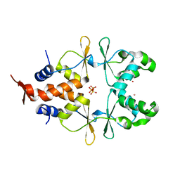
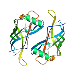
7CSV
 | | Pseudomonas aeruginosa antitoxin HigA | | 分子名称: | HTH cro/C1-type domain-containing protein | | 著者 | Song, Y.J, Luo, G.H, Bao, R. | | 登録日 | 2020-08-17 | | 公開日 | 2021-01-13 | | 最終更新日 | 2023-11-29 | | 実験手法 | X-RAY DIFFRACTION (1.71 Å) | | 主引用文献 | Pseudomonas aeruginosa antitoxin HigA functions as a diverse regulatory factor by recognizing specific pseudopalindromic DNA motifs.
Environ.Microbiol., 23, 2021
|
|
7CSY
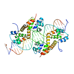 | | Pseudomonas aeruginosa antitoxin HigA with higBA promoter | | 分子名称: | DNA (28-MER), DNA (29-MER), HTH cro/C1-type domain-containing protein, ... | | 著者 | Song, Y.J, Luo, G.H, Bao, R. | | 登録日 | 2020-08-17 | | 公開日 | 2021-01-13 | | 最終更新日 | 2024-05-29 | | 実験手法 | X-RAY DIFFRACTION (2.29 Å) | | 主引用文献 | Pseudomonas aeruginosa antitoxin HigA functions as a diverse regulatory factor by recognizing specific pseudopalindromic DNA motifs.
Environ.Microbiol., 23, 2021
|
|
4YT4
 | |
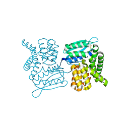
3DW8
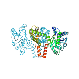
 | | Structure of a Protein Phosphatase 2A Holoenzyme with B55 subunit | | 分子名称: | MANGANESE (II) ION, Serine/threonine-protein phosphatase 2A 55 kDa regulatory subunit B alpha isoform, Serine/threonine-protein phosphatase 2A 65 kDa regulatory subunit A alpha isoform, ... | | 著者 | Xu, Y, Chen, Y, Zhang, P, Jeffrey, P.D, Shi, Y. | | 登録日 | 2008-07-21 | | 公開日 | 2008-10-07 | | 最終更新日 | 2024-04-03 | | 実験手法 | X-RAY DIFFRACTION (2.85 Å) | | 主引用文献 | Structure of a protein phosphatase 2A holoenzyme: insights into B55-mediated Tau dephosphorylation.
Mol.Cell, 31, 2008
|
|
3A1W
 | |
1FJM
 | | Protein serine/threonine phosphatase-1 (alpha isoform, type 1) complexed with microcystin-LR toxin | | 分子名称: | BETA-MERCAPTOETHANOL, MANGANESE (II) ION, PROTEIN SERINE/THREONINE PHOSPHATASE-1 (ALPHA ISOFORM, ... | | 著者 | Goldberg, J, Nairn, A.C, Kuriyan, J. | | 登録日 | 1995-12-17 | | 公開日 | 1996-06-20 | | 最終更新日 | 2023-11-15 | | 実験手法 | X-RAY DIFFRACTION (2.1 Å) | | 主引用文献 | Three-dimensional structure of the catalytic subunit of protein serine/threonine phosphatase-1.
Nature, 376, 1995
|
|
3K1K
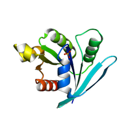
 | | Green fluorescent protein bound to enhancer nanobody | | 分子名称: | Enhancer, Green Fluorescent Protein | | 著者 | Kirchhofer, A, Helma, J, Schmidthals, K, Frauer, C, Cui, S, Karcher, A, Pellis, M, Muyldermans, S, Delucci, C.C, Cardoso, M.C, Leonhardt, H, Hopfner, K.-P, Rothbauer, U. | | 登録日 | 2009-09-28 | | 公開日 | 2009-12-08 | | 最終更新日 | 2023-11-15 | | 実験手法 | X-RAY DIFFRACTION (2.15 Å) | | 主引用文献 | Modulation of protein properties in living cells using nanobodies
Nat.Struct.Mol.Biol., 17, 2010
|
|


