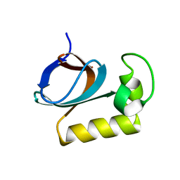
1VEK
 
 | | Solution Structure of RSGI RUH-011, a UBA Domain from Arabidopsis cDNA | | 分子名称: | ubiquitin-specific protease 14, putative | | 著者 | Higuchi, Y, Abe, T, Hirota, H, Saito, K, Koshiba, S, Kigawa, T, Yokoyama, S, RIKEN Structural Genomics/Proteomics Initiative (RSGI) | | 登録日 | 2004-03-31 | | 公開日 | 2004-09-30 | | 最終更新日 | 2023-12-27 | | 実験手法 | SOLUTION NMR | | 主引用文献 | Solution Structure of RSGI RUH-011, a UBA Domain from Arabidopsis cDNA
To be Published
|
|
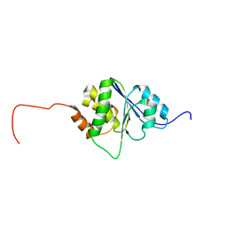
1VCS
 | | Solution Structure of RSGI RUH-009, an N-Terminal Domain of Vti1a [Mus musculus] | | 分子名称: | Vesicle transport through interaction with t-SNAREs homolog 1A | | 著者 | Abe, T, Hirota, H, Tomizawa, T, Koshiba, S, Kigawa, T, Yokoyama, S, RIKEN Structural Genomics/Proteomics Initiative (RSGI) | | 登録日 | 2004-03-10 | | 公開日 | 2005-05-03 | | 最終更新日 | 2023-12-27 | | 実験手法 | SOLUTION NMR | | 主引用文献 | Solution Structure of RSGI RUH-009, an N-Terminal Domain of Vti1a [Mus musculus]
To be Published
|
|
1T3O
 | | Solution structure of CsrA, a bacterial carbon storage regulatory protein | | 分子名称: | Carbon storage regulator | | 著者 | Koharudin, L.M.I, Georgiou, T, Kleanthous, C, Geoffrey, R, Kaptein, R, Boelens, R. | | 登録日 | 2004-04-27 | | 公開日 | 2005-10-18 | | 最終更新日 | 2024-05-22 | | 実験手法 | SOLUTION NMR | | 主引用文献 | A model for RNA binding by the bacterial carbon storage regulatory protein, CsrA
To be Published
|
|
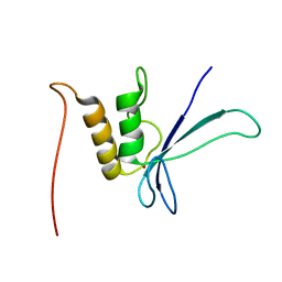
1VEH
 | | Solution structure of RSGI RUH-018, a NifU-like domain of hirip5 protein from mouse cDNA | | 分子名称: | NifU-like protein HIRIP5 | | 著者 | Ohashi, W, Hirota, H, Yamazaki, T, Yokoyama, S, RIKEN Structural Genomics/Proteomics Initiative (RSGI) | | 登録日 | 2004-03-31 | | 公開日 | 2004-09-30 | | 最終更新日 | 2023-12-27 | | 実験手法 | SOLUTION NMR | | 主引用文献 | Solution structure of RSGI RUH-018, a NifU-like domain of hirip5 protein from mouse cDNA
To be published
|
|
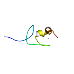
1VEJ
 | | Solution Structure of RSGI RUH-016, a UBA Domain from mouse cDNA | | 分子名称: | RIKEN cDNA 4931431F19 | | 著者 | Higuchi, Y, Abe, T, Hirota, H, Hayashi, F, Yokoyama, S, RIKEN Structural Genomics/Proteomics Initiative (RSGI) | | 登録日 | 2004-03-31 | | 公開日 | 2005-05-31 | | 最終更新日 | 2023-12-27 | | 実験手法 | SOLUTION NMR | | 主引用文献 | Solution Structure of RSGI RUH-016, a UBA Domain from mouse cDNA
To be Published
|
|
2XDF
 | | Solution Structure of the Enzyme I Dimer Complexed with HPr Using Residual Dipolar Couplings and Small Angle X-Ray Scattering | | 分子名称: | PHOSPHOCARRIER PROTEIN HPR, PHOSPHOENOLPYRUVATE-PROTEIN PHOSPHOTRANSFERASE | | 著者 | Schwieters, C.D, Suh, J.-Y, Grishaev, A, Guirlando, R, Takayama, Y, Clore, G.M. | | 登録日 | 2010-04-30 | | 公開日 | 2010-09-22 | | 最終更新日 | 2024-05-15 | | 実験手法 | SOLUTION NMR, SOLUTION SCATTERING | | 主引用文献 | Solution Structure of the 128 kDa Enzyme I Dimer from Escherichia Coli and its 146 kDa Complex with Hpr Using Residual Dipolar Couplings and Small- and Wide-Angle X-Ray Scattering.
J.Am.Chem.Soc., 132, 2010
|
|
2L21
 | | chicken IGF2R domain 11 | | 分子名称: | Cation-independent mannose-6-phosphate receptor | | 著者 | Williams, C, Hoppe, H, Strickland, M, Frago, S, Ellis, R.Z, Wattana-Amorn, P, Prince, S.N, Zaccheo, O.J, Forbes, B, Jones, E.Y, Rezgui, D.Z, Crump, M.P, Hassan, A.B. | | 登録日 | 2010-08-10 | | 公開日 | 2012-02-15 | | 最終更新日 | 2023-06-14 | | 実験手法 | SOLUTION NMR | | 主引用文献 | CD loop dependency of the IGF2: M6P/IGF2 receptor binding interaction predates imprinting
To be Published
|
|
1EJQ
 | | SOLUTION STRUCTURE OF THE SYNDECAN-4 WHOLE CYTOPLASMIC DOMAIN IN THE PRESENCE OF PHOSPHATIDYLINOSITOL 4,5-BISPHOSPHATE | | 分子名称: | SYNDECAN-4 | | 著者 | Shin, J, Oh, E.S, Lee, D, Couchman, J.R, Lee, W. | | 登録日 | 2000-03-04 | | 公開日 | 2001-03-07 | | 最終更新日 | 2024-05-29 | | 実験手法 | SOLUTION NMR | | 主引用文献 | SOLUTION STRUCTURE OF THE SYNDECAN-4 WHOLE CYTOPLASMIC DOMAIN IN THE PRESENCE OF PHOSPHATIDYLINOSITOL 4,5-BISPHOSPHATE
To be Published
|
|
1EMW
 | | SOLUTION STRUCTURE OF THE RIBOSOMAL PROTEIN S16 FROM THERMUS THERMOPHILUS | | 分子名称: | S16 RIBOSOMAL PROTEIN | | 著者 | Allard, P, Rak, A.V, Wimberly, B.T, Clemons Jr, W.M, Kalinin, A, Helgstrand, M, Garber, M.B, Ramakrishnan, V, Hard, T. | | 登録日 | 2000-03-20 | | 公開日 | 2000-08-09 | | 最終更新日 | 2024-05-22 | | 実験手法 | SOLUTION NMR | | 主引用文献 | Another piece of the ribosome: solution structure of S16 and its location in the 30S subunit.
Structure Fold.Des., 8, 2000
|
|
7V5E
 | |
7V5F
 | |
7VOZ
 | |
7VU7
 | |
1OY2
 | | Coupling of Folding and Binding in the PTB Domain of the Signaling Protein Shc | | 分子名称: | SHC transforming protein | | 著者 | Farooq, A, Zeng, L, Yan, K.S, Ravichandran, K.S, Zhou, M.-M. | | 登録日 | 2003-04-03 | | 公開日 | 2004-04-06 | | 最終更新日 | 2024-05-22 | | 実験手法 | SOLUTION NMR | | 主引用文献 | Coupling of Folding and Binding in the PTB Domain of the Signaling Protein Shc
Structure, 11, 2003
|
|
1U3N
 | | A SOD-like protein from B. subtilis, unstructured in solution, becomes ordered in the crystal: implications for function and for fibrillogenesis | | 分子名称: | Hypothetical superoxide dismutase-like protein yojM | | 著者 | Banci, L, Bertini, I, Calderone, V, Cramaro, F, Del Conte, R, Fantoni, A, Mangani, S, Quattrone, A, Viezzoli, M.S. | | 登録日 | 2004-07-22 | | 公開日 | 2005-05-03 | | 最終更新日 | 2022-03-02 | | 実験手法 | SOLUTION NMR | | 主引用文献 | A prokaryotic superoxide dismutase paralog lacking two Cu ligands: from largely unstructured in solution to ordered in the crystal.
Proc.Natl.Acad.Sci.Usa, 102, 2005
|
|
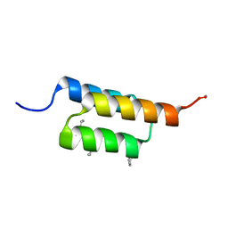
1S4T
 | | Solution structure of synthetic 21mer peptide spanning region 135-155 (in human numbering) of sheep prion protein | | 分子名称: | Major prion protein | | 著者 | Kozin, S.A, Lepage, C, Hui Bon Hoa, G, Rabesona, H, Mazur, A.K, Blond, A, Cheminant, M, Haertle, T, Debey, P, Rebuffat, S. | | 登録日 | 2004-01-18 | | 公開日 | 2004-01-27 | | 最終更新日 | 2024-05-22 | | 実験手法 | SOLUTION NMR | | 主引用文献 | Specific recognition between surface loop 2 (132-143) and helix 1 (144-154)
within sheep prion protein from in vitro studies of synthetic peptides
To be Published
|
|
1N8X
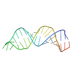 | | Solution structure of HIV-1 Stem Loop SL1 | | 分子名称: | HIV-1 STEM LOOP SL1 MONOMERIC RNA | | 著者 | Lawrence, D.C, Stover, C.C, Noznitsky, J, Wu, Z, Summers, M.F. | | 登録日 | 2002-11-21 | | 公開日 | 2003-04-08 | | 最終更新日 | 2024-05-22 | | 実験手法 | SOLUTION NMR | | 主引用文献 | Structure of the Intact Stem and Bulge of HIV-1 Psi-RNA Stem-Loop SL1
J.Mol.Biol., 326, 2003
|
|
7URJ
 | |
7UGC
 | | Structure of the N-terminal domain of ViaA | | 分子名称: | Protein ViaA | | 著者 | Lemak, A, Reichheld, S, Bhandari, V, Houliston, S, Arrowsmith, C.H, Sharpe, S, Houry, W.A. | | 登録日 | 2022-03-24 | | 公開日 | 2023-03-29 | | 最終更新日 | 2024-05-15 | | 実験手法 | SOLUTION NMR | | 主引用文献 | Structure of the N-terminal domain of ViaA
To Be Published
|
|
1NY8
 | | Solution structure of Protein yrbA from Escherichia Coli: Northeast Structural Genomics Consortium target ER115 | | 分子名称: | Protein yrbA | | 著者 | Swapna, G.V.T, Huang, J.Y, Acton, T.B, Shastry, R, Chiang, Y.-W, Montelione, G.T, Northeast Structural Genomics Consortium (NESG) | | 登録日 | 2003-02-11 | | 公開日 | 2004-06-15 | | 最終更新日 | 2024-05-22 | | 実験手法 | SOLUTION NMR | | 主引用文献 | Solution structure of Protein yrbA from Escherichia Coli: Northeast Structural Genomics Consortium target ER115
To be Published
|
|
7X31
 | |
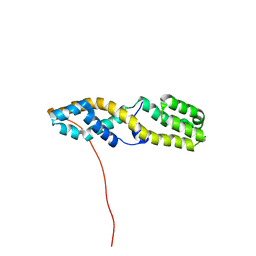
1R6H
 | | Solution Structure of human PRL-3 | | 分子名称: | protein tyrosine phosphatase type IVA, member 3 isoform 1 | | 著者 | Kozlov, G, Gehring, K, Ekiel, I. | | 登録日 | 2003-10-15 | | 公開日 | 2004-01-13 | | 最終更新日 | 2024-05-22 | | 実験手法 | SOLUTION NMR | | 主引用文献 | Structural Insights into Molecular Function of the Metastasis-associated Phosphatase PRL-3.
J.Biol.Chem., 279, 2004
|
|
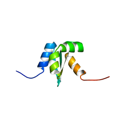
1V9J
 | | Solution structure of a BolA-like protein from Mus musculus | | 分子名称: | BolA-like protein RIKEN cDNA 1110025L05 | | 著者 | Kasai, T, Inoue, M, Koshiba, S, Yabuki, T, Aoki, M, Nunokawa, E, Seki, E, Matsuda, T, Matsuda, N, Tomo, Y, Shirouzu, M, Terada, T, Obayashi, N, Hamana, H, Shinya, N, Tatsuguchi, A, Yasuda, S, Yoshida, M, Hirota, H, Matsuo, Y, Tani, K, Suzuki, H, Arakawa, T, Carninci, P, Kawai, J, Hayashizaki, Y, Kigawa, T, Yokoyama, S, RIKEN Structural Genomics/Proteomics Initiative (RSGI) | | 登録日 | 2004-01-26 | | 公開日 | 2004-02-10 | | 最終更新日 | 2023-12-27 | | 実験手法 | SOLUTION NMR | | 主引用文献 | Solution structure of a BolA-like protein from Mus musculus
Protein Sci., 13, 2004
|
|
1UW0
 | |
7Y7L
 | | Solution structure of zinc finger domain 2 of human ZFAND1 | | 分子名称: | AN1-type zinc finger protein 1, ZINC ION | | 著者 | Fang, P.J, Lai, C.H, Ko, K.T, Chang, C.F, Hsu, S.T.D. | | 登録日 | 2022-06-22 | | 公開日 | 2023-06-28 | | 最終更新日 | 2024-05-15 | | 実験手法 | SOLUTION NMR | | 主引用文献 | Structural basis of p97 recognition by human ZFAND1
To Be Published
|
|


