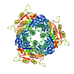
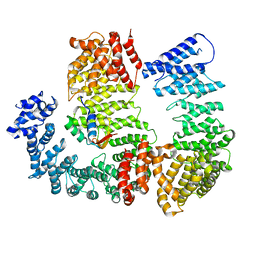
4DH5
 
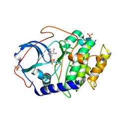 | | Room temperature X-ray structure of cAMP dependent Protein Kinase A catalytic subunit with high Mg2+, ADP, Phosphate, and IP20 | | 分子名称: | ADENOSINE-5'-DIPHOSPHATE, MAGNESIUM ION, PHOSPHATE ION, ... | | 著者 | Kovalevsky, A.Y, Langan, P. | | 登録日 | 2012-01-27 | | 公開日 | 2012-06-27 | | 最終更新日 | 2024-11-27 | | 実験手法 | X-RAY DIFFRACTION (2.2 Å) | | 主引用文献 | Low- and room-temperature X-ray structures of protein kinase A ternary complexes shed new light on its activity.
Acta Crystallogr.,Sect.D, 68, 2012
|
|
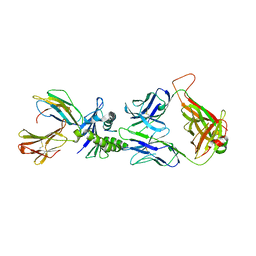
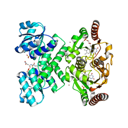
1TZ2
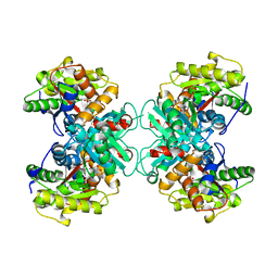 | | Crystal structure of 1-aminocyclopropane-1-carboyxlate deaminase complexed with ACC | | 分子名称: | 1-AMINOCYCLOPROPANECARBOXYLIC ACID, 1-aminocyclopropane-1-carboxylate deaminase, PYRIDOXAL-5'-PHOSPHATE | | 著者 | Karthikeyan, S, Zhou, Q, Zhao, Z, Kao, C.L, Tao, Z, Robinson, H, Liu, H.W, Zhang, H. | | 登録日 | 2004-07-09 | | 公開日 | 2004-11-02 | | 最終更新日 | 2023-08-23 | | 実験手法 | X-RAY DIFFRACTION (2.1 Å) | | 主引用文献 | Structural Analysis of Pseudomonas 1-Aminocyclopropane-1-carboxylate Deaminase Complexes: Insight into the Mechanism of a Unique Pyridoxal-5'-phosphate Dependent Cyclopropane Ring-Opening Reaction
Biochemistry, 43, 2004
|
|
1U6Z
 | |
3L9W
 | |
1UCG
 | | Crystal structure of Ribonuclease MC1 N71T mutant | | 分子名称: | MANGANESE (II) ION, Ribonuclease MC | | 著者 | Suzuki, A, Numata, T, Yao, M, Tanaka, I, Kimura, M. | | 登録日 | 2003-04-14 | | 公開日 | 2003-04-29 | | 最終更新日 | 2024-11-06 | | 実験手法 | X-RAY DIFFRACTION (1.65 Å) | | 主引用文献 | Crystal structures of the ribonuclease MC1 mutants N71T and N71S in complex with 5'-GMP: structural basis for alterations in substrate specificity
Biochemistry, 42, 2003
|
|
2LFY
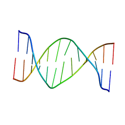 | | Structure of the duplex when (5'S)-8,5'-cyclo-2'-deoxyguanosine is placed opposite dA | | 分子名称: | DNA (5'-D(*AP*CP*AP*AP*AP*CP*AP*AP*GP*CP*AP*C)-3'), DNA (5'-D(*GP*TP*GP*CP*(2LF)P*TP*GP*TP*TP*TP*GP*T)-3') | | 著者 | Huang, H, Das, R.S, Basu, A, Stone, M.P. | | 登録日 | 2011-07-18 | | 公開日 | 2012-06-27 | | 最終更新日 | 2024-05-15 | | 実験手法 | SOLUTION NMR | | 主引用文献 | Structures of (5'S)-8,5'-Cyclo-2'-deoxyguanosine Mismatched with dA or dT.
Chem.Res.Toxicol., 25, 2012
|
|
1DDF
 | | FAS DEATH DOMAIN, NMR, MINIMIZED AVERAGE STRUCTURE | | 分子名称: | FAS | | 著者 | Huang, B, Eberstadt, M, Olejniczak, E, Meadows, R.P, Fesik, S. | | 登録日 | 1996-11-08 | | 公開日 | 1997-11-12 | | 最終更新日 | 2024-05-22 | | 実験手法 | SOLUTION NMR | | 主引用文献 | NMR structure and mutagenesis of the Fas (APO-1/CD95) death domain.
Nature, 384, 1996
|
|
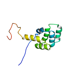
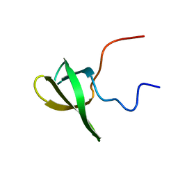
2L6L
 | | Solution structure of human J-protein co-chaperone, Dph4 | | 分子名称: | DnaJ homolog subfamily C member 24, ZINC ION | | 著者 | Thakur, A, Chitoor, B.S, Atreya, H.S, Silva, P.D. | | 登録日 | 2010-11-23 | | 公開日 | 2011-12-07 | | 最終更新日 | 2024-05-01 | | 実験手法 | SOLUTION NMR | | 主引用文献 | Structure and mechanistic insights into novel iron-mediated moonlighting functions of human J-protein cochaperone, Dph4.
J.Biol.Chem., 287, 2012
|
|
2L8R
 | |
1DOM
 | |
7A1R
 | |
7A1D
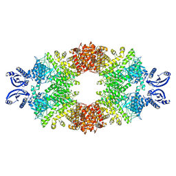 | | Cryo-EM map of the large glutamate dehydrogenase composed of 180 kDa subunits from Mycobacterium smegmatis (open conformation) | | 分子名称: | NAD-specific glutamate dehydrogenase | | 著者 | Lazaro, M, Melero, R, Huet, C, Lopez-Alonso, J.P, Delgado, S, Dodu, A, Bruch, E.M, Abriata, L.A, Alzari, P.M, Valle, M, Lisa, M.N. | | 登録日 | 2020-08-12 | | 公開日 | 2021-06-09 | | 最終更新日 | 2025-05-14 | | 実験手法 | ELECTRON MICROSCOPY (4.19 Å) | | 主引用文献 | 3D architecture and structural flexibility revealed in the subfamily of large glutamate dehydrogenases by a mycobacterial enzyme.
Commun Biol, 4, 2021
|
|
7NXX
 | | Structure of Superoxide Dismutase 1 (SOD1) in complex with nanobody 2 (Nb2). | | 分子名称: | COPPER (II) ION, Superoxide dismutase [Cu-Zn], ZINC ION, ... | | 著者 | Gallardo, R, Rousseau, F, Schymkowitz, J, Ulens, C. | | 登録日 | 2021-03-19 | | 公開日 | 2022-09-28 | | 最終更新日 | 2024-10-23 | | 実験手法 | X-RAY DIFFRACTION (2.189 Å) | | 主引用文献 | Identification and rational improvement of a nanobody that suppresses aggregation of mutant SOD1
To Be Published
|
|
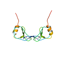
1HRJ
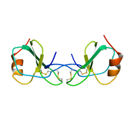 | | HUMAN RANTES, NMR, 13 STRUCTURES | | 分子名称: | HUMAN REGULATED UPON ACTIVATION NORMAL T-CELL EXPRESSED AND SECRETED | | 著者 | Chung, C, Cooke, R.M, Proudfoot, A.E.I, Wells, T.N.C. | | 登録日 | 1995-08-18 | | 公開日 | 1996-10-14 | | 最終更新日 | 2024-10-16 | | 実験手法 | SOLUTION NMR | | 主引用文献 | The three-dimensional solution structure of RANTES.
Biochemistry, 34, 1995
|
|
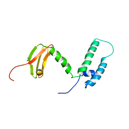
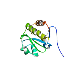
2L8D
 | | Structure/function of the LBR Tudor domain | | 分子名称: | Lamin-B receptor | | 著者 | Liokatis, S, Edlich, C, Soupsana, K, Giannios, I, Sattler, M, Georgatos, S.D, Politou, A.S. | | 登録日 | 2011-01-10 | | 公開日 | 2011-11-09 | | 最終更新日 | 2024-05-15 | | 実験手法 | SOLUTION NMR | | 主引用文献 | Solution structure and molecular interactions of lamin B receptor tudor domain.
J.Biol.Chem., 287, 2012
|
|
7VRR
 | |
2LGR
 | |
2LJW
 | | Solution NMR structure of Alr2454 protein from Nostoc sp. strain PCC 7120, Northeast Structural Genomics Consortium Target NsR264 | | 分子名称: | Alr2454 protein | | 著者 | Aramini, J.M, Lee, D, Ciccosanti, C, Janjua, H, Rost, B, Acton, T.B, Xiao, R, Everett, J.K, Montelione, G.T, Northeast Structural Genomics Consortium (NESG) | | 登録日 | 2011-09-29 | | 公開日 | 2011-10-19 | | 最終更新日 | 2024-05-15 | | 実験手法 | SOLUTION NMR | | 主引用文献 | Solution NMR structure of Alr2454 from Nostoc sp. PCC 7120, the first structural representative of Pfam domain family PF11267.
J.Struct.Funct.Genom., 13, 2012
|
|
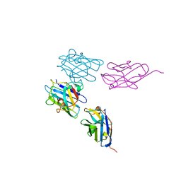
5VTB
 | | Crystal structure of RBBP4 bound to BCL11a peptide | | 分子名称: | B-cell lymphoma/leukemia 11A, GLYCEROL, Histone-binding protein RBBP4 | | 著者 | Meagher, J.L, Stuckey, J.A. | | 登録日 | 2017-05-16 | | 公開日 | 2017-12-27 | | 最終更新日 | 2023-10-04 | | 実験手法 | X-RAY DIFFRACTION (2.4 Å) | | 主引用文献 | Probing the interaction between the histone methyltransferase/deacetylase subunit RBBP4/7 and the transcription factor BCL11A in epigenetic complexes.
J. Biol. Chem., 293, 2018
|
|
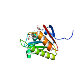
2J4J
 | | Crystal structure of uridylate kinase from Sulfolobus solfataricus in complex with UMP and AMPPCP to 2.1 Angstrom resolution | | 分子名称: | COBALT (II) ION, MAGNESIUM ION, P1-(5'-ADENOSINE)P4-(5'-URIDINE)-BETA,GAMMA-METHYLENE TETRAPHOSPHATE, ... | | 著者 | Jensen, K.S, Johansson, E, Jensen, K.F. | | 登録日 | 2006-09-01 | | 公開日 | 2007-02-27 | | 最終更新日 | 2023-12-13 | | 実験手法 | X-RAY DIFFRACTION (2.1 Å) | | 主引用文献 | Structural and Enzymatic Investigation of the Sulfolobus Solfataricus Uridylate Kinase Shows Competitive Utp Inhibition and the Lack of GTP Stimulation
Biochemistry, 46, 2007
|
|
2J8U
 | | Large CDR3a loop alteration as a function of MHC mutation. | | 分子名称: | AHIII TCR ALPHA CHAIN, AHIII TCR BETA CHAIN, Beta-2-microglobulin, ... | | 著者 | Miller, P.J, Benhar, Y.P, Biddison, W, Collins, E.J. | | 登録日 | 2006-10-27 | | 公開日 | 2007-10-16 | | 最終更新日 | 2024-11-13 | | 実験手法 | X-RAY DIFFRACTION (2.88 Å) | | 主引用文献 | Single MHC mutation eliminates enthalpy associated with T cell receptor binding.
J. Mol. Biol., 373, 2007
|
|
2J4L
 | |
2JKZ
 | | SACCHAROMYCES CEREVISIAE HYPOXANTHINE-GUANINE PHOSPHORIBOSYLTRANSFERASE IN COMPLEX WITH GMP (GUANOSINE 5'- MONOPHOSPHATE) (ORTHORHOMBIC CRYSTAL FORM) | | 分子名称: | GUANOSINE-5'-MONOPHOSPHATE, HYPOXANTHINE-GUANINE PHOSPHORIBOSYLTRANSFERASE, SULFATE ION | | 著者 | Moynie, L, Giraud, M.F, Breton, A, Boissier, F, Daignan-Fornier, B, Dautant, A. | | 登録日 | 2008-09-02 | | 公開日 | 2009-11-17 | | 最終更新日 | 2023-12-13 | | 実験手法 | X-RAY DIFFRACTION (3.45 Å) | | 主引用文献 | Functional Significance of Four Successive Glycine Residues in the Pyrophosphate Binding Loop of Fungal 6-Oxopurine Phosphoribosyltransferases.
Protein Sci., 21, 2012
|
|
6N88
 | |
3L9X
 | | KefC C-terminal domain in complex with KefF and ESG | | 分子名称: | FLAVIN MONONUCLEOTIDE, Glutathione-regulated potassium-efflux system protein kefC, linker, ... | | 著者 | Roosild, T.P. | | 登録日 | 2010-01-05 | | 公開日 | 2010-11-17 | | 最終更新日 | 2023-09-06 | | 実験手法 | X-RAY DIFFRACTION (2.1 Å) | | 主引用文献 | Mechanism of ligand-gated potassium efflux in bacterial pathogens.
Proc.Natl.Acad.Sci.USA, 107, 2010
|
|


