8FL4
 
 | |
8FL7
 | |
8FLE
 | |
8FL3
 | |
8FLB
 | |
8FLF
 | |
8CLK
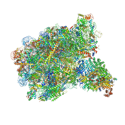
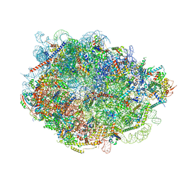
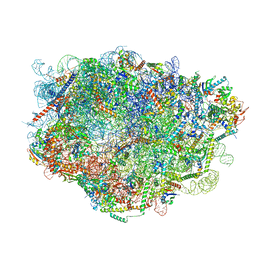
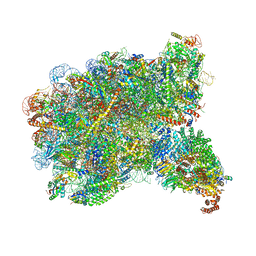
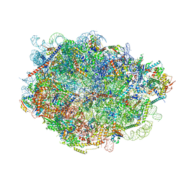
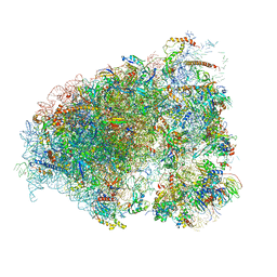
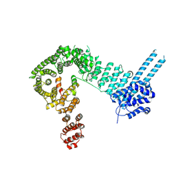
 | | TFIIIC TauA complex | | 分子名称: | General transcription factor 3C polypeptide 1, General transcription factor 3C polypeptide 3, General transcription factor 3C polypeptide 5, ... | | 著者 | Seifert-Davila, W, Girbig, M, Hauptmann, L, Hoffmann, T, Eustermann, S, Mueller, C.W. | | 登録日 | 2023-02-16 | | 公開日 | 2023-06-21 | | 最終更新日 | 2024-07-24 | | 実験手法 | ELECTRON MICROSCOPY (3.5 Å) | | 主引用文献 | Structural insights into human TFIIIC promoter recognition.
Sci Adv, 9, 2023
|
|
8FLC
 | |
6OM7
 | |
2ZU6
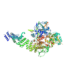
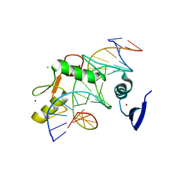
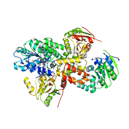 | | crystal structure of the eIF4A-PDCD4 complex | | 分子名称: | 1,2-ETHANEDIOL, ACETIC ACID, Eukaryotic initiation factor 4A-I, ... | | 著者 | Cho, Y, Chang, J.H, Sohn, S.Y. | | 登録日 | 2008-10-13 | | 公開日 | 2009-02-24 | | 最終更新日 | 2011-07-13 | | 実験手法 | X-RAY DIFFRACTION (2.8 Å) | | 主引用文献 | Crystal structure of the eIF4A-PDCD4 complex
Proc.Natl.Acad.Sci.Usa, 106, 2009
|
|
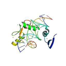
2YYN
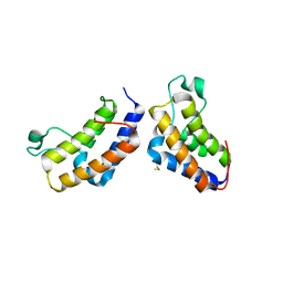 | | Crystal structure of human bromodomain protein | | 分子名称: | Transcription intermediary factor 1-alpha | | 著者 | Kishishita, S, Uchikubo-Kamo, T, Murayama, K, Terada, T, Shirouzu, M, Yokoyama, S, RIKEN Structural Genomics/Proteomics Initiative (RSGI) | | 登録日 | 2007-04-30 | | 公開日 | 2008-05-06 | | 最終更新日 | 2022-12-21 | | 実験手法 | X-RAY DIFFRACTION (2.5 Å) | | 主引用文献 | Crystal structure of human bromodomain protein
To be Published
|
|
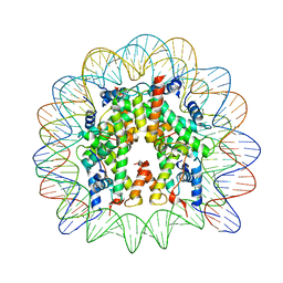
3AV2
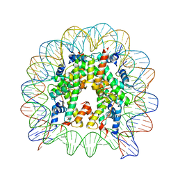 | | The human nucleosome structure containing the histone variant H3.3 | | 分子名称: | 146-MER DNA, Histone H2A type 1-B/E, Histone H2B type 1-J, ... | | 著者 | Tachiwana, H, Osakabe, A, Shiga, T, Miya, M, Kimura, H, Kagawa, W, Kurumizaka, H. | | 登録日 | 2011-02-18 | | 公開日 | 2011-06-01 | | 最終更新日 | 2023-11-01 | | 実験手法 | X-RAY DIFFRACTION (2.8 Å) | | 主引用文献 | Structures of human nucleosomes containing major histone H3 variants
Acta Crystallogr.,Sect.D, 67, 2011
|
|
6QNX
 | | Structure of the SA2/SCC1/CTCF complex | | 分子名称: | Cohesin subunit SA-2, Double-strand-break repair protein rad21 homolog, Transcriptional repressor CTCF | | 著者 | Li, Y, Muir, K.W, Panne, D. | | 登録日 | 2019-02-12 | | 公開日 | 2020-01-22 | | 最終更新日 | 2024-05-15 | | 実験手法 | X-RAY DIFFRACTION (2.7 Å) | | 主引用文献 | The structural basis for cohesin-CTCF-anchored loops.
Nature, 578, 2020
|
|
6R0C
 | | Human-D02 Nucleosome Core Particle with biotin-streptavidin label | | 分子名称: | DNA (142-MER), Histone H2A type 1, Histone H2B type 1-C/E/F/G/I, ... | | 著者 | Pye, V.E, Wilson, M.D, Cherepanov, P, Costa, A. | | 登録日 | 2019-03-12 | | 公開日 | 2019-09-25 | | 最終更新日 | 2024-05-15 | | 実験手法 | ELECTRON MICROSCOPY (4.2 Å) | | 主引用文献 | Retroviral integration into nucleosomes through DNA looping and sliding along the histone octamer.
Nat Commun, 10, 2019
|
|
5VMY
 | | Kaiso (ZBTB33) zinc finger DNA binding domain in complex with a hemi CpG-methylated DNA resembling the specific Kaiso binding sequence (KBS) | | 分子名称: | CHLORIDE ION, DNA (5'-D(*CP*GP*TP*TP*AP*TP*TP*CP*GP*CP*GP*GP*GP*AP*AP*GP*CP*A)-3'), DNA (5'-D(*TP*GP*CP*TP*TP*CP*CP*(5CM)P*GP*(5CM)P*GP*AP*AP*TP*AP*AP*CP*G)-3'), ... | | 著者 | Nikolova, E.N, Stanfield, R.L, Martinez-Yamout, M.A, Dyson, H.J, Wright, P.E. | | 登録日 | 2017-04-28 | | 公開日 | 2018-04-04 | | 最終更新日 | 2023-10-04 | | 実験手法 | X-RAY DIFFRACTION (2.002 Å) | | 主引用文献 | CH···O Hydrogen Bonds Mediate Highly Specific Recognition of Methylated CpG Sites by the Zinc Finger Protein Kaiso.
Biochemistry, 57, 2018
|
|
6R94
 | | Cryo-EM structure of NCP_THF2(-3) | | 分子名称: | Histone H2A type 1-B/E, Histone H2B type 1-J, Histone H3.1, ... | | 著者 | Matsumoto, S, Cavadini, S, Bunker, R.D, Thoma, N.H. | | 登録日 | 2019-04-02 | | 公開日 | 2019-06-12 | | 最終更新日 | 2024-05-22 | | 実験手法 | ELECTRON MICROSCOPY (3.5 Å) | | 主引用文献 | DNA damage detection in nucleosomes involves DNA register shifting.
Nature, 571, 2019
|
|
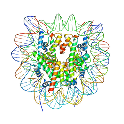
3AV1
 | | The human nucleosome structure containing the histone variant H3.2 | | 分子名称: | 146-MER DNA, Histone H2A type 1-B/E, Histone H2B type 1-J, ... | | 著者 | Tachiwana, H, Osakabe, A, Shiga, T, Miya, Y, Kimura, H, Kagawa, W, Kurumizaka, H. | | 登録日 | 2011-02-18 | | 公開日 | 2011-06-01 | | 最終更新日 | 2023-11-01 | | 実験手法 | X-RAY DIFFRACTION (2.5 Å) | | 主引用文献 | Structures of human nucleosomes containing major histone H3 variants
Acta Crystallogr.,Sect.D, 67, 2011
|
|
6R6P
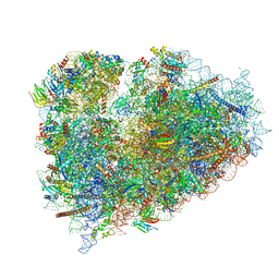 | | Structure of XBP1u-paused ribosome nascent chain complex (rotated state) | | 分子名称: | 18S rRNA, 28S ribosomal RNA, 40S ribosomal protein S12, ... | | 著者 | Shanmuganathan, V, Cheng, J, Berninghausen, O, Beckmann, R. | | 登録日 | 2019-03-27 | | 公開日 | 2019-07-10 | | 最終更新日 | 2024-05-22 | | 実験手法 | ELECTRON MICROSCOPY (3.1 Å) | | 主引用文献 | Structural and mutational analysis of the ribosome-arresting human XBP1u.
Elife, 8, 2019
|
|
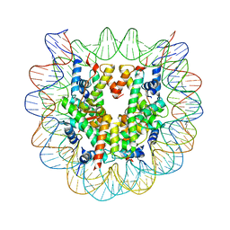
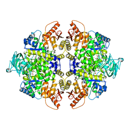
2Z9S
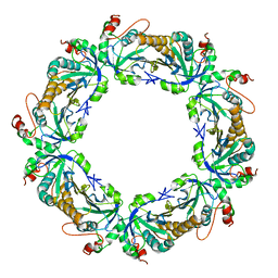 | | Crystal Structure Analysis of rat HBP23/Peroxiredoxin I, Cys52Ser mutant | | 分子名称: | Peroxiredoxin-1 | | 著者 | Matsumura, T, Okamoto, K, Nishino, T, Abe, Y. | | 登録日 | 2007-09-25 | | 公開日 | 2007-11-20 | | 最終更新日 | 2021-11-10 | | 実験手法 | X-RAY DIFFRACTION (2.9 Å) | | 主引用文献 | Dimer-Oligomer Interconversion of Wild-type and Mutant Rat 2-Cys Peroxiredoxin: DISULFIDE FORMATION AT DIMER-DIMER INTERFACES IS NOT ESSENTIAL FOR DECAMERIZATION
J.Biol.Chem., 283, 2008
|
|
5VMW
 | | Kaiso (ZBTB33) zinc finger DNA binding domain in complex with a double CpG-methylated DNA resembling the specific Kaiso binding sequence (KBS) | | 分子名称: | CHLORIDE ION, DNA (5'-D(*CP*GP*TP*TP*AP*TP*TP*(5CM)P*GP*(5CM)P*GP*GP*GP*AP*AP*GP*CP*A)-3'), DNA (5'-D(*TP*GP*CP*TP*TP*CP*CP*(5CM)P*GP*(5CM)P*GP*AP*AP*TP*AP*AP*CP*G)-3'), ... | | 著者 | Nikolova, E.N, Stanfield, R.L, Martinez-Yamout, M.A, Dyson, H.J, Wright, P.E. | | 登録日 | 2017-04-28 | | 公開日 | 2018-04-04 | | 最終更新日 | 2023-10-04 | | 実験手法 | X-RAY DIFFRACTION (2.397 Å) | | 主引用文献 | CH···O Hydrogen Bonds Mediate Highly Specific Recognition of Methylated CpG Sites by the Zinc Finger Protein Kaiso.
Biochemistry, 57, 2018
|
|
3AYW
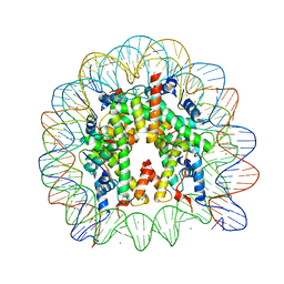 | | Crystal Structure of Human Nucleosome Core Particle Containing H3K56Q mutation | | 分子名称: | 146-MER DNA, CHLORIDE ION, Histone H2A type 1-B/E, ... | | 著者 | Iwasaki, W, Tachiwana, H, Kawaguchi, K, Shibata, T, Kagawa, W, Kurumizaka, H. | | 登録日 | 2011-05-19 | | 公開日 | 2011-09-21 | | 最終更新日 | 2023-11-01 | | 実験手法 | X-RAY DIFFRACTION (2.9 Å) | | 主引用文献 | Comprehensive Structural Analysis of Mutant Nucleosomes Containing Lysine to Glutamine (KQ) Substitutions in the H3 and H4 Histone-Fold Domains
Biochemistry, 50, 2011
|
|
3AZI
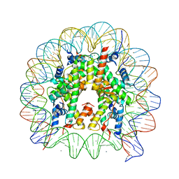 | | Crystal Structure of Human Nucleosome Core Particle Containing H4K31Q mutation | | 分子名称: | 146-MER DNA, CHLORIDE ION, Histone H2A type 1-B/E, ... | | 著者 | Iwasaki, W, Tachiwana, H, Kawaguchi, K, Shibata, T, Kagawa, W, Kurumizaka, H. | | 登録日 | 2011-05-25 | | 公開日 | 2011-09-21 | | 最終更新日 | 2023-11-01 | | 実験手法 | X-RAY DIFFRACTION (2.7 Å) | | 主引用文献 | Comprehensive Structural Analysis of Mutant Nucleosomes Containing Lysine to Glutamine (KQ) Substitutions in the H3 and H4 Histone-Fold Domains
Biochemistry, 50, 2011
|
|
3AZL
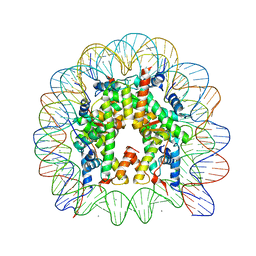 | | Crystal Structure of Human Nucleosome Core Particle Containing H4K77Q mutation | | 分子名称: | 146-MER DNA, CHLORIDE ION, Histone H2A type 1-B/E, ... | | 著者 | Iwasaki, W, Tachiwana, H, Kawaguchi, K, Shibata, T, Kagawa, W, Kurumizaka, H. | | 登録日 | 2011-05-25 | | 公開日 | 2011-09-21 | | 最終更新日 | 2023-11-01 | | 実験手法 | X-RAY DIFFRACTION (2.7 Å) | | 主引用文献 | Comprehensive Structural Analysis of Mutant Nucleosomes Containing Lysine to Glutamine (KQ) Substitutions in the H3 and H4 Histone-Fold Domains
Biochemistry, 50, 2011
|
|
3AZN
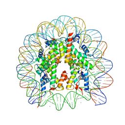 | | Crystal Structure of Human Nucleosome Core Particle Containing H4K91Q mutation | | 分子名称: | 146-MER DNA, CHLORIDE ION, Histone H2A type 1-B/E, ... | | 著者 | Iwasaki, W, Tachiwana, H, Kawaguchi, K, Shibata, T, Kagawa, W, Kurumizaka, H. | | 登録日 | 2011-05-25 | | 公開日 | 2011-09-21 | | 最終更新日 | 2023-11-01 | | 実験手法 | X-RAY DIFFRACTION (3 Å) | | 主引用文献 | Comprehensive Structural Analysis of Mutant Nucleosomes Containing Lysine to Glutamine (KQ) Substitutions in the H3 and H4 Histone-Fold Domains
Biochemistry, 50, 2011
|
|
3BJF
 | |


