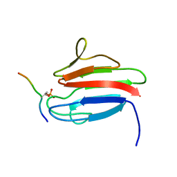
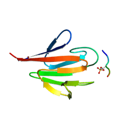
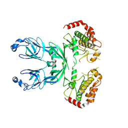
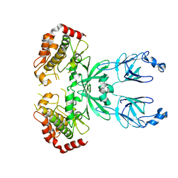
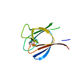
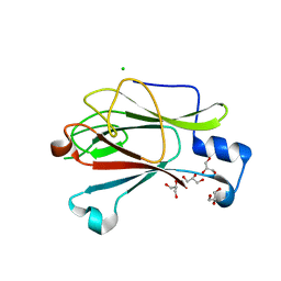
4EGX
 
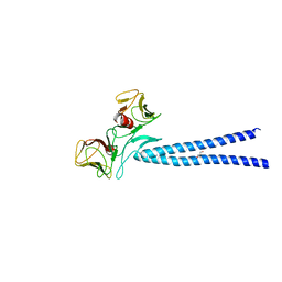 | | Crystal structure of KIF1A CC1-FHA tandem | | 分子名称: | DI(HYDROXYETHYL)ETHER, GLYCEROL, Kinesin-like protein KIF1A | | 著者 | Yu, J, Huo, L, Yue, Y, Xu, T, Zhang, M, Feng, W. | | 登録日 | 2012-04-02 | | 公開日 | 2012-10-03 | | 最終更新日 | 2023-11-08 | | 実験手法 | X-RAY DIFFRACTION (2.51 Å) | | 主引用文献 | The CC1-FHA Tandem as a Central Hub for Controlling the Dimerization and Activation of Kinesin-3 KIF1A
Structure, 20, 2012
|
|
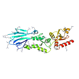
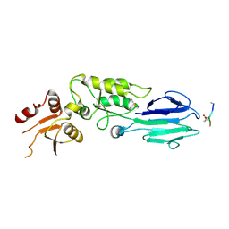
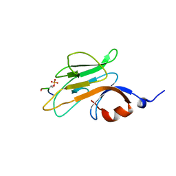
3VPY
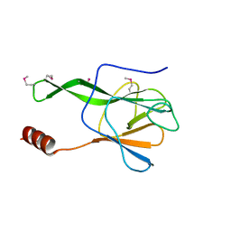 | | Crystal structure of Arabidopsis DDL FHA domain | | 分子名称: | FHA domain-containing protein DDL | | 著者 | Yuan, Y.A, Machida, S. | | 登録日 | 2012-03-15 | | 公開日 | 2013-02-06 | | 実験手法 | X-RAY DIFFRACTION (1.7 Å) | | 主引用文献 | Crystal Structure of Arabidopsis thaliana Dawdle Forkhead-Associated Domain reveals a conserved phospho-threonine recognition cleft for Dicer-like1 binding.
Mol Plant, 2013
|
|
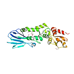
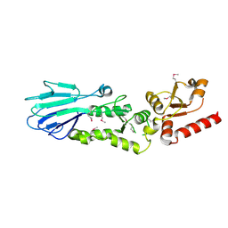
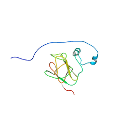
3VA4
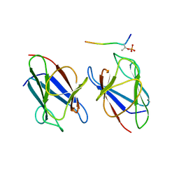 | | Crystal structure of the mammalian MDC1 FHA domain complexed with CHK2 pThr68 peptide | | 分子名称: | Mediator of DNA damage checkpoint protein 1, Serine/threonine-protein kinase Chk2 | | 著者 | Wu, H.H, Wu, P.Y, Huang, K.F, Kao, Y.Y, Tsai, M.D. | | 登録日 | 2011-12-28 | | 公開日 | 2012-02-01 | | 実験手法 | X-RAY DIFFRACTION (1.54 Å) | | 主引用文献 | Structural Delineation of MDC1-FHA Domain Binding with CHK2-pThr68.
Biochemistry, 2012
|
|
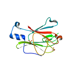
3VA1
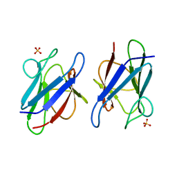 | | Crystal structure of the mammalian MDC1 FHA domain | | 分子名称: | Mediator of DNA damage checkpoint protein 1, SULFATE ION | | 著者 | Wu, H.H, Wu, P.Y, Huang, K.F, Kao, Y.Y, Tsai, M.D. | | 登録日 | 2011-12-28 | | 公開日 | 2012-02-01 | | 最終更新日 | 2024-03-20 | | 実験手法 | X-RAY DIFFRACTION (1.74 Å) | | 主引用文献 | Structural Delineation of MDC1-FHA Domain Binding with CHK2-pThr68.
Biochemistry, 2012
|
|
3UOT
 | | Crystal Structure of MDC1 FHA Domain in Complex with a Phosphorylated Peptide from the MDC1 N-terminus | | 分子名称: | Mediator of DNA damage checkpoint protein 1 | | 著者 | Clapperton, J.A, Lloyd, J, Haire, L.F, Li, J, Smerdon, S.J. | | 登録日 | 2011-11-17 | | 公開日 | 2011-12-28 | | 最終更新日 | 2012-07-18 | | 実験手法 | X-RAY DIFFRACTION (1.8 Å) | | 主引用文献 | The molecular basis of ATM-dependent dimerization of the Mdc1 DNA damage checkpoint mediator.
Nucleic Acids Res., 40, 2012
|
|
3UNN
 | |
3UNM
 | | Crystal Structure of The Human MDC1 FHA Domain | | 分子名称: | Mediator of DNA damage checkpoint protein 1 | | 著者 | Luo, S, Ye, K. | | 登録日 | 2011-11-16 | | 公開日 | 2012-01-25 | | 最終更新日 | 2023-11-01 | | 実験手法 | X-RAY DIFFRACTION (1.8 Å) | | 主引用文献 | Structural mechanism of the phosphorylation-dependent dimerization of the MDC1 forkhead-associated domain
Nucleic Acids Res., 40, 2012
|
|
3UN0
 | | Crystal Structure of MDC1 FHA Domain | | 分子名称: | Mediator of DNA damage checkpoint protein 1, SULFATE ION | | 著者 | Clapperton, J.A, Lloyd, J, Haire, L.F, Li, J, Smerdon, S.J. | | 登録日 | 2011-11-15 | | 公開日 | 2011-12-28 | | 最終更新日 | 2024-02-28 | | 実験手法 | X-RAY DIFFRACTION (2.3 Å) | | 主引用文献 | The molecular basis of ATM-dependent dimerization of the Mdc1 DNA damage checkpoint mediator.
Nucleic Acids Res., 40, 2012
|
|
3UMZ
 | | Crystal Structure of the human MDC1 FHA Domain | | 分子名称: | Mediator of DNA damage checkpoint protein 1 | | 著者 | Luo, S, Ye, K. | | 登録日 | 2011-11-15 | | 公開日 | 2012-01-25 | | 最終更新日 | 2024-03-20 | | 実験手法 | X-RAY DIFFRACTION (1.65 Å) | | 主引用文献 | Structural mechanism of the phosphorylation-dependent dimerization of the MDC1 forkhead-associated domain
Nucleic Acids Res., 40, 2012
|
|
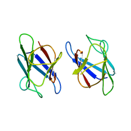
3POA
 | |
3PO8
 | |
3OUN
 | |
3MDB
 | | Crystal structure of the ternary complex of full length centaurin alpha-1, KIF13B FHA domain, and IP4 | | 分子名称: | (2R)-3-{[(R)-{[(1S,2S,3R,4S,5S,6S)-2,6-dihydroxy-3,4,5-tris(phosphonooxy)cyclohexyl]oxy}(hydroxy)phosphoryl]oxy}propane -1,2-diyl dioctanoate, Arf-GAP with dual PH domain-containing protein 1, Kinesin-like protein KIF13B, ... | | 著者 | Shen, L, Tong, Y, Tempel, W, MacKenzie, F, Arrowsmith, C.H, Edwards, A.M, Bountra, C, Weigelt, J, Bochkarev, A, Park, H, Structural Genomics Consortium (SGC) | | 登録日 | 2010-03-30 | | 公開日 | 2010-08-04 | | 最終更新日 | 2023-09-06 | | 実験手法 | X-RAY DIFFRACTION (2.952 Å) | | 主引用文献 | Crystal structure of the ternary complex of full length centaurin alpha-1, KIF13B FHA domain, and IP4
to be published
|
|
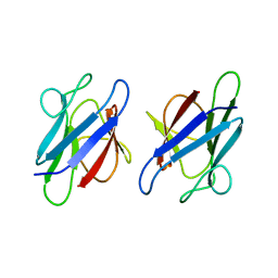
3I6W
 | |
3I6U
 | |
3I0N
 | | Structure of the S. pombe Nbs1 FHA/BRCT-repeat domain | | 分子名称: | DNA repair and telomere maintenance protein nbs1, GLYCEROL | | 著者 | Clapperton, J.A, Lloyd, J, Chapman, J.R, Jackson, S.P, Smerdon, S.J. | | 登録日 | 2009-06-25 | | 公開日 | 2009-10-13 | | 最終更新日 | 2023-11-01 | | 実験手法 | X-RAY DIFFRACTION (2.3 Å) | | 主引用文献 | A supramodular FHA/BRCT-repeat architecture mediates Nbs1 adaptor function in response to DNA damage
Cell(Cambridge,Mass.), 139, 2009
|
|
3I0M
 | | Structure of the S. pombe Nbs1 FHA/BRCT-repeat domain | | 分子名称: | DNA repair and telomere maintenance protein nbs1, GLYCEROL | | 著者 | Clapperton, J.A, Lloyd, J, Chapman, J.R, Jackson, S.P, Smerdon, S.J. | | 登録日 | 2009-06-25 | | 公開日 | 2009-10-13 | | 最終更新日 | 2012-05-02 | | 実験手法 | X-RAY DIFFRACTION (2.6 Å) | | 主引用文献 | A supramodular FHA/BRCT-repeat architecture mediates Nbs1 adaptor function in response to DNA damage
Cell(Cambridge,Mass.), 139, 2009
|
|
3HX1
 | | Crystal structure of the Slr1951 protein from Synechocystis sp. Northeast Structural Genomics Consortium Target SgR167A | | 分子名称: | Slr1951 protein | | 著者 | Vorobiev, S, Chen, Y, Seetharaman, J, Janjua, J, Xiao, R, Ciccosanti, C, Belote, R.L, Everett, J.K, Nair, R, Acton, T.B, Rost, B, Montelione, G.T, Hunt, J.F, Tong, L, Northeast Structural Genomics Consortium (NESG) | | 登録日 | 2009-06-19 | | 公開日 | 2009-06-30 | | 最終更新日 | 2019-07-24 | | 実験手法 | X-RAY DIFFRACTION (2.5 Å) | | 主引用文献 | Crystal structure of the Slr1951 protein from Synechocystis sp.
To be Published
|
|
3HUF
 | | Structure of the S. pombe Nbs1-Ctp1 complex | | 分子名称: | DNA repair and telomere maintenance protein nbs1, Double-strand break repair protein ctp1, THIOCYANATE ION | | 著者 | Williams, R.S, Guenther, G, Tainer, J.A. | | 登録日 | 2009-06-13 | | 公開日 | 2009-10-13 | | 最終更新日 | 2011-07-13 | | 実験手法 | X-RAY DIFFRACTION (2.15 Å) | | 主引用文献 | Nbs1 flexibly tethers Ctp1 and Mre11-Rad50 to coordinate DNA double-strand break processing and repair.
Cell(Cambridge,Mass.), 139, 2009
|
|
3HUE
 | |
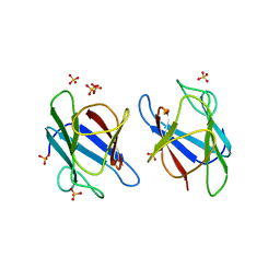
3FM8
 | | Crystal structure of full length centaurin alpha-1 bound with the FHA domain of KIF13B (CAPRI target) | | 分子名称: | Centaurin-alpha-1, Kinesin-like protein KIF13B, SULFATE ION, ... | | 著者 | Shen, L, Tong, Y, Tempel, W, MacKenzie, F, Arrowsmith, C.H, Edwards, A.M, Bountra, C, Weigelt, J, Bochkarev, A, Park, H, Structural Genomics Consortium (SGC) | | 登録日 | 2008-12-19 | | 公開日 | 2009-08-25 | | 最終更新日 | 2024-04-03 | | 実験手法 | X-RAY DIFFRACTION (2.3 Å) | | 主引用文献 | Phosphorylation-independent dual-site binding of the FHA domain of KIF13 mediates phosphoinositide transport via centaurin alpha1.
Proc.Natl.Acad.Sci.USA, 107, 2010
|
|
3ELV
 | | Crystal Structure of Full-Length Yeast Pml1p | | 分子名称: | Pre-mRNA leakage protein 1, SULFATE ION | | 著者 | Trowitzsch, S, Weber, G, L hrmann, R, Wahl, M.C. | | 登録日 | 2008-09-23 | | 公開日 | 2009-02-10 | | 最終更新日 | 2024-02-21 | | 実験手法 | X-RAY DIFFRACTION (2.4 Å) | | 主引用文献 | Crystal structure of the Pml1p subunit of the yeast precursor mRNA retention and splicing complex.
J.Mol.Biol., 385, 2009
|
|
3ELS
 | | Crystal Structure of Yeast Pml1p, Residues 51-204 | | 分子名称: | CHLORIDE ION, GLYCEROL, MAGNESIUM ION, ... | | 著者 | Trowitzsch, S, Weber, G, Luehrmann, R, Wahl, M.C. | | 登録日 | 2008-09-23 | | 公開日 | 2009-02-10 | | 最終更新日 | 2024-02-21 | | 実験手法 | X-RAY DIFFRACTION (1.8 Å) | | 主引用文献 | Crystal structure of the Pml1p subunit of the yeast precursor mRNA retention and splicing complex.
J.Mol.Biol., 385, 2009
|
|
2PIE
 | |
2N84
 | |


