Loading
PDBj
メニューPDBj@FacebookPDBj@TwitterPDBj@YouTubewwPDB FoundationwwPDB
RCSB PDBPDBeBMRBAdv. SearchSearch help

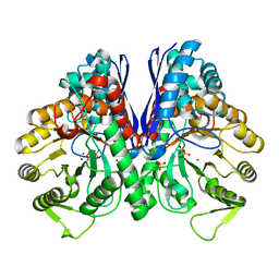
7UGH
DownloadVisualize
BU of 7ugh by Molmil
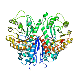
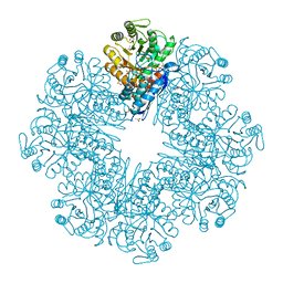
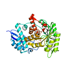
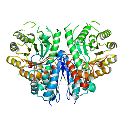
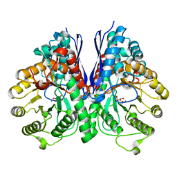
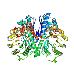
Crystal Structure of enolase family protein from Naegleria fowleri with bound 2-phosphoglyceric acid
分子名称: 1,2-ETHANEDIOL, 2-PHOSPHOGLYCERIC ACID, ACETATE ION, ...
著者Seattle Structural Genomics Center for Infectious Disease (SSGCID)
登録日2022-03-24
公開日2022-04-13
最終更新日2023-10-25
実験手法X-RAY DIFFRACTION (1.95 Å)
主引用文献Crystal Structure of enolase family protein from Naegleria fowleri with bound 2-phosphoglyceric acid
to be published
8UEL
DownloadVisualize
BU of 8uel by Molmil
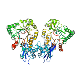
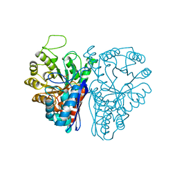
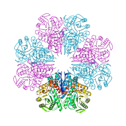
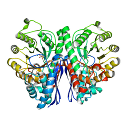
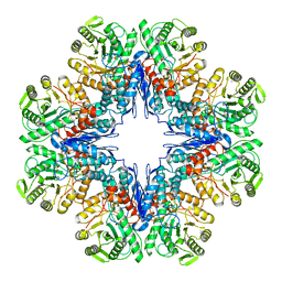
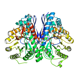
Crystal structure of enolase from Litopenaeus vannamei
分子名称: Enolase, MAGNESIUM ION, PHOSPHOENOLPYRUVATE, ...
著者Chang, X, Zhao, G.
登録日2023-10-01
公開日2023-12-13
最終更新日2023-12-27
実験手法X-RAY DIFFRACTION (2.49 Å)
主引用文献Characterization and Structural Analyses of Enolase from Shrimp ( Litopenaeus vannamei ).
J.Agric.Food Chem., 71, 2023
8UOP
DownloadVisualize
BU of 8uop by Molmil
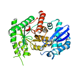
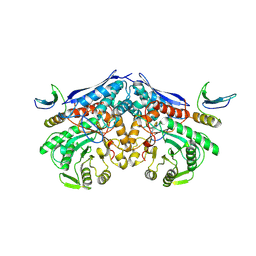
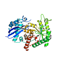
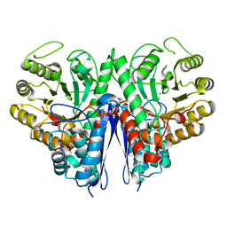
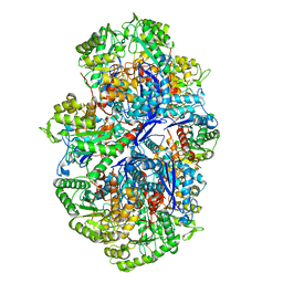
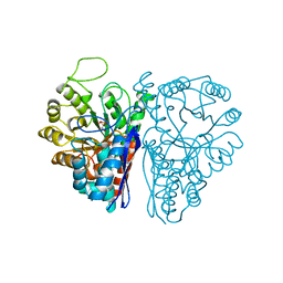
Major interface of Streptococcal surface enolase dimer from AP53 group A streptococcus bound to a lipid vesicle
分子名称: Enolase
著者Tjia-Fleck, S, Readnour, B.M, Castellino, F.J.
登録日2023-10-20
公開日2023-12-27
実験手法ELECTRON MICROSCOPY (3.8 Å)
主引用文献Streptococcus surface alpha enolase exposed dimers were found to be the active form on lipid surface that binds to human plasminogen
To Be Published
8UOY
DownloadVisualize
BU of 8uoy by Molmil
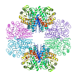
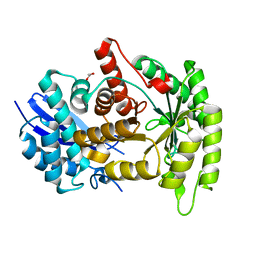
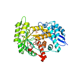
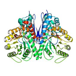
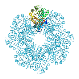
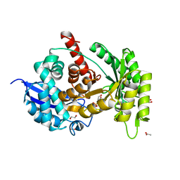
Major interface of Streptococcal surface enolase dimer from AP53 group A streptococcus bound to a lipid vesicle
分子名称: Enolase
著者Tjia-Fleck, S, Readnour, B.M, Castellino, F.J.
登録日2023-10-20
公開日2023-12-27
実験手法ELECTRON MICROSCOPY (3.4 Å)
主引用文献Streptococcus surface alpha enolase exposed dimers were found to be the active form on lipid surface that binds to human plasminogen
To Be Published
7CKP
DownloadVisualize
BU of 7ckp by Molmil
Mycobacterium tuberculosis Enolase
分子名称: (4S)-2-METHYL-2,4-PENTANEDIOL, Enolase, MAGNESIUM ION
著者Biswal, B.K, Ahmad, M, Jha, B.
登録日2020-07-18
公開日2021-07-21
最終更新日2023-11-29
実験手法X-RAY DIFFRACTION (2.9 Å)
主引用文献Structural snapshots of Mycobacterium tuberculosis enolase reveal dual mode of 2PG binding and its implication in enzyme catalysis.
Iucrj, 10, 2023
7CLL
DownloadVisualize
BU of 7cll by Molmil
Mycobacterium tubeculosis enolase in complex with 2-Phosphoglycerate
分子名称: 2-PHOSPHOGLYCERIC ACID, ACETATE ION, CHLORIDE ION, ...
著者Ahmad, M, Jha, B, Tiwari, S, Pal, R.K, Biswal, B.K.
登録日2020-07-21
公開日2021-07-28
最終更新日2023-11-29
実験手法X-RAY DIFFRACTION (1.99 Å)
主引用文献Structural snapshots of Mycobacterium tuberculosis enolase reveal dual mode of 2PG binding and its implication in enzyme catalysis.
Iucrj, 10, 2023
7CLK
DownloadVisualize
BU of 7clk by Molmil
Mycobacterium tuberculosis enolase in complex with alternate 2-phosphoglycerate
分子名称: 1,2-ETHANEDIOL, 2-PHOSPHOGLYCERIC ACID, ACETATE ION, ...
著者Ahmad, M, Jha, B, Tiwari, S, Pal, R.K, Biswal, B.K.
登録日2020-07-21
公開日2022-01-26
最終更新日2023-11-29
実験手法X-RAY DIFFRACTION (2.15 Å)
主引用文献Structural snapshots of Mycobacterium tuberculosis enolase reveal dual mode of 2PG binding and its implication in enzyme catalysis.
Iucrj, 10, 2023
4ENL
DownloadVisualize
BU of 4enl by Molmil
CRYSTAL STRUCTURE OF HOLOENZYME REFINED AT 1.9 ANGSTROMS RESOLUTION: TRIGONAL-BIPYRAMIDAL GEOMETRY OF THE CATION BINDING SITE
分子名称: ENOLASE, SULFATE ION, ZINC ION
著者Lebioda, L, Stec, B.
登録日1990-11-13
公開日1992-04-15
最終更新日2024-02-28
実験手法X-RAY DIFFRACTION (1.9 Å)
主引用文献Crystal Structure of Holoenzyme Refined at 1.9 Angstroms Resolution: Trigonal-Bipyramidal Geometry of the Cation Binding Site
J.Am.Chem.Soc., 111, 1989
7XML
DownloadVisualize
BU of 7xml by Molmil
Cryo-EM structure of PEIP-Bs_enolase complex
分子名称: Enolase, MAGNESIUM ION, Putative gene 60 protein
著者Li, S, Zhang, K.
登録日2022-04-26
公開日2022-07-27
最終更新日2024-07-03
実験手法ELECTRON MICROSCOPY (3.2 Å)
主引用文献Bacteriophage protein PEIP is a potent Bacillus subtilis enolase inhibitor.
Cell Rep, 40, 2022
4G7F
DownloadVisualize
BU of 4g7f by Molmil
Crystal Structure of Enolase from Trypanosoma Cruzi
分子名称: 1,2-ETHANEDIOL, Enolase, MAGNESIUM ION
著者Craig, T.K, Edwards, T.E, Staker, B, Seattle Structural Genomics Center for Infectious Disease (SSGCID)
登録日2012-07-20
公開日2012-08-29
最終更新日2023-09-13
実験手法X-RAY DIFFRACTION (2.4 Å)
主引用文献Crystal Structure of Enolase from Trypanosoma Cruzi
To be Published
3ZLF
DownloadVisualize
BU of 3zlf by Molmil
Structure of group A Streptococcal enolase K312A mutant
分子名称: ENOLASE, PHOSPHATE ION
著者Cork, A.J, Ericsson, D.J, Law, R.H.P, Casey, L.W, Valkov, E, Bertozzi, C, Stamp, A, Aquilina, J.A, Whisstock, J.C, Walker, M.J, Kobe, B.
登録日2013-01-31
公開日2014-02-05
最終更新日2023-12-20
実験手法X-RAY DIFFRACTION (2.15 Å)
主引用文献Stability of the Octameric Structure Affects Plasminogen-Binding Capacity of Streptococcal Enolase.
Plos One, 10, 2015
4A3R
DownloadVisualize
BU of 4a3r by Molmil
Crystal structure of Enolase from Bacillus subtilis.
分子名称: CITRIC ACID, ENOLASE, SODIUM ION
著者Newman, J.A, Hewitt, L, Rodrigues, C, Solovyova, A.S, Harwood, C.R, Lewis, R.J.
登録日2011-10-04
公開日2012-08-15
最終更新日2023-12-20
実験手法X-RAY DIFFRACTION (2.2 Å)
主引用文献Dissection of the Network of Interactions that Links RNA Processing with Glycolysis in the Bacillus Subtilis Degradosome.
J.Mol.Biol., 416, 2012
3ZLH
DownloadVisualize
BU of 3zlh by Molmil
Structure of group A Streptococcal enolase
分子名称: ENOLASE
著者Cork, A.J, Ericsson, D.J, Law, R.H.P, Casey, L.W, Valkov, E, Bertozzi, C, Stamp, A, Aquilina, J.A, Whisstock, J.C, Walker, M.J, Kobe, B.
登録日2013-01-31
公開日2014-02-05
最終更新日2023-12-20
実験手法X-RAY DIFFRACTION (2.9 Å)
主引用文献Stability of the Octameric Structure Affects Plasminogen-Binding Capacity of Streptococcal Enolase.
Plos One, 10, 2015
3ZLG
DownloadVisualize
BU of 3zlg by Molmil
Structure of group A Streptococcal enolase K362A mutant
分子名称: ENOLASE, PHOSPHATE ION
著者Cork, A.J, Ericsson, D.J, Law, R.H.P, Casey, L.W, Valkov, E, Bertozzi, C, Stamp, A, Aquilina, J.A, Whisstock, J.C, Walker, M.J, Kobe, B.
登録日2013-01-31
公開日2014-02-05
最終更新日2023-12-20
実験手法X-RAY DIFFRACTION (2.1 Å)
主引用文献Stability of the Octameric Structure Affects Plasminogen-Binding Capacity of Streptococcal Enolase.
Plos One, 10, 2015
4YWS
DownloadVisualize
BU of 4yws by Molmil
Thermostable enolase from Chloroflexus aurantiacus
分子名称: Enolase, MAGNESIUM ION
著者Zadvornyy, O.A, Peters, J.W.
登録日2015-03-20
公開日2015-07-01
最終更新日2023-09-27
実験手法X-RAY DIFFRACTION (2.45 Å)
主引用文献Biochemical and Structural Characterization of Enolase from Chloroflexus aurantiacus: Evidence for a Thermophilic Origin.
Front Bioeng Biotechnol, 3, 2015
4ZCW
DownloadVisualize
BU of 4zcw by Molmil
Structure of Human Enolase 2 in complex with SF2312
分子名称: Gamma-enolase, MAGNESIUM ION, [(3S,5S)-1,5-dihydroxy-2-oxopyrrolidin-3-yl]phosphonic acid
著者Leonard, P.G, Maxwell, D, Czako, B, Muller, F.L.
登録日2015-04-16
公開日2016-07-20
最終更新日2023-09-27
実験手法X-RAY DIFFRACTION (1.992 Å)
主引用文献SF2312 is a natural phosphonate inhibitor of enolase.
Nat.Chem.Biol., 12, 2016
4Z1Y
DownloadVisualize
BU of 4z1y by Molmil
Thermostable enolase from Chloroflexus aurantiacus with substrate 2-phosphoglycerate
分子名称: 2-PHOSPHOGLYCERIC ACID, Enolase, MAGNESIUM ION
著者Zadvornyy, O.A, Peters, J.W.
登録日2015-03-27
公開日2015-07-01
最終更新日2024-03-06
実験手法X-RAY DIFFRACTION (2.53 Å)
主引用文献Biochemical and Structural Characterization of Enolase from Chloroflexus aurantiacus: Evidence for a Thermophilic Origin.
Front Bioeng Biotechnol, 3, 2015
4Z17
DownloadVisualize
BU of 4z17 by Molmil
Thermostable enolase from Chloroflexus aurantiacus
分子名称: Enolase, MAGNESIUM ION, PHOSPHOENOLPYRUVATE
著者Zadvornyy, O.A, Peters, J.W.
登録日2015-03-26
公開日2015-07-01
最終更新日2023-09-27
実験手法X-RAY DIFFRACTION (2.65 Å)
主引用文献Biochemical and Structural Characterization of Enolase from Chloroflexus aurantiacus: Evidence for a Thermophilic Origin.
Front Bioeng Biotechnol, 3, 2015
4ZA0
DownloadVisualize
BU of 4za0 by Molmil
Structure of Human Enolase 2 in complex with Phosphonoacetohydroxamate
分子名称: Gamma-enolase, MAGNESIUM ION, PHOSPHONOACETOHYDROXAMIC ACID
著者Leonard, P.G, Maxwell, D, Czako, B, Muller, F.L.
登録日2015-04-13
公開日2016-04-13
最終更新日2023-09-27
実験手法X-RAY DIFFRACTION (2.31 Å)
主引用文献SF2312 is a natural phosphonate inhibitor of enolase.
Nat.Chem.Biol., 12, 2016
7E51
DownloadVisualize
BU of 7e51 by Molmil
Structure of PEP bound Enolase from Mycobacterium tuberculosis
分子名称: Enolase, MAGNESIUM ION, PHOSPHOENOLPYRUVATE
著者Bose, S, Vinothkumar, K.R.
登録日2021-02-16
公開日2022-02-16
最終更新日2023-11-08
実験手法ELECTRON MICROSCOPY (3.23 Å)
主引用文献Structural snapshots of Mycobacterium tuberculosis enolase reveal dual mode of 2PG binding and its implication in enzyme catalysis.
Iucrj, 10, 2023
7E4X
DownloadVisualize
BU of 7e4x by Molmil
Structure of Enolase from Mycobacterium tuberculosis
分子名称: Enolase
著者Bose, S, Vinothkumar, K.R.
登録日2021-02-15
公開日2022-02-16
最終更新日2023-11-08
実験手法ELECTRON MICROSCOPY (3.08 Å)
主引用文献Structural snapshots of Mycobacterium tuberculosis enolase reveal dual mode of 2PG binding and its implication in enzyme catalysis.
Iucrj, 10, 2023
7E4F
DownloadVisualize
BU of 7e4f by Molmil
Mycobacterium tuberculosis enolase mutant - E204A complex with phosphoenolpyruvate
分子名称: 1,2-ETHANEDIOL, ACETATE ION, DI(HYDROXYETHYL)ETHER, ...
著者Ahmad, M, Pal, R.K, Biswal, B.K.
登録日2021-02-11
公開日2022-02-16
最終更新日2023-11-29
実験手法X-RAY DIFFRACTION (2.3 Å)
主引用文献Structural snapshots of Mycobacterium tuberculosis enolase reveal dual mode of 2PG binding and its implication in enzyme catalysis.
Iucrj, 10, 2023
2XH4
DownloadVisualize
BU of 2xh4 by Molmil
Engineering the enolase active site pocket: Crystal structure of the S39A D321A mutant of yeast enolase 1
分子名称: 2-PHOSPHOGLYCERIC ACID, ENOLASE 1, MAGNESIUM ION
著者Schreier, B, Hocker, B.
登録日2010-06-09
公開日2010-08-25
最終更新日2023-12-20
実験手法X-RAY DIFFRACTION (1.7 Å)
主引用文献Engineering the Enolase Magnesium II Binding Site -Implications for its Evolution.
Biochemistry, 49, 2010
2XH7
DownloadVisualize
BU of 2xh7 by Molmil
Engineering the enolase active site pocket: Crystal structure of the D321A mutant of yeast enolase 1
分子名称: 2-PHOSPHOGLYCERIC ACID, ENOLASE 1, MAGNESIUM ION
著者Schreier, B, Hocker, B.
登録日2010-06-09
公開日2010-08-25
最終更新日2023-12-20
実験手法X-RAY DIFFRACTION (1.8 Å)
主引用文献Engineering the Enolase Magnesium II Binding Site -Implications for its Evolution.
Biochemistry, 49, 2010
7ENL
DownloadVisualize
BU of 7enl by Molmil
MECHANISM OF ENOLASE: THE CRYSTAL STRUCTURE OF ENOLASE-MG2+-PHOSPHOGLYCERATE(SLASH) PHOSPHOENOLPYRUVATE COMPLEX AT 2.2-ANGSTROMS RESOLUTION
分子名称: 2-PHOSPHOGLYCERIC ACID, ENOLASE, MAGNESIUM ION
著者Lebioda, L, Stec, B.
登録日1990-11-13
公開日1992-04-15
最終更新日2024-03-06
実験手法X-RAY DIFFRACTION (2.2 Å)
主引用文献Mechanism of enolase: the crystal structure of enolase-Mg2(+)-2-phosphoglycerate/phosphoenolpyruvate complex at 2.2-A resolution.
Biochemistry, 30, 1991

222624

件を2024-07-17に公開中

PDB statisticsPDBj update infoContact PDBjnumon