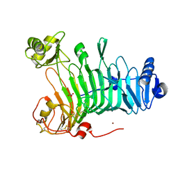
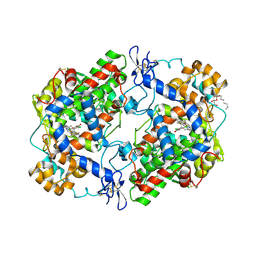
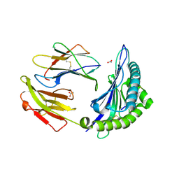
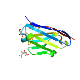
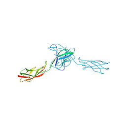
3LMS
 
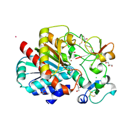 | | Structure of human activated thrombin-activatable fibrinolysis inhibitor, TAFIa, in complex with tick-derived funnelin inhibitor, TCI. | | 分子名称: | CALCIUM ION, CHLORIDE ION, Carboxypeptidase B2, ... | | 著者 | Sanglas, L, Arolas, J.L, Valnickova, Z, Aviles, F.X, Enghild, J.J, Gomis-Ruth, F.X. | | 登録日 | 2010-02-01 | | 公開日 | 2010-02-16 | | 最終更新日 | 2024-11-13 | | 実験手法 | X-RAY DIFFRACTION (2.5 Å) | | 主引用文献 | Insights into the molecular inactivation mechanism of human activated thrombin-activatable fibrinolysis inhibitor
J.Thromb.Haemost., 8, 2010
|
|
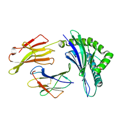
3LMT
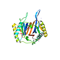 | | Crystal structure of DTD from Plasmodium falciparum | | 分子名称: | D-tyrosyl-tRNA(Tyr) deacylase, IODIDE ION | | 著者 | Manickam, Y, Bhatt, T.K, Khan, S, Sharma, A. | | 登録日 | 2010-02-01 | | 公開日 | 2010-03-02 | | 最終更新日 | 2025-05-28 | | 実験手法 | X-RAY DIFFRACTION (2.75 Å) | | 主引用文献 | Structure of D-tyrosyl-tRNATyr deacylase using home-source Cu Kalpha and moderate-quality iodide-SAD data: structural polymorphism and HEPES-bound enzyme states
Acta Crystallogr.,Sect.D, 66, 2010
|
|
3LMU
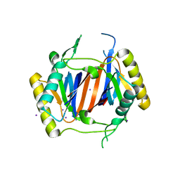 | | Crystal structure of DTD from Plasmodium falciparum | | 分子名称: | D-tyrosyl-tRNA(Tyr) deacylase, IODIDE ION | | 著者 | Manickam, Y, Bhatt, T.K, Khan, S, Sharma, A. | | 登録日 | 2010-02-01 | | 公開日 | 2010-03-02 | | 最終更新日 | 2024-03-20 | | 実験手法 | X-RAY DIFFRACTION (3.3 Å) | | 主引用文献 | Structure of D-tyrosyl-tRNATyr deacylase using home-source Cu Kalpha and moderate-quality iodide-SAD data: structural polymorphism and HEPES-bound enzyme states
Acta Crystallogr.,Sect.D, 66, 2010
|
|
3LMV
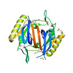 | | D-Tyr-tRNA(Tyr) Deacylase from plasmodium falciparum in complex with hepes | | 分子名称: | 4-(2-HYDROXYETHYL)-1-PIPERAZINE ETHANESULFONIC ACID, D-tyrosyl-tRNA(Tyr) deacylase, SULFITE ION | | 著者 | Manickam, Y, Khan, S, Bhatt, T.K, Sharma, A. | | 登録日 | 2010-02-01 | | 公開日 | 2010-03-02 | | 最終更新日 | 2023-11-01 | | 実験手法 | X-RAY DIFFRACTION (2.833 Å) | | 主引用文献 | Structure of D-tyrosyl-tRNATyr deacylase using home-source Cu Kalpha and moderate-quality iodide-SAD data: structural polymorphism and HEPES-bound enzyme states
Acta Crystallogr.,Sect.D, 66, 2010
|
|
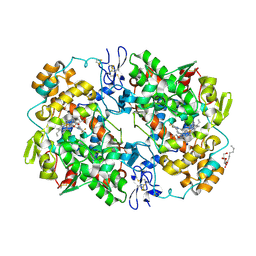
3LMW
 | | Crystal structure of iota-carrageenase family GH82 from A. fortis in absence of chloride ions | | 分子名称: | CALCIUM ION, Iota-carrageenase, CgiA, ... | | 著者 | Rebuffet, E, Barbeyron, T, Jeudy, A, Czjzek, M, Michel, G. | | 登録日 | 2010-02-01 | | 公開日 | 2010-08-18 | | 最終更新日 | 2024-11-13 | | 実験手法 | X-RAY DIFFRACTION (2.6 Å) | | 主引用文献 | Identification of catalytic residues and mechanistic analysis of family GH82 iota-carrageenases
Biochemistry, 49, 2010
|
|
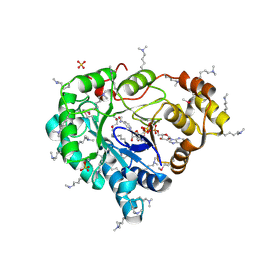
3LMX
 | | Tyrosine 447 of Protocatechuate 34,-Dioxygenase Controls Efficient Progress Through Catalysis | | 分子名称: | 3,4-DIHYDROXYBENZOIC ACID, BETA-MERCAPTOETHANOL, CHLORIDE ION, ... | | 著者 | Purpero, V.M, Lipscomb, J.D, Shi, K. | | 登録日 | 2010-02-01 | | 公開日 | 2011-02-16 | | 最終更新日 | 2024-02-21 | | 実験手法 | X-RAY DIFFRACTION (2.2 Å) | | 主引用文献 | Tyrosine 447 of Protocatechuate 34,-Dioxygenase Controls Efficient Progress Through Catalysis
To be Published
|
|
3LMY
 | | The Crystal Structure of beta-hexosaminidase B in complex with Pyrimethamine | | 分子名称: | 2-acetamido-2-deoxy-alpha-D-glucopyranose, 2-acetamido-2-deoxy-beta-D-glucopyranose, 2-acetamido-2-deoxy-beta-D-glucopyranose-(1-4)-2-acetamido-2-deoxy-beta-D-glucopyranose, ... | | 著者 | Bateman, K.S, Cherney, M.M, Withers, S.G, Mahuran, D.J, Tropak, M, James, M.N.G. | | 登録日 | 2010-02-01 | | 公開日 | 2011-02-16 | | 最終更新日 | 2024-10-30 | | 実験手法 | X-RAY DIFFRACTION (2.8 Å) | | 主引用文献 | The Crystal Structure of beta-hexosaminidase B in complex with Pyrimethamine
To be Published
|
|
3LMZ
 | |
3LN0
 | | Structure of compound 5c-S bound at the active site of COX-2 | | 分子名称: | (2S)-6,8-dichloro-2-(trifluoromethyl)-2H-chromene-3-carboxylic acid, 2-acetamido-2-deoxy-beta-D-glucopyranose, 2-acetamido-2-deoxy-beta-D-glucopyranose-(1-4)-2-acetamido-2-deoxy-beta-D-glucopyranose-(1-4)-2-acetamido-2-deoxy-beta-D-glucopyranose, ... | | 著者 | Kiefer, J.R, Kurumbail, R.G, Stallings, W.C, Pawlitz, J.L. | | 登録日 | 2010-02-01 | | 公開日 | 2010-10-27 | | 最終更新日 | 2024-11-20 | | 実験手法 | X-RAY DIFFRACTION (2.2 Å) | | 主引用文献 | The novel benzopyran class of selective cyclooxygenase-2 inhibitors. Part 2: The second clinical candidate having a shorter and favorable human half-life.
Bioorg.Med.Chem.Lett., 20, 2010
|
|
3LN1
 | | Structure of celecoxib bound at the COX-2 active site | | 分子名称: | 2-acetamido-2-deoxy-beta-D-glucopyranose, 2-acetamido-2-deoxy-beta-D-glucopyranose-(1-4)-2-acetamido-2-deoxy-beta-D-glucopyranose-(1-4)-2-acetamido-2-deoxy-beta-D-glucopyranose, 4-[5-(4-METHYLPHENYL)-3-(TRIFLUOROMETHYL)-1H-PYRAZOL-1-YL]BENZENESULFONAMIDE, ... | | 著者 | Kiefer, J.R, Kurumbail, R.G, Stallings, W.C, Pawlitz, J.L. | | 登録日 | 2010-02-01 | | 公開日 | 2010-10-27 | | 最終更新日 | 2024-10-16 | | 実験手法 | X-RAY DIFFRACTION (2.4 Å) | | 主引用文献 | The novel benzopyran class of selective cyclooxygenase-2 inhibitors. Part 2: The second clinical candidate having a shorter and favorable human half-life.
Bioorg.Med.Chem.Lett., 20, 2010
|
|
3LN2
 | |
3LN3
 | |
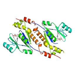
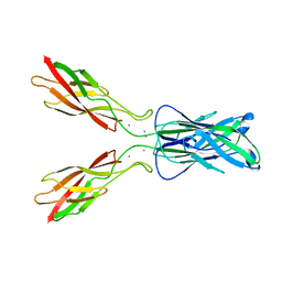
3LN4
 | | Crystal structure of HLA-B*4103 in complex with a 16mer self-peptide derived from heterogeneous nuclear ribonucleoproteins C1/C2 | | 分子名称: | 16-mer peptide from Heterogeneous nuclear ribonucleoproteins C1/C2, ACETATE ION, Beta-2-microglobulin, ... | | 著者 | Theodossis, A, Gras, S, Rossjohn, J. | | 登録日 | 2010-02-01 | | 公開日 | 2010-10-20 | | 最終更新日 | 2024-10-30 | | 実験手法 | X-RAY DIFFRACTION (1.296 Å) | | 主引用文献 | The impact of human leukocyte antigen (HLA) micropolymorphism on ligand specificity within the HLA-B*41 allotypic family
Haematologica, 96, 2011
|
|
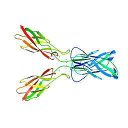
3LN5
 | | Crystal structure of HLA-B*4104 in complex with a 11mer self-peptide derived from S-methyl-5-thioadenosine phosphorylase | | 分子名称: | 11-mer peptide from S-methyl-5'-thioadenosine phosphorylase, Beta-2-microglobulin, HLA class I histocompatibility antigen, ... | | 著者 | Theodossis, A, Gras, S, Rossjohn, J. | | 登録日 | 2010-02-02 | | 公開日 | 2010-10-20 | | 最終更新日 | 2024-11-06 | | 実験手法 | X-RAY DIFFRACTION (1.9 Å) | | 主引用文献 | The impact of human leukocyte antigen (HLA) micropolymorphism on ligand specificity within the HLA-B*41 allotypic family
Haematologica, 96, 2011
|
|
3LN6
 | |
3LN7
 | |
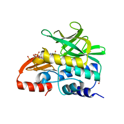
3LN8
 | | The X-ray structure of Zf-RNase-1 from a new crystal form at pH 7.3 | | 分子名称: | HYDROLASE, SULFATE ION | | 著者 | Russo Krauss, I, Merlino, A, Mazzarella, L, Sica, F. | | 登録日 | 2010-02-02 | | 公開日 | 2010-12-08 | | 最終更新日 | 2024-11-27 | | 実験手法 | X-RAY DIFFRACTION (1.61 Å) | | 主引用文献 | A new RNase sheds light on the RNase/angiogenin subfamily from zebrafish.
Biochem.J., 433, 2010
|
|
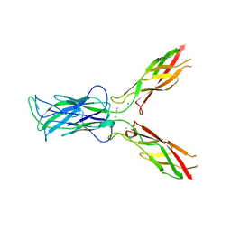
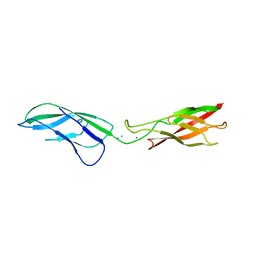
3LN9
 | | Crystal structure of the fibril-specific B10 antibody fragment | | 分子名称: | CITRATE ANION, GLYCEROL, Immunoglobulin heavy chain antibody variable domain B10, ... | | 著者 | Parthier, C, Morgado, I, Stubbs, M.T, Faendrich, M. | | 登録日 | 2010-02-02 | | 公開日 | 2010-12-15 | | 最終更新日 | 2024-10-30 | | 実験手法 | X-RAY DIFFRACTION (1.8 Å) | | 主引用文献 | Amyloid Fibril Recognition with the Conformational B10 Antibody Fragment Depends on Electrostatic Interactions.
J.Mol.Biol., 2010
|
|
3LNB
 | | Crystal Structure Analysis of Arylamine N-acetyltransferase C from Bacillus anthracis | | 分子名称: | COENZYME A, FORMIC ACID, N-acetyltransferase family protein | | 著者 | Li de la Sierra-Gallay, I, Pluvinage, B, Rodrigues-Lima, F. | | 登録日 | 2010-02-02 | | 公開日 | 2011-01-26 | | 最終更新日 | 2023-11-01 | | 実験手法 | X-RAY DIFFRACTION (2.01 Å) | | 主引用文献 | The Bacillus anthracis arylamine N-acetyltransferase ((BACAN)NAT1) that inactivates sulfamethoxazole, reveals unusual structural features compared with the other NAT isoenzymes.
Febs Lett., 585, 2011
|
|
3LNC
 | |
3LND
 | |
3LNE
 | |
3LNF
 | |
3LNG
 | |
3LNH
 | |


