Loading
PDBj
メニューPDBj@FacebookPDBj@TwitterPDBj@YouTubewwPDB FoundationwwPDB
RCSB PDBPDBeBMRBAdv. SearchSearch help

6UN6
DownloadVisualize
BU of 6un6 by Molmil
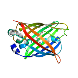
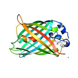
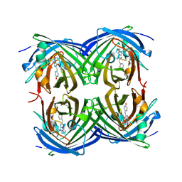
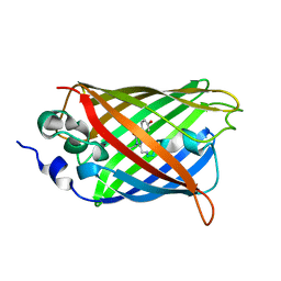
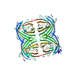
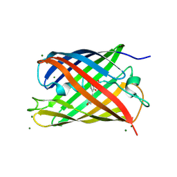
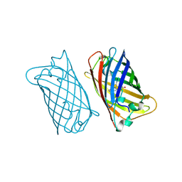
Crystal structure of green fluorescent protein (GFP); S65T, Y66(3-NO2Y); ih circular permutant (50-51)
分子名称: Green fluorescent protein
著者Lin, C.-Y, Boxer, S.G.
登録日2019-10-10
公開日2020-10-14
最終更新日2023-11-15
実験手法X-RAY DIFFRACTION (1.5 Å)
主引用文献Unusual Spectroscopic and Electric Field Sensitivity of Chromophores with Short Hydrogen Bonds: GFP and PYP as Model Systems.
J.Phys.Chem.B, 124, 2020
6UHL
DownloadVisualize
BU of 6uhl by Molmil
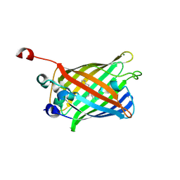
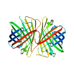
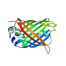
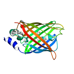
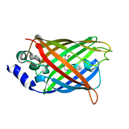
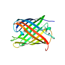
Crystal Structure of C148 mGFP-scDNA-1
分子名称: C148 mGFP-scDNA-1, UNKNOWN LIGAND
著者Winegar, P.W, Hayes, O.G, McMillan, J.R, Figg, C.A, Focia, P.J, Mirkin, C.A.
登録日2019-09-27
公開日2020-03-18
最終更新日2023-11-15
実験手法X-RAY DIFFRACTION (1.91 Å)
主引用文献DNA-Directed Protein Packing within Single Crystals.
Chem, 6, 2020
3PJ5
DownloadVisualize
BU of 3pj5 by Molmil
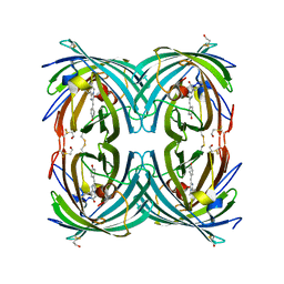
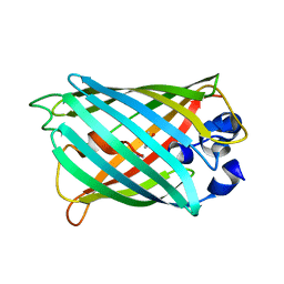
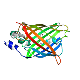
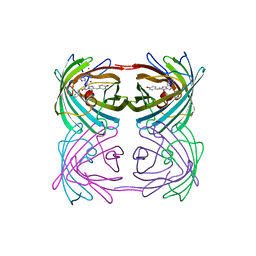
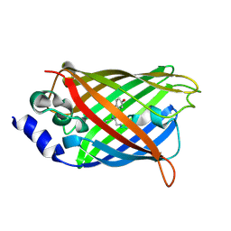
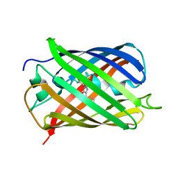
Crystal structure of far-red fluorescent protein Katushka crystallized at pH 5.0
分子名称: Red fluorescent protein eqFP578, SULFATE ION
著者Pletnev, S, Pletneva, N.V, Pletnev, V.Z.
登録日2010-11-08
公開日2011-05-25
最終更新日2012-02-22
実験手法X-RAY DIFFRACTION (1.6 Å)
主引用文献Crystallographic study of red fluorescent protein eqFP578 and its far-red variant Katushka reveals opposite pH-induced isomerization of chromophore.
Protein Sci., 20, 2011
6UHM
DownloadVisualize
BU of 6uhm by Molmil
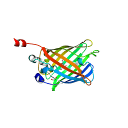
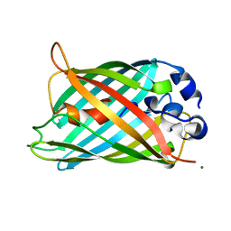
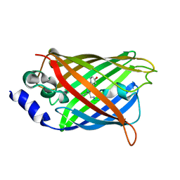
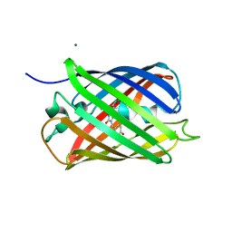
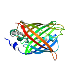
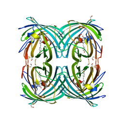
Crystal Structure of a Physical Mixture of C148 mGFP and scDNA-1
分子名称: C148 mGFP
著者Winegar, P.W, Hayes, O.G, McMillan, J.R, Figg, C.A, Focia, P.J, Mirkin, C.A.
登録日2019-09-27
公開日2020-03-18
最終更新日2023-11-15
実験手法X-RAY DIFFRACTION (2.1 Å)
主引用文献DNA-Directed Protein Packing within Single Crystals.
Chem, 6, 2020
6UN4
DownloadVisualize
BU of 6un4 by Molmil
Crystal structure of rsEGFP2, Y67(3-ClY), Y107(3-ClY)
分子名称: Green fluorescent protein, SULFATE ION
著者Lin, C.-Y, Romei, M.G, Boxer, S.G, Chang, J.
登録日2019-10-10
公開日2020-07-15
最終更新日2023-11-15
実験手法X-RAY DIFFRACTION (1.499 Å)
主引用文献Structural and spectroscopic characterization of photoactive yellow protein and photoswitchable fluorescent protein constructs containing heavy atoms.
J Photochem Photobiol A Chem, 401, 2020
6UHO
DownloadVisualize
BU of 6uho by Molmil
Crystal Structure of C148 mGFP-cDNA-2
分子名称: C148 mGFP-cDNA-2, UNKNOWN LIGAND
著者Winegar, P.W, Hayes, O.G, McMillan, J.R, Figg, C.A, Focia, P.J, Mirkin, C.A.
登録日2019-09-27
公開日2020-03-18
最終更新日2023-11-15
実験手法X-RAY DIFFRACTION (1.95 Å)
主引用文献DNA-Directed Protein Packing within Single Crystals.
Chem, 6, 2020
6VAM
DownloadVisualize
BU of 6vam by Molmil
Cryo-EM structure of octameric chicken CALHM1
分子名称: Green fluorescent protein,CALHM1 chimera
著者Syrjanen, J.L, Chou, T.H, Furukawa, H.
登録日2019-12-17
公開日2020-01-29
最終更新日2020-11-11
実験手法ELECTRON MICROSCOPY (3.63 Å)
主引用文献Structure and assembly of calcium homeostasis modulator proteins.
Nat.Struct.Mol.Biol., 27, 2020
6VIO
DownloadVisualize
BU of 6vio by Molmil
Crystal structure of eYFP His148Ser
分子名称: Green fluorescent protein GFP
著者Lieberman, R.L, Hill, S.E, Patterson-Orazem, A.C.
登録日2020-01-13
公開日2021-01-13
最終更新日2023-11-15
実験手法X-RAY DIFFRACTION (3.6 Å)
主引用文献Optically Modulated and Optically Activated Delayed Fluorescent Proteins through Dark State Engineering
J.Phys.Chem.B, 125, 2021
3RWT
DownloadVisualize
BU of 3rwt by Molmil
Crystal structure of circular permutated Red Fluorescent Protein mKate(cp 154-153)
分子名称: Fluorescent protein FP480,Fluorescent protein FP480, MAGNESIUM ION
著者Wang, Q, Sondermann, H.
登録日2011-05-09
公開日2011-06-15
最終更新日2017-11-08
実験手法X-RAY DIFFRACTION (3 Å)
主引用文献Circular permutation of red fluorescent proteins.
Plos One, 6, 2011
3PIB
DownloadVisualize
BU of 3pib by Molmil
Crystal structure of red fluorescent protein eqFP578 crystallized at pH 5.5
分子名称: GLYCEROL, eqFP578 fluorescent protein
著者Pletnev, S, Pletneva, N.V, Pletnev, V.Z.
登録日2010-11-05
公開日2011-05-25
最終更新日2012-02-22
実験手法X-RAY DIFFRACTION (1.154 Å)
主引用文献Crystallographic study of red fluorescent protein eqFP578 and its far-red variant Katushka reveals opposite pH-induced isomerization of chromophore.
Protein Sci., 20, 2011
3PJ7
DownloadVisualize
BU of 3pj7 by Molmil
Crystal structure of far-red fluorescent protein Katushka crystallized at pH 8.5
分子名称: Red fluorescent protein eqFP578
著者Pletnev, S, Pletneva, N.V, Pletnev, V.Z.
登録日2010-11-08
公開日2011-05-25
最終更新日2012-02-22
実験手法X-RAY DIFFRACTION (1.85 Å)
主引用文献Crystallographic study of red fluorescent protein eqFP578 and its far-red variant Katushka reveals opposite pH-induced isomerization of chromophore.
Protein Sci., 20, 2011
3PJB
DownloadVisualize
BU of 3pjb by Molmil
Crystal structure of red fluorescent protein eqFP578 crystallized at pH 4.0
分子名称: GLYCEROL, Red fluorescent protein eqFP578
著者Pletnev, S, Pletneva, N.V, Pletnev, V.Z.
登録日2010-11-09
公開日2011-05-25
最終更新日2018-01-24
実験手法X-RAY DIFFRACTION (1.75 Å)
主引用文献Crystallographic study of red fluorescent protein eqFP578 and its far-red variant Katushka reveals opposite pH-induced isomerization of chromophore.
Protein Sci., 20, 2011
6WVE
DownloadVisualize
BU of 6wve by Molmil
Chicken SPCS1
分子名称: Green fluorescent protein,Chicken Signal Peptidase Complex Subunit 1,Green fluorescent protein
著者Liu, S, Li, W.
登録日2020-05-05
公開日2021-01-13
最終更新日2023-11-15
実験手法X-RAY DIFFRACTION (2.429 Å)
主引用文献Termini restraining of small membrane proteins enables structure determination at near-atomic resolution.
Sci Adv, 6, 2020
3RWA
DownloadVisualize
BU of 3rwa by Molmil
Crystal structure of circular-permutated mKate
分子名称: Fluorescent protein FP480
著者Wang, Q, Byrnes, L, Sondermann, H.
登録日2011-05-08
公開日2011-06-15
最終更新日2019-07-17
実験手法X-RAY DIFFRACTION (1.67 Å)
主引用文献Circular permutation of red fluorescent proteins.
Plos One, 6, 2011
3S05
DownloadVisualize
BU of 3s05 by Molmil
mEos2 Fluorescent Protein-Green Form
分子名称: Green to red photoconvertible GPF-like protein EosFP
著者Zhang, M.S, Wu, L.J, Xu, P.Y, Liu, Y.F, Xu, T.
登録日2011-05-13
公開日2011-11-16
最終更新日2023-11-15
実験手法X-RAY DIFFRACTION (2.204 Å)
主引用文献mEos2 Fluorescent Protein-Green Form
To be Published
3SRY
DownloadVisualize
BU of 3sry by Molmil
Engineered high-affinity halide-binding protein derived from YFP: halide-free
分子名称: Green fluorescent protein
著者Wang, W, Grimley, J.S, Beese, L.S, Hellinga, H.W.
登録日2011-07-07
公開日2012-07-11
最終更新日2023-11-15
実験手法X-RAY DIFFRACTION (1.159 Å)
主引用文献Determination of engineered chloride-binding site structures in fluorescent proteins reveals principles of halide recognition
To be Published
3SVB
DownloadVisualize
BU of 3svb by Molmil
Engineered medium-affinity halide-binding protein derived from YFP: fluoride complex
分子名称: 1,2-ETHANEDIOL, Green fluorescent protein
著者Wang, W, Grimley, J.S, Beese, L.S, Hellinga, H.W.
登録日2011-07-12
公開日2012-07-18
最終更新日2023-11-15
実験手法X-RAY DIFFRACTION (1.3 Å)
主引用文献Determination of engineered chloride-binding site structures in fluorescent proteins reveals principles of halide recognition
To be Published
3SST
DownloadVisualize
BU of 3sst by Molmil
Engineered low-affinity halide-binding protein derived from YFP: chloride complex
分子名称: CHLORIDE ION, Green fluorescent protein
著者Wang, W, Grimley, J.S, Beese, L.S, Hellinga, H.W.
登録日2011-07-08
公開日2012-07-11
最終更新日2023-11-15
実験手法X-RAY DIFFRACTION (1.403 Å)
主引用文献Determination of engineered chloride-binding site structures in fluorescent proteins reveals principles of halide recognition
To be Published
3SV5
DownloadVisualize
BU of 3sv5 by Molmil
Engineered medium-affinity halide-binding protein derived from YFP: iodide complex
分子名称: 1,2-ETHANEDIOL, FORMIC ACID, Green fluorescent protein, ...
著者Wang, W, Grimley, J.S, Beese, L.S, Hellinga, H.W.
登録日2011-07-12
公開日2012-07-18
最終更新日2023-11-15
実験手法X-RAY DIFFRACTION (1.53 Å)
主引用文献Visualization of Synaptic Inhibition with an Optogenetic Sensor Developed by Cell-Free Protein Engineering Automation.
J.Neurosci., 33, 2013
3SVE
DownloadVisualize
BU of 3sve by Molmil
Engineered low-affinity halide-binding protein derived from YFP: bromide complex
分子名称: BROMIDE ION, Green fluorescent protein
著者Wang, W, Grimley, J.S, Beese, L.S, Hellinga, H.W.
登録日2011-07-12
公開日2012-07-18
最終更新日2023-11-15
実験手法X-RAY DIFFRACTION (1.492 Å)
主引用文献Determination of engineered chloride-binding site structures in fluorescent proteins reveals principles of halide recognition
To be Published
3SS0
DownloadVisualize
BU of 3ss0 by Molmil
Engineered high-affinity halide-binding protein derived from YFP: fluoride complex
分子名称: Green fluorescent protein
著者Wang, W, Grimley, J.S, Beese, L.S, Hellinga, H.W.
登録日2011-07-07
公開日2012-07-11
最終更新日2023-11-15
実験手法X-RAY DIFFRACTION (1.492 Å)
主引用文献Determination of engineered chloride-binding site structures in fluorescent proteins reveals principles of halide recognition
To be Published
3SSH
DownloadVisualize
BU of 3ssh by Molmil
Engineered high-affinity halide-binding protein derived from YFP: chloride complex
分子名称: CHLORIDE ION, Green fluorescent protein
著者Wang, W, Grimley, J.S, Beese, L.S, Hellinga, H.W.
登録日2011-07-08
公開日2012-07-11
最終更新日2023-11-15
実験手法X-RAY DIFFRACTION (1.277 Å)
主引用文献Determination of engineered chloride-binding site structures in fluorescent proteins reveals principles of halide recognition
To be Published
3SSK
DownloadVisualize
BU of 3ssk by Molmil
Engineered high-affinity halide-binding protein derived from YFP: bromide complex
分子名称: BROMIDE ION, Green fluorescent protein
著者Wang, W, Grimley, J.S, Beese, L.S, Hellinga, H.W.
登録日2011-07-08
公開日2012-07-11
最終更新日2023-11-15
実験手法X-RAY DIFFRACTION (1.361 Å)
主引用文献Determination of engineered chloride-binding site structures in fluorescent proteins reveals principles of halide recognition
To be Published
3SVC
DownloadVisualize
BU of 3svc by Molmil
Engineered medium-affinity halide-binding protein derived from YFP: chloride complex
分子名称: 1,2-ETHANEDIOL, CHLORIDE ION, Green fluorescent protein
著者Wang, W, Grimley, J.S, Beese, L.S, Hellinga, H.W.
登録日2011-07-12
公開日2012-07-18
最終更新日2023-11-15
実験手法X-RAY DIFFRACTION (1.31 Å)
主引用文献Determination of engineered chloride-binding site structures in fluorescent proteins reveals principles of halide recognition
To be Published
3SSL
DownloadVisualize
BU of 3ssl by Molmil
Engineered high-affinity halide-binding protein derived from YFP: iodide complex
分子名称: Green fluorescent protein, IODIDE ION
著者Wang, W, Grimley, J.S, Beese, L.S, Hellinga, H.W.
登録日2011-07-08
公開日2012-07-11
最終更新日2023-11-15
実験手法X-RAY DIFFRACTION (1.449 Å)
主引用文献Determination of engineered chloride-binding site structures in fluorescent proteins reveals principles of halide recognition
To be published

221051

件を2024-06-12に公開中

PDB statisticsPDBj update infoContact PDBjnumon