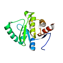
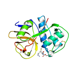
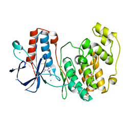
7FOJ
 
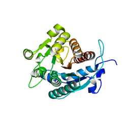
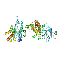 | | PanDDA analysis group deposition -- Aar2/RNaseH in complex with fragment P08B08 from the F2X-Universal Library | | 分子名称: | (2S)-N-(3-chlorophenyl)-2-[(2-hydroxyethyl)sulfanyl]propanamide, A1 cistron-splicing factor AAR2, Pre-mRNA-splicing factor 8 | | 著者 | Barthel, T, Wollenhaupt, J, Lima, G.M.A, Wahl, M.C, Weiss, M.S. | | 登録日 | 2022-08-26 | | 公開日 | 2022-11-02 | | 最終更新日 | 2024-05-22 | | 実験手法 | X-RAY DIFFRACTION (1.65 Å) | | 主引用文献 | Large-Scale Crystallographic Fragment Screening Expedites Compound Optimization and Identifies Putative Protein-Protein Interaction Sites.
J.Med.Chem., 65, 2022
|
|
5WBZ
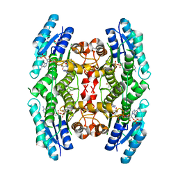
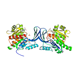 | | Structure of human Ketohexokinase complexed with hits from fragment screening | | 分子名称: | 6-[(3S,4S)-3,4-dihydroxypyrrolidin-1-yl]-2-[(3S)-3-hydroxy-3-methylpyrrolidin-1-yl]-4-(trifluoromethyl)pyridine-3-carbonitrile, CITRIC ACID, Ketohexokinase, ... | | 著者 | Pandit, J. | | 登録日 | 2017-06-29 | | 公開日 | 2017-09-13 | | 最終更新日 | 2023-10-04 | | 実験手法 | X-RAY DIFFRACTION (2.4 Å) | | 主引用文献 | Discovery of Fragment-Derived Small Molecules for in Vivo Inhibition of Ketohexokinase (KHK).
J. Med. Chem., 60, 2017
|
|
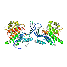
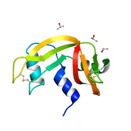
7FO2
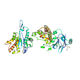 | | PanDDA analysis group deposition -- Aar2/RNaseH in complex with fragment P07G04 from the F2X-Universal Library | | 分子名称: | 3-[(3-bromophenyl)methanesulfonyl]propanoic acid, A1 cistron-splicing factor AAR2, DIMETHYL SULFOXIDE, ... | | 著者 | Barthel, T, Wollenhaupt, J, Lima, G.M.A, Wahl, M.C, Weiss, M.S. | | 登録日 | 2022-08-26 | | 公開日 | 2022-11-02 | | 最終更新日 | 2024-05-22 | | 実験手法 | X-RAY DIFFRACTION (2.01 Å) | | 主引用文献 | Large-Scale Crystallographic Fragment Screening Expedites Compound Optimization and Identifies Putative Protein-Protein Interaction Sites.
J.Med.Chem., 65, 2022
|
|
7FRA
 | |
7FR1
 | |
7FR7
 | |
1P19
 | |
7M1D
 | |
3QPP
 | | Structure of PDE10-inhibitor complex | | 分子名称: | 7-methoxy-4-[(3S)-3-phenylpiperidin-1-yl]-6-[2-(quinolin-2-yl)ethoxy]quinazoline, DIMETHYL SULFOXIDE, MAGNESIUM ION, ... | | 著者 | Pandit, J, Marr, E.S. | | 登録日 | 2011-02-14 | | 公開日 | 2011-06-15 | | 最終更新日 | 2024-02-21 | | 実験手法 | X-RAY DIFFRACTION (1.8 Å) | | 主引用文献 | Use of Structure-Based Design to Discover a Potent, Selective, In Vivo Active Phosphodiesterase 10A Inhibitor Lead Series for the Treatment of Schizophrenia.
J.Med.Chem., 54, 2011
|
|
2AIM
 | |
1YZV
 | |
4DK2
 | |
5WBQ
 | |
5RA2
 | |
5WBM
 | |
3GN2
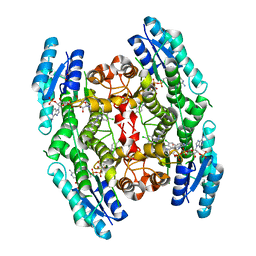 | | Structure of Pteridine Reductase 1 (PTR1) from TRYPANOSOMA BRUCEI in ternary complex with cofactor (NADP+) and inhibitor (DDD00066730) | | 分子名称: | 1-(3,4-dichlorobenzyl)-1H-benzimidazol-2-amine, ACETATE ION, NADP NICOTINAMIDE-ADENINE-DINUCLEOTIDE PHOSPHATE, ... | | 著者 | Tulloch, L.B, Brenk, R, Hunter, W.N. | | 登録日 | 2009-03-16 | | 公開日 | 2009-12-29 | | 最終更新日 | 2024-02-21 | | 実験手法 | X-RAY DIFFRACTION (1.6 Å) | | 主引用文献 | One scaffold, three binding modes: novel and selective pteridine reductase 1 inhibitors derived from fragment hits discovered by virtual screening.
J.Med.Chem., 52, 2009
|
|
3U4B
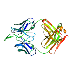 | | CH04H/CH02L Fab P4 | | 分子名称: | CH02 Light chain, CH04 Heavy chain | | 著者 | Pancera, M, Louder, R, Mclellan, J.S, KWong, P.D. | | 登録日 | 2011-10-07 | | 公開日 | 2011-11-30 | | 最終更新日 | 2011-12-21 | | 実験手法 | X-RAY DIFFRACTION (2.893 Å) | | 主引用文献 | Structure of HIV-1 gp120 V1/V2 domain with broadly neutralizing antibody PG9.
Nature, 480, 2011
|
|
5R9I
 | |
3GN1
 | | Structure of Pteridine Reductase 1 (PTR1) from TRYPANOSOMA BRUCEI in ternary complex with cofactor (NADP+) and inhibitor (DDD00067116) | | 分子名称: | 1H-benzimidazol-2-amine, ACETATE ION, NADP NICOTINAMIDE-ADENINE-DINUCLEOTIDE PHOSPHATE, ... | | 著者 | Tulloch, L.B, Brenk, R, Hunter, W.N. | | 登録日 | 2009-03-16 | | 公開日 | 2009-12-29 | | 最終更新日 | 2011-09-07 | | 実験手法 | X-RAY DIFFRACTION (2 Å) | | 主引用文献 | One scaffold, three binding modes: novel and selective pteridine reductase 1 inhibitors derived from fragment hits discovered by virtual screening.
J.Med.Chem., 52, 2009
|
|
2A0M
 | |
6ETL
 | |
6ETM
 | |
6ETR
 | |
6ETP
 | |
6ETK
 | |


