Loading
PDBj
メニューPDBj@FacebookPDBj@TwitterPDBj@YouTubewwPDB FoundationwwPDB
RCSB PDBPDBeBMRBAdv. SearchSearch help

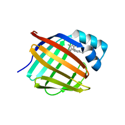
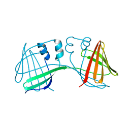
6NNX
DownloadVisualize
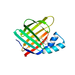
BU of 6nnx by Molmil
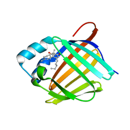
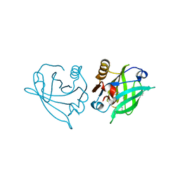
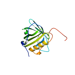
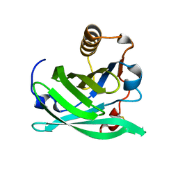
Crystal Structure of the All-Trans Retinal-Bound R111K:Y134F:T54V:R132Q:P39Y:R59Y:L121M Mutant of Human Cellular Retinoic Acid Binding Protein II in the Dark at 1.87 Angstrom Resolution
分子名称: Cellular retinoic acid-binding protein 2, RETINAL
著者Ghanbarpour, A, Geiger, J.
登録日2019-01-15
公開日2019-01-30
最終更新日2023-10-11
実験手法X-RAY DIFFRACTION (1.87 Å)
主引用文献Crystal Structure of the All-Trans Retinal-Bound R111K:Y134F:T54V:R132Q:P39Y:R59Y:L121M Mutant of Human Cellular Retionic Acid Binding Protein II in the Dark at 1.87 Angstrom Resolution
To Be Published
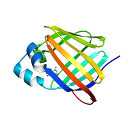
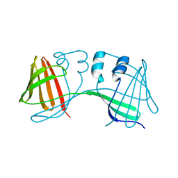
6NNY
DownloadVisualize
BU of 6nny by Molmil
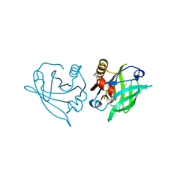
Crystal Structure of the All-Trans Retinal-Bound R111K:Y134F:T54V:R132Q:P39E:R59Y:L121E Mutant of Human Cellular Retinoic Acid Binding Protein II in the Dark at 1.67 Angstrom Resolution
分子名称: Cellular retinoic acid-binding protein 2, RETINAL
著者Ghanbarpour, A, Geiger, J.
登録日2019-01-15
公開日2019-01-30
最終更新日2023-10-11
実験手法X-RAY DIFFRACTION (1.668 Å)
主引用文献Crystal Structure of the All-Trans Retinal-Bound R111K:Y134F:T54V:R132Q:P39E:R59Y:L121E Mutant of Human Cellular Retinoic Acid Binding Protein II in the Dark at 1.68 Angstrom Resolution
To Be Published
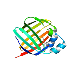
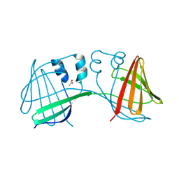
6E6M
DownloadVisualize
BU of 6e6m by Molmil
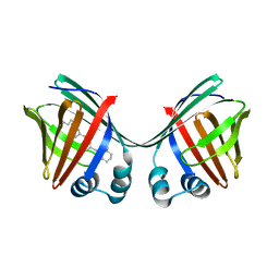
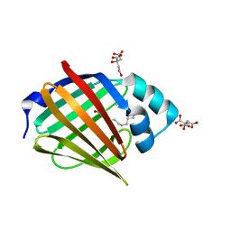
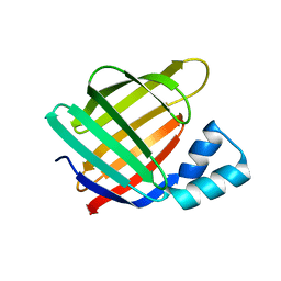
Crystal structure of human cellular retinol-binding protein 1 in complex with cannabidiorcin (CBDO)
分子名称: (1'R,2'R)-4,5'-dimethyl-2'-(prop-1-en-2-yl)-1',2',3',4'-tetrahydro[1,1'-biphenyl]-2,6-diol, Retinol-binding protein 1
著者Silvaroli, J.A, Horwitz, S, Banerjee, S, Kiser, P.D, Golczak, M.
登録日2018-07-25
公開日2019-02-13
最終更新日2024-03-13
実験手法X-RAY DIFFRACTION (1.55 Å)
主引用文献Abnormal Cannabidiol Modulates Vitamin A Metabolism by Acting as a Competitive Inhibitor of CRBP1.
Acs Chem.Biol., 14, 2019
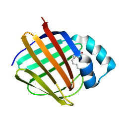
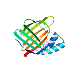
6E5T
DownloadVisualize
BU of 6e5t by Molmil
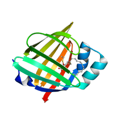
Crystal structure of human cellular retinol binding protein 1 in complex with abnormal-cannabidiorcin (Abn-CBDO)
分子名称: (1'R,2'R)-5',6-dimethyl-2'-(prop-1-en-2-yl)-1',2',3',4'-tetrahydro[1,1'-biphenyl]-2,4-diol, Retinol-binding protein 1
著者Silvaroli, J.A, Horwitz, S, Banerjee, S, Kiser, P.D, Golczak, M.
登録日2018-07-23
公開日2019-02-13
最終更新日2023-10-11
実験手法X-RAY DIFFRACTION (1.55 Å)
主引用文献Abnormal Cannabidiol Modulates Vitamin A Metabolism by Acting as a Competitive Inhibitor of CRBP1.
Acs Chem.Biol., 14, 2019
6E5W
DownloadVisualize
BU of 6e5w by Molmil
Crystal structure of human cellular retinol binding protein 3 in complex with abnormal-cannabidiol (abn-CBD)
分子名称: (1'R,2'R)-5'-methyl-6-pentyl-2'-(prop-1-en-2-yl)-1',2',3',4'-tetrahydro[1,1'-biphenyl]-2,4-diol, GLYCEROL, Retinol-binding protein 5
著者Silvaroli, J.A, Banerjee, S, Kiser, P.D, Golczak, M.
登録日2018-07-23
公開日2019-02-13
最終更新日2023-10-11
実験手法X-RAY DIFFRACTION (2.5 Å)
主引用文献Abnormal Cannabidiol Modulates Vitamin A Metabolism by Acting as a Competitive Inhibitor of CRBP1.
Acs Chem.Biol., 14, 2019
6E6K
DownloadVisualize
BU of 6e6k by Molmil
Crystal structure of human cellular retinol-binding protein 4 in complex with abnormal-cannabidiol (abn-CBD)
分子名称: (1'R,2'R)-5'-methyl-6-pentyl-2'-(prop-1-en-2-yl)-1',2',3',4'-tetrahydro[1,1'-biphenyl]-2,4-diol, Retinoid-binding protein 7
著者Silvaroli, J.A, Banerjee, S, Kiser, P.D, Golczak, M.
登録日2018-07-25
公開日2019-02-13
最終更新日2023-10-11
実験手法X-RAY DIFFRACTION (1.3 Å)
主引用文献Abnormal Cannabidiol Modulates Vitamin A Metabolism by Acting as a Competitive Inhibitor of CRBP1.
Acs Chem.Biol., 14, 2019
6E5L
DownloadVisualize
BU of 6e5l by Molmil
Crystal structure of human cellular retinol binding protein 1 in complex with abnormal-cannabidiol (abn-CBD)
分子名称: (1'R,2'R)-5'-methyl-6-pentyl-2'-(prop-1-en-2-yl)-1',2',3',4'-tetrahydro[1,1'-biphenyl]-2,4-diol, Retinol-binding protein 1
著者Silvaroli, J.A, Banerjee, S, Kiser, P.D, Golczak, M.
登録日2018-07-20
公開日2019-02-13
最終更新日2023-10-11
実験手法X-RAY DIFFRACTION (1.17 Å)
主引用文献Abnormal Cannabidiol Modulates Vitamin A Metabolism by Acting as a Competitive Inhibitor of CRBP1.
Acs Chem.Biol., 14, 2019
6QPD
DownloadVisualize
BU of 6qpd by Molmil
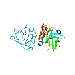
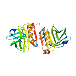
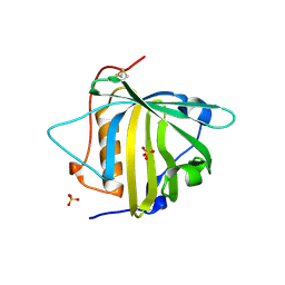
Engineered beta-lactoglobulin: variant I56F
分子名称: Beta-lactoglobulin, GLYCEROL, SULFATE ION
著者Loch, J.I, Kaczor, K, Leiwnski, K.
登録日2019-02-13
公開日2019-02-27
最終更新日2024-01-24
実験手法X-RAY DIFFRACTION (2 Å)
主引用文献Structure-based design approach to rational site-directed mutagenesis of beta-lactoglobulin.
J.Struct.Biol., 210, 2020
6QPE
DownloadVisualize
BU of 6qpe by Molmil
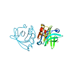
Engineered beta-lactoglobulin: variant L58F
分子名称: ACETATE ION, Beta-lactoglobulin
著者Loch, J.I, Kaczor, K, Leiwnski, K.
登録日2019-02-13
公開日2019-02-27
最終更新日2024-01-24
実験手法X-RAY DIFFRACTION (2.2 Å)
主引用文献Structure-based design approach to rational site-directed mutagenesis of beta-lactoglobulin.
J.Struct.Biol., 210, 2020
6RWP
DownloadVisualize
BU of 6rwp by Molmil
Engineered beta-lactoglobulin: variant L58F in complex with myristic acid
分子名称: Beta-lactoglobulin, MYRISTIC ACID, SULFATE ION
著者Loch, J.I, Kaczor, K, Lewinski, K.
登録日2019-06-05
公開日2019-06-19
最終更新日2024-01-24
実験手法X-RAY DIFFRACTION (2.1 Å)
主引用文献Structure-based design approach to rational site-directed mutagenesis of beta-lactoglobulin.
J.Struct.Biol., 210, 2020
6RWQ
DownloadVisualize
BU of 6rwq by Molmil
Engineered beta-lactoglobulin: variant F105L in complex with myristic acid
分子名称: 1,2-ETHANEDIOL, Beta-lactoglobulin, MYRISTIC ACID
著者Loch, J.I, Gotkowski, M, Lewinski, K.
登録日2019-06-05
公開日2019-06-19
最終更新日2024-01-24
実験手法X-RAY DIFFRACTION (2.05 Å)
主引用文献Structure-based design approach to rational site-directed mutagenesis of beta-lactoglobulin.
J.Struct.Biol., 210, 2020
6RWR
DownloadVisualize
BU of 6rwr by Molmil
Engineered beta-lactoglobulin: variant M107L in complex with myristic acid
分子名称: Beta-lactoglobulin, MYRISTIC ACID
著者Loch, J.I, Gotkowski, M, Lewinski, K.
登録日2019-06-05
公開日2019-06-19
最終更新日2024-01-24
実験手法X-RAY DIFFRACTION (2.1 Å)
主引用文献Structure-based design approach to rational site-directed mutagenesis of beta-lactoglobulin.
J.Struct.Biol., 210, 2020
6RYT
DownloadVisualize
BU of 6ryt by Molmil
Engineered beta-lactoglobulin: variant M107L
分子名称: Beta-lactoglobulin, GLYCEROL, PHOSPHATE ION
著者Loch, J.I, Kurpiewska, K, Lewinski, K.
登録日2019-06-11
公開日2019-06-26
最終更新日2024-01-24
実験手法X-RAY DIFFRACTION (2.1 Å)
主引用文献Structure-based design approach to rational site-directed mutagenesis of beta-lactoglobulin.
J.Struct.Biol., 210, 2020
6E5S
DownloadVisualize
BU of 6e5s by Molmil
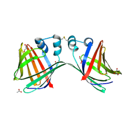
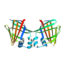
Crystal structure of holo retinal-bound domain-swapped dimer Q108K:T51D mutant of human Cellular Retinol Binding Protein II
分子名称: RETINAL, Retinol-binding protein 2
著者Nosrati, M, Ghanbarpour, A, Geiger, J.
登録日2018-07-22
公開日2019-07-24
最終更新日2023-10-11
実験手法X-RAY DIFFRACTION (2.061 Å)
主引用文献Engineering the hCRBPII Domain-Swapped Dimer into a New Class of Protein Switches.
J.Am.Chem.Soc., 141, 2019
6E5R
DownloadVisualize
BU of 6e5r by Molmil
Crystal structure of the apo domain-swapped dimer Q108K:T51D:A28C mutant of human Cellular Retinol Binding Protein II
分子名称: ACETATE ION, GLYCEROL, Retinol-binding protein 2
著者Ghanbarpour, A, Geiger, J.
登録日2018-07-22
公開日2019-08-07
最終更新日2023-10-11
実験手法X-RAY DIFFRACTION (2.592 Å)
主引用文献Engineering the hCRBPII Domain-Swapped Dimer into a New Class of Protein Switches.
J.Am.Chem.Soc., 141, 2019
6QMU
DownloadVisualize
BU of 6qmu by Molmil
A tetrahedral boronic acid diester formed by a non-natural amino acid in the ligand pocket of an engineered lipocalin
分子名称: 3-nitrophenol, Neutrophil gelatinase-associated lipocalin
著者Skerra, A, Eichinger, A.
登録日2019-02-08
公開日2019-08-28
最終更新日2024-01-24
実験手法X-RAY DIFFRACTION (1.98 Å)
主引用文献A Tetrahedral Boronic Acid Diester Formed by an Unnatural Amino Acid in the Ligand Pocket of an Engineered Lipocalin.
Chembiochem, 21, 2020
6S2S
DownloadVisualize
BU of 6s2s by Molmil
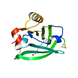
Hydrogenated human myelin protein P2 at 0.86-A resolution
分子名称: CITRIC ACID, Myelin P2 protein, PALMITIC ACID, ...
著者Laulumaa, S, Kursula, P.
登録日2019-06-21
公開日2019-08-28
最終更新日2024-01-24
実験手法X-RAY DIFFRACTION (0.86 Å)
主引用文献Sub-Atomic Resolution Crystal Structures Reveal Conserved Geometric Outliers at Functional Sites.
Molecules, 24, 2019
6S2M
DownloadVisualize
BU of 6s2m by Molmil
Perdeuterated human myelin protein P2 at 0.72-A resolution
分子名称: Myelin P2 protein, PALMITIC ACID, VACCENIC ACID
著者Laulumaa, S, Kursula, P.
登録日2019-06-21
公開日2019-08-28
最終更新日2024-01-24
実験手法X-RAY DIFFRACTION (0.72 Å)
主引用文献Sub-Atomic Resolution Crystal Structures Reveal Conserved Geometric Outliers at Functional Sites.
Molecules, 24, 2019
6O5D
DownloadVisualize
BU of 6o5d by Molmil
PYOCHELIN
分子名称: GLYCEROL, Neutrophil gelatinase-associated lipocalin, SULFATE ION
著者Rupert, P.B, Strong, R.K, Clifton, M.C, Edwards, T.E, Seattle Structural Genomics Center for Infectious Disease (SSGCID)
登録日2019-03-01
公開日2019-09-11
最終更新日2020-07-29
実験手法X-RAY DIFFRACTION (2.4 Å)
主引用文献Parsing the functional specificity of Siderocalin/Lipocalin 2/NGAL for siderophores and related small-molecule ligands.
J Struct Biol X, 2, 2019
6NRE
DownloadVisualize
BU of 6nre by Molmil
Monomeric Lipocalin Can F 6
分子名称: Lipocalin-Can f 6 allergen
著者Clayton G, M, Kappler J, W, Chan, S.
登録日2019-01-23
公開日2019-09-25
実験手法X-RAY DIFFRACTION (2.06 Å)
主引用文献Structural characteristics of lipocalin allergens: Crystal structure of the immunogenic dog allergen Can f 6.
Plos One, 14, 2019
6MOW
DownloadVisualize
BU of 6mow by Molmil
Crystal Structure of the apo R111K:Y134F:T54V:R132Q:P39Y:R59Y:L121Q mutant of Human Cellular Retinoic Acid Binding Protein II at 2.3 Angstrom Resolution
分子名称: Cellular retinoic acid-binding protein 2
著者Ghanbarpour, A, Geiger, J.
登録日2018-10-04
公開日2019-10-09
最終更新日2023-10-11
実験手法X-RAY DIFFRACTION (2.354 Å)
主引用文献Crystal Structure of the apo R111K:Y134F:T54V:R132Q:P39Y:R59Y:L121Q mutant of Human Cellular Retinoic Acid Binding Protein II at 2.3 Angstrom Resolution
To Be Published
6ON5
DownloadVisualize
BU of 6on5 by Molmil
Crystal Structure of the Zn-bound Domain-Swapped Dimer Q108K:T51D:A28C:L36C:F57H Mutant of Human Cellular Retinol Binding Protein II
分子名称: Retinol-binding protein 2, ZINC ION
著者Ghanbarpour, A, Geiger, J.
登録日2019-04-20
公開日2019-10-16
最終更新日2023-10-11
実験手法X-RAY DIFFRACTION (1.638 Å)
主引用文献Engineering the hCRBPII Domain-Swapped Dimer into a New Class of Protein Switches.
J.Am.Chem.Soc., 141, 2019
6ON8
DownloadVisualize
BU of 6on8 by Molmil
Crystal Structure of the Reduced Form of Apo Domain-Swapped Dimer Q108K:T51D:A28C:L36C:F57H Mutant of Human Cellular Retinol Binding Protein II
分子名称: Retinol-binding protein 2
著者Ghanbarpour, A, Geiger, J.
登録日2019-04-20
公開日2019-10-16
最終更新日2023-10-11
実験手法X-RAY DIFFRACTION (2.395 Å)
主引用文献Engineering the hCRBPII Domain-Swapped Dimer into a New Class of Protein Switches.
J.Am.Chem.Soc., 141, 2019
6E51
DownloadVisualize
BU of 6e51 by Molmil
Crystal structure of the apo domain-swapped dimer Q108K:K40L:T51W mutant of human cellular retinol binding protein II
分子名称: Retinol-binding protein 2
著者Ghanbarpour, A, Geiger, J.
登録日2018-07-19
公開日2019-10-16
最終更新日2023-10-11
実験手法X-RAY DIFFRACTION (2.256 Å)
主引用文献Engineering the hCRBPII Domain-Swapped Dimer into a New Class of Protein Switches.
J.Am.Chem.Soc., 141, 2019
6E50
DownloadVisualize
BU of 6e50 by Molmil
Crystal structure of the apo domain-swapped dimer Q108K:K40L:T51F mutant of human Cellular Retinol Binding Protein II
分子名称: ACETATE ION, Retinol-binding protein 2
著者Ghanbarpour, A, Geiger, J.
登録日2018-07-18
公開日2019-10-16
最終更新日2023-10-11
実験手法X-RAY DIFFRACTION (1.965 Å)
主引用文献Engineering the hCRBPII Domain-Swapped Dimer into a New Class of Protein Switches.
J.Am.Chem.Soc., 141, 2019

222415

件を2024-07-10に公開中

PDB statisticsPDBj update infoContact PDBjnumon