4LV5
 
 | |
4LVS
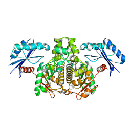
 | | DNA polymerase beta mismatched substrate complex with Mn2+, 2.5 min | | 分子名称: | 2'-DEOXYADENOSINE 5'-TRIPHOSPHATE, DNA (5'-D(*CP*CP*GP*AP*CP*GP*GP*CP*GP*CP*AP*TP*CP*AP*GP*C)-3'), DNA (5'-D(*GP*CP*TP*GP*AP*TP*GP*CP*GP*C)-3'), ... | | 著者 | Freudenthal, B.D, Beard, W.A, Shock, D.D, Wilson, S.H. | | 登録日 | 2013-07-26 | | 公開日 | 2013-09-04 | | 最終更新日 | 2023-09-20 | | 実験手法 | X-RAY DIFFRACTION (2.002 Å) | | 主引用文献 | Observing a DNA polymerase choose right from wrong.
Cell(Cambridge,Mass.), 154, 2013
|
|
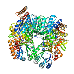
4LYY
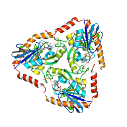
 | | Crystal structure of hypoxanthine phosphoribosyltransferase from Shewanella pealeana ATCC 700345, NYSGRC Target 029677. | | 分子名称: | Hypoxanthine phosphoribosyltransferase, PHOSPHATE ION | | 著者 | Malashkevich, V.N, Bhosle, R, Toro, R, Hillerich, B, Gizzi, A, Garforth, S, Kar, A, Chan, M.K, Lafluer, J, Patel, H, Matikainen, B, Chamala, S, Lim, S, Celikgil, A, Villegas, G, Evans, B, Love, J, Fiser, A, Khafizov, K, Seidel, R, Bonanno, J.B, Almo, S.C, New York Structural Genomics Research Consortium (NYSGRC) | | 登録日 | 2013-07-31 | | 公開日 | 2013-08-14 | | 最終更新日 | 2023-12-06 | | 実験手法 | X-RAY DIFFRACTION (1.86 Å) | | 主引用文献 | Crystal structure of hypoxanthine phosphoribosyltransferase from Shewanella pealeana ATCC 700345, NYSGRC Target 029677.
To be Published
|
|
4D9Y
 | | The crystal structure of Chelerythrine bound to DNA d(CGTACG) | | 分子名称: | 1,2-dimethoxy-12-methyl[1,3]benzodioxolo[5,6-c]phenanthridin-12-ium, CALCIUM ION, DNA (5'-D(*CP*GP*TP*AP*CP*G)-3') | | 著者 | Ferraroni, M, Bazzicalupi, C, Gratteri, P, Bilia, A.R. | | 登録日 | 2012-01-12 | | 公開日 | 2013-01-23 | | 最終更新日 | 2023-09-13 | | 実験手法 | X-RAY DIFFRACTION (2.1 Å) | | 主引用文献 | The crystal structure of Chelerythrine bound to DNA d(CGTACG)
To be Published
|
|
4M0E
 | | Structure of human acetylcholinesterase in complex with dihydrotanshinone I | | 分子名称: | 1,2-ETHANEDIOL, 2-acetamido-2-deoxy-beta-D-glucopyranose, 2-acetamido-2-deoxy-beta-D-glucopyranose-(1-4)-[alpha-L-fucopyranose-(1-6)]2-acetamido-2-deoxy-beta-D-glucopyranose, ... | | 著者 | Cheung, J, Gary, E.N, Shiomi, K, Rosenberry, T.L. | | 登録日 | 2013-08-01 | | 公開日 | 2013-10-16 | | 最終更新日 | 2023-09-20 | | 実験手法 | X-RAY DIFFRACTION (2 Å) | | 主引用文献 | Structures of human acetylcholinesterase bound to dihydrotanshinone I and territrem B show peripheral site flexibility.
ACS Med Chem Lett, 4, 2013
|
|
4D90
 | | Crystal Structure of Del-1 EGF domains | | 分子名称: | 2-acetamido-2-deoxy-beta-D-galactopyranose, 2-acetamido-2-deoxy-beta-D-glucopyranose, CALCIUM ION, ... | | 著者 | Chen, Q, Schurpf, T, Springer, T, Wang, J. | | 登録日 | 2012-01-11 | | 公開日 | 2012-05-30 | | 最終更新日 | 2023-09-13 | | 実験手法 | X-RAY DIFFRACTION (2.601 Å) | | 主引用文献 | The RGD finger of Del-1 is a unique structural feature critical for integrin binding.
Faseb J., 26, 2012
|
|
4M3B
 | | Rapid and efficient design of new inhibitors of Mycobacterium tuberculosis transcriptional repressor EthR using fragment growing, merging and linking approaches | | 分子名称: | 4-(2-methyl-1,3-thiazol-4-yl)-N-(3,3,3-trifluoropropyl)benzamide, HTH-type transcriptional regulator EthR | | 著者 | Villemagne, B, Flipo, M, Blondiaux, N, Crauste, C, Malaquin, S, Leroux, F, Piveteau, C, Villeret, V, Brodin, P, Villoutreix, B, Sperandio, O, Wohlkonig, A, Wintjens, R, Deprez, B, Baulard, A, Willand, N. | | 登録日 | 2013-08-06 | | 公開日 | 2014-06-25 | | 最終更新日 | 2024-02-28 | | 実験手法 | X-RAY DIFFRACTION (2.001 Å) | | 主引用文献 | Ligand Efficiency Driven Design of New Inhibitors of Mycobacterium tuberculosis Transcriptional Repressor EthR Using Fragment Growing, Merging, and Linking Approaches.
J.Med.Chem., 57, 2014
|
|
4KWH
 | | The crystal structure of angucycline C-6 ketoreductase LanV with bound NADP | | 分子名称: | ACETIC ACID, DI(HYDROXYETHYL)ETHER, NADP NICOTINAMIDE-ADENINE-DINUCLEOTIDE PHOSPHATE, ... | | 著者 | Paananen, P, Patrikainen, P, Kallio, P, Mantsala, P, Niemi, J, Niiranen, L, Metsa-Ketela, M. | | 登録日 | 2013-05-24 | | 公開日 | 2013-07-31 | | 最終更新日 | 2023-09-20 | | 実験手法 | X-RAY DIFFRACTION (1.7 Å) | | 主引用文献 | Structural and functional analysis of angucycline C-6 ketoreductase LanV involved in landomycin biosynthesis.
Biochemistry, 52, 2013
|
|
4D9X
 | | The crystal structure of Coptisine bound to DNA d(CGTACG) | | 分子名称: | 6,7-dihydro[1,3]dioxolo[4,5-g][1,3]dioxolo[7,8]isoquino[3,2-a]isoquinolin-5-ium, CALCIUM ION, DNA (5'-D(*CP*GP*TP*AP*CP*G)-3') | | 著者 | Ferraroni, M, Bazzicalupi, C, Gratteri, P, Bilia, A.R. | | 登録日 | 2012-01-12 | | 公開日 | 2013-01-23 | | 最終更新日 | 2023-09-13 | | 実験手法 | X-RAY DIFFRACTION (2.44 Å) | | 主引用文献 | The crystal structure of Coptisine bound to DNA d(CGTACG)
to be published
|
|
4L82
 | |
4L8M
 | | Human p38 MAP kinase in complex with a Dibenzoxepinone | | 分子名称: | Mitogen-activated protein kinase 14, N-[2-fluoro-5-({9-[2-(morpholin-4-yl)ethoxy]-11-oxo-6,11-dihydrodibenzo[b,e]oxepin-3-yl}amino)phenyl]benzamide, octyl beta-D-glucopyranoside | | 著者 | Richters, A, Mayer-Wrangowski, S.C, Gruetter, C, Rauh, D. | | 登録日 | 2013-06-17 | | 公開日 | 2013-10-30 | | 最終更新日 | 2023-09-20 | | 実験手法 | X-RAY DIFFRACTION (2.1 Å) | | 主引用文献 | Metabolically Stable Dibenzo[b,e]oxepin-11(6H)-ones as Highly Selective p38 MAP Kinase Inhibitors: Optimizing Anti-Cytokine Activity in Human Whole Blood.
J.Med.Chem., 56, 2013
|
|
4DBG
 | | Crystal structure of HOIL-1L-UBL complexed with a HOIP-UBA derivative | | 分子名称: | RING finger protein 31, RanBP-type and C3HC4-type zinc finger-containing protein 1 | | 著者 | Yagi, H, Hiromoto, T, Mizushima, T, Kurimoto, E, Kato, K. | | 登録日 | 2012-01-15 | | 公開日 | 2012-04-04 | | 最終更新日 | 2012-05-16 | | 実験手法 | X-RAY DIFFRACTION (2.71 Å) | | 主引用文献 | A non-canonical UBA-UBL interaction forms the linear-ubiquitin-chain assembly complex
Embo Rep., 13, 2012
|
|
4L9G
 | | Structure of PpsR N-Q-PAS1 from Rb. sphaeroides | | 分子名称: | 2-(N-MORPHOLINO)-ETHANESULFONIC ACID, SULFATE ION, Transcriptional regulator, ... | | 著者 | Heintz, U, Meinhart, A, Schlichting, I, Winkler, A. | | 登録日 | 2013-06-18 | | 公開日 | 2014-02-12 | | 最終更新日 | 2024-04-03 | | 実験手法 | X-RAY DIFFRACTION (2.2 Å) | | 主引用文献 | Multi-PAS domain-mediated protein oligomerization of PpsR from Rhodobacter sphaeroides.
Acta Crystallogr.,Sect.D, 70, 2014
|
|
4DQ8
 | |
4L9Y
 | | Crystal Structure of Rhodobacter sphaeroides malyl-CoA lyase in complex with magnesium, glyoxylate, and propionyl-CoA | | 分子名称: | CHLORIDE ION, GLYOXYLIC ACID, MAGNESIUM ION, ... | | 著者 | Zarzycki, J, Kerfeld, C.A. | | 登録日 | 2013-06-18 | | 公開日 | 2013-12-04 | | 最終更新日 | 2023-09-20 | | 実験手法 | X-RAY DIFFRACTION (2.102 Å) | | 主引用文献 | The crystal structures of the tri-functional Chloroflexus aurantiacus and bi-functional Rhodobacter sphaeroides malyl-CoA lyases and comparison with CitE-like superfamily enzymes and malate synthases.
Bmc Struct.Biol., 13, 2013
|
|
4L5A
 | | Methylthioadenosine phosphorylase from Schistosoma mansoni in complex with tubercidin | | 分子名称: | '2-(4-AMINO-PYRROLO[2,3-D]PYRIMIDIN-7-YL)-5-HYDROXYMETHYL-TETRAHYDRO-FURAN-3,4-DIOL, S-methyl-5'-thioadenosine phosphorylase, SULFATE ION | | 著者 | Torini, J.R, DeMarco, R, Brandao-Neto, J, Pereira, H.M. | | 登録日 | 2013-06-10 | | 公開日 | 2014-06-11 | | 最終更新日 | 2024-02-28 | | 実験手法 | X-RAY DIFFRACTION (2.2993 Å) | | 主引用文献 | Crystal Structure of Schistosoma mansoni Adenosine Phosphorylase/5'-Methylthioadenosine Phosphorylase and Its Importance on Adenosine Salvage Pathway.
Plos Negl Trop Dis, 10, 2016
|
|
4LBE
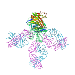 | | Structure of KcsA with R122A mutation | | 分子名称: | DIACYL GLYCEROL, Fab heavy chain, Fab light chain, ... | | 著者 | Nimigean, C.M, Posson, D.J, McCoy, J.M. | | 登録日 | 2013-06-20 | | 公開日 | 2013-10-30 | | 最終更新日 | 2017-11-15 | | 実験手法 | X-RAY DIFFRACTION (2.751 Å) | | 主引用文献 | Molecular interactions involved in proton-dependent gating in KcsA potassium channels.
J.Gen.Physiol., 142, 2013
|
|
4LC0
 | | Identifying ligand binding hot spots in proteins using brominated fragments | | 分子名称: | AMMONIUM ION, Elongation factor Tu-A, MAGNESIUM ION, ... | | 著者 | Groftehauge, M.K, Therkelsen, M, Taaning, R, Skrydstrup, T, Morth, J.P, Nissen, P. | | 登録日 | 2013-06-21 | | 公開日 | 2013-09-11 | | 最終更新日 | 2023-09-20 | | 実験手法 | X-RAY DIFFRACTION (2.221 Å) | | 主引用文献 | Identifying ligand-binding hot spots in proteins using brominated fragments.
Acta Crystallogr.,Sect.F, 69, 2013
|
|
4LCE
 | | CtBP1 in complex with substrate MTOB | | 分子名称: | 4-(METHYLSULFANYL)-2-OXOBUTANOIC ACID, C-terminal-binding protein 1, NICOTINAMIDE-ADENINE-DINUCLEOTIDE | | 著者 | Hilbert, B.J, Schiffer, C.A, Royer Jr, W.E. | | 登録日 | 2013-06-21 | | 公開日 | 2014-03-19 | | 最終更新日 | 2024-02-28 | | 実験手法 | X-RAY DIFFRACTION (2.38 Å) | | 主引用文献 | Crystal structures of human CtBP in complex with substrate MTOB reveal active site features useful for inhibitor design.
Febs Lett., 588, 2014
|
|
4LCM
 | | Simvastatin Synthase (LOVD), from Aspergillus Terreus, LovD9 mutant (simh9014) | | 分子名称: | Transesterase | | 著者 | Gao, X, Sawaya, M.R, Yeates, T.O, Tang, Y. | | 登録日 | 2013-06-21 | | 公開日 | 2014-04-02 | | 最終更新日 | 2023-09-20 | | 実験手法 | X-RAY DIFFRACTION (3.19 Å) | | 主引用文献 | The role of distant mutations and allosteric regulation on LovD active site dynamics.
Nat.Chem.Biol., 10, 2014
|
|
4L75
 | |
4DHO
 | | Small-molecule inhibitors of 14-3-3 protein-protein interactions from virtual screening | | 分子名称: | (2-{2-[(3-methoxyphenyl)amino]-2-oxoethoxy}phenyl)phosphonic acid, 14-3-3 protein sigma, CHLORIDE ION, ... | | 著者 | Thiel, P, Roeglin, L, Kohlbacher, O, Ottmann, C. | | 登録日 | 2012-01-30 | | 公開日 | 2013-07-31 | | 最終更新日 | 2024-02-28 | | 実験手法 | X-RAY DIFFRACTION (1.7 Å) | | 主引用文献 | Virtual screening and experimental validation reveal novel small-molecule inhibitors of 14-3-3 protein-protein interactions.
Chem.Commun.(Camb.), 49, 2013
|
|
4DI9
 | | CRYSTAL STRUCTURE OF THE D248A mutant of 2-PYRONE-4,6-DICARBOXYLIC ACID HYDROLASE FROM SPHINGOMONAS PAUCIMOBILIS complexed with substrate at pH 6.5 | | 分子名称: | (1E,3Z)-4-hydroxybuta-1,3-diene-1,2,4-tricarboxylic acid, 2-pyrone-4,6-dicarbaxylate hydrolase, ACETATE ION | | 著者 | Malashkevich, V.N, Toro, R, Hobbs, M.E, Raushel, F.M, Almo, S.C. | | 登録日 | 2012-01-11 | | 公開日 | 2012-10-03 | | 最終更新日 | 2024-02-28 | | 実験手法 | X-RAY DIFFRACTION (1.35 Å) | | 主引用文献 | Structure and Catalytic Mechanism of LigI: Insight into the Amidohydrolase Enzymes of cog3618 and Lignin Degradation.
Biochemistry, 51, 2012
|
|
4DR0
 | | Crystal structure of Bacillus subtilis dimanganese(II) NrdF | | 分子名称: | MANGANESE (II) ION, Ribonucleoside-diphosphate reductase subunit beta, SULFATE ION | | 著者 | Boal, A.K, Cotruvo Jr, J.A, Stubbe, J, Rosenzweig, A.C. | | 登録日 | 2012-02-16 | | 公開日 | 2012-04-11 | | 最終更新日 | 2024-02-28 | | 実験手法 | X-RAY DIFFRACTION (1.9 Å) | | 主引用文献 | The Dimanganese(II) Site of Bacillus subtilis Class Ib Ribonucleotide Reductase.
Biochemistry, 51, 2012
|
|
4LBL
 | |


