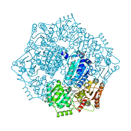
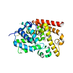
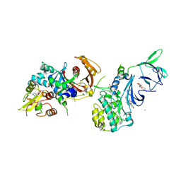
6PCV
 
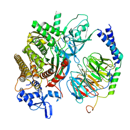 | | Single Particle Reconstruction of Phosphatidylinositol (3,4,5) trisphosphate-dependent Rac exchanger 1 bound to G protein beta gamma subunits | | 分子名称: | Guanine nucleotide-binding protein G(I)/G(S)/G(O) subunit gamma-2, Guanine nucleotide-binding protein G(I)/G(S)/G(T) subunit beta-1, Phosphatidylinositol (3,4,5) trisphosphate-dependent Rac exchanger 1 | | 著者 | Cash, J.N, Cianfrocco, M.A, Tesmer, J.J.G. | | 登録日 | 2019-06-18 | | 公開日 | 2019-10-23 | | 最終更新日 | 2024-03-20 | | 実験手法 | ELECTRON MICROSCOPY (3.2 Å) | | 主引用文献 | Cryo-electron microscopy structure and analysis of the P-Rex1-G beta gamma signaling scaffold.
Sci Adv, 5, 2019
|
|
6ZP6
 | | Yeast 20S proteasome in complex with glidobactin-like natural product HB334 | | 分子名称: | CHLORIDE ION, MAGNESIUM ION, Probable proteasome subunit alpha type-7, ... | | 著者 | Zhao, L, Le Chapelain, C, Brachmann, A.O, Kaiser, M, Groll, M, Bode, H.B. | | 登録日 | 2020-07-08 | | 公開日 | 2021-05-19 | | 最終更新日 | 2024-11-13 | | 実験手法 | X-RAY DIFFRACTION (2.8 Å) | | 主引用文献 | Activation, Structure, Biosynthesis and Bioactivity of Glidobactin-like Proteasome Inhibitors from Photorhabdus laumondii.
Chembiochem, 22, 2021
|
|
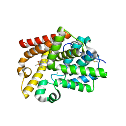
1K6D
 | | CRYSTAL STRUCTURE OF ACETATE COA-TRANSFERASE ALPHA SUBUNIT | | 分子名称: | ACETATE COA-TRANSFERASE ALPHA SUBUNIT, MAGNESIUM ION | | 著者 | Korolev, S, Koroleva, O, Petterson, K, Collart, F, Dementieva, I, Joachimiak, A, Midwest Center for Structural Genomics (MCSG) | | 登録日 | 2001-10-15 | | 公開日 | 2002-06-26 | | 最終更新日 | 2024-02-07 | | 実験手法 | X-RAY DIFFRACTION (1.9 Å) | | 主引用文献 | Autotracing of Escherichia coli acetate CoA-transferase alpha-subunit structure using 3.4 A MAD and 1.9 A native data.
Acta Crystallogr.,Sect.D, 58, 2002
|
|
7VT6
 | | Crystal structure of CBM deleted MtGlu5 in complex with BGC. | | 分子名称: | Endoglucanase H, GLYCEROL, TRYPTOPHAN, ... | | 著者 | Ye, T.J, Ko, P.T, Huang, K.F, Wu, S.H. | | 登録日 | 2021-10-28 | | 公開日 | 2022-09-07 | | 最終更新日 | 2023-11-29 | | 実験手法 | X-RAY DIFFRACTION (1.53 Å) | | 主引用文献 | Synergic action of an inserted carbohydrate-binding module in a glycoside hydrolase family 5 endoglucanase.
Acta Crystallogr D Struct Biol, 78, 2022
|
|
6DER
 | | Crystal structure of Candida albicans acetohydroxyacid synthase in complex with the herbicide metosulam | | 分子名称: | (3Z)-4-{[(4-AMINO-2-METHYLPYRIMIDIN-5-YL)METHYL]AMINO}-3-MERCAPTOPENT-3-EN-1-YL TRIHYDROGEN DIPHOSPHATE, 2-{3-[(4-AMINO-2-METHYLPYRIMIDIN-5-YL)METHYL]-4-METHYL-2-OXO-2,3-DIHYDRO-1,3-THIAZOL-5-YL}ETHYL TRIHYDROGEN DIPHOSPHATE, Acetolactate synthase, ... | | 著者 | Garcia, M.D, Guddat, L.W. | | 登録日 | 2018-05-12 | | 公開日 | 2018-09-26 | | 最終更新日 | 2023-10-11 | | 実験手法 | X-RAY DIFFRACTION (2.126 Å) | | 主引用文献 | Commercial AHAS-inhibiting herbicides are promising drug leads for the treatment of human fungal pathogenic infections.
Proc. Natl. Acad. Sci. U.S.A., 115, 2018
|
|
5CAS
 | | EGFR kinase domain mutant "TMLR" with compound 41a | | 分子名称: | (1R)-1-{6-({2-[(3R,4S)-3-fluoro-4-methoxypiperidin-1-yl]pyrimidin-4-yl}amino)-1-[(2S)-1,1,1-trifluoropropan-2-yl]-1H-imidazo[4,5-c]pyridin-2-yl}ethanol, Epidermal growth factor receptor, SULFATE ION | | 著者 | Eigenbrot, C, Yu, C. | | 登録日 | 2015-06-29 | | 公開日 | 2015-10-28 | | 最終更新日 | 2024-03-06 | | 実験手法 | X-RAY DIFFRACTION (2.1 Å) | | 主引用文献 | Noncovalent Mutant Selective Epidermal Growth Factor Receptor Inhibitors: A Lead Optimization Case Study.
J.Med.Chem., 58, 2015
|
|
5SHY
 | | CRYSTAL STRUCTURE OF HUMAN PHOSPHODIESTERASE 10 IN COMPLEX WITH n1c(scc1Nc2ncc(c3ccc(nc23)CO)c4cccnc4)C, micromolar IC50=0.297 | | 分子名称: | CHLORIDE ION, MAGNESIUM ION, POTASSIUM ION, ... | | 著者 | Joseph, C, Benz, J, Flohr, A, Schnider, P, Rudolph, M.G. | | 登録日 | 2022-02-01 | | 公開日 | 2022-10-12 | | 最終更新日 | 2024-10-16 | | 実験手法 | X-RAY DIFFRACTION (2.3 Å) | | 主引用文献 | Crystal Structure of a human phosphodiesterase 10 complex
To be published
|
|
4BBH
 | | Plasmodium vivax N-myristoyltransferase with a bound benzothiophene inhibitor | | 分子名称: | 2-oxopentadecyl-CoA, 3-methoxybenzyl 3-(piperidin-4-yloxy)-1-benzothiophene-2-carboxylate, CHLORIDE ION, ... | | 著者 | Rackham, M.D, Brannigan, J.A, Moss, D.K, Yu, Z, Wilkinson, A.J, Holder, A.A, Tate, E.W, Leatherbarrow, R.J. | | 登録日 | 2012-09-23 | | 公開日 | 2012-12-05 | | 最終更新日 | 2024-05-08 | | 実験手法 | X-RAY DIFFRACTION (1.63 Å) | | 主引用文献 | Discovery of Novel and Ligand-Efficient Inhibitors of Plasmodium Falciparum and Plasmodium Vivax N-Myristoyltransferase.
J.Med.Chem., 56, 2013
|
|
1TU9
 | | Crystal Structure of a Protein PA3967, a Structurally Highly Homologous to a Human Hemoglobin, from Pseudomonas aeruginosa PAO1 | | 分子名称: | 1,2-ETHANEDIOL, PROPANOIC ACID, PROTOPORPHYRIN IX CONTAINING FE, ... | | 著者 | Kim, Y, Joachimiak, A, Skarina, T, Egorova, O, Bochkarev, A, Savchenko, A, Edwards, A, Midwest Center for Structural Genomics (MCSG) | | 登録日 | 2004-06-24 | | 公開日 | 2004-08-10 | | 最終更新日 | 2024-02-14 | | 実験手法 | X-RAY DIFFRACTION (1.2 Å) | | 主引用文献 | Crystal Structure of PA3967 from Pseudomonas aeruginosa PAO1, a Hypothetical Protein which is highly homologous to human Hemoglobin in structure.
To be Published
|
|
5SJO
 | | CRYSTAL STRUCTURE OF HUMAN PHOSPHODIESTERASE 10 IN COMPLEX WITH C1=CN(N=C(C1=O)c2ccnn2c3ccc(cc3F)F)c4cc(ccc4)OC(F)(F)F, micromolar IC50=0.041355 | | 分子名称: | 3-[1-(2,4-difluorophenyl)-1H-pyrazol-5-yl]-1-[3-(trifluoromethoxy)phenyl]pyridazin-4(1H)-one, CHLORIDE ION, GLYCEROL, ... | | 著者 | Joseph, C, Benz, J, Flohr, A, Rogers-Evans, M, Rudolph, M.G. | | 登録日 | 2022-02-01 | | 公開日 | 2022-10-12 | | 最終更新日 | 2024-10-16 | | 実験手法 | X-RAY DIFFRACTION (2.1 Å) | | 主引用文献 | Crystal Structure of a human phosphodiesterase 10 complex
To be published
|
|
4Y2N
 | | Structure of CFA/I pili major subunit CfaB trimer | | 分子名称: | 1,2-ETHANEDIOL, CFA/I fimbrial subunit B, MAGNESIUM ION | | 著者 | Bao, R, Xia, D. | | 登録日 | 2015-02-10 | | 公開日 | 2016-08-10 | | 最終更新日 | 2023-09-27 | | 実験手法 | X-RAY DIFFRACTION (2.4 Å) | | 主引用文献 | Off-pathway assembly of fimbria subunits is prevented by chaperone CfaA of CFA/I fimbriae from enterotoxigenic E. coli.
Mol. Microbiol., 102, 2016
|
|
4I9B
 | | Structure of aminoaldehyde dehydrogenase 1 from Solanum lycopersium (SlAMADH1) with a thiohemiacetal intermediate | | 分子名称: | (2-hydroxyethoxy)acetaldehyde, 1,2-ETHANEDIOL, DI(HYDROXYETHYL)ETHER, ... | | 著者 | Morera, S, Vigouroux, A, Kopecny, D. | | 登録日 | 2012-12-05 | | 公開日 | 2013-02-20 | | 最終更新日 | 2024-11-20 | | 実験手法 | X-RAY DIFFRACTION (1.9 Å) | | 主引用文献 | Plant ALDH10 family: identifying critical residues for substrate specificity and trapping a thiohemiacetal intermediate.
J.Biol.Chem., 288, 2013
|
|
9HNL
 | | Structure of the (6-4) photolyase of Caulobacter crescentus in its oxidized state at room temperture-synchrotron | | 分子名称: | 1-deoxy-1-(6,7-dimethyl-2,4-dioxo-3,4-dihydropteridin-8(2H)-yl)-D-ribitol, Cryptochrome/photolyase family protein, FLAVIN-ADENINE DINUCLEOTIDE, ... | | 著者 | Po Hsun, W, Maestre-Reyna, M, Essen, L.-O. | | 登録日 | 2024-12-11 | | 公開日 | 2025-05-07 | | 最終更新日 | 2025-05-21 | | 実験手法 | X-RAY DIFFRACTION (2.2 Å) | | 主引用文献 | Redox-State-Dependent Structural Changes within a Prokaryotic 6-4 Photolyase.
J.Am.Chem.Soc., 147, 2025
|
|
3OMS
 | | Putative 3-demethylubiquinone-9 3-methyltransferase, PhnB protein, from Bacillus cereus. | | 分子名称: | 1,2-ETHANEDIOL, PhnB protein | | 著者 | Osipiuk, J, Li, H, Abdullah, J, Joachimiak, A, Midwest Center for Structural Genomics (MCSG) | | 登録日 | 2010-08-27 | | 公開日 | 2010-09-08 | | 最終更新日 | 2024-11-06 | | 実験手法 | X-RAY DIFFRACTION (1.9 Å) | | 主引用文献 | X-ray crystal structure of putative 3-demethylubiquinone-9
3-methyltransferase, PhnB protein, from Bacillus cereus.
To be Published
|
|
9HNN
 | | Structure of the (6-4) photolyase of Caulobacter crescentus in its fully reduced state determined by serial femtosecond crystallography | | 分子名称: | 1-deoxy-1-(6,7-dimethyl-2,4-dioxo-3,4-dihydropteridin-8(2H)-yl)-D-ribitol, Cryptochrome/photolyase family protein, FLAVIN-ADENINE DINUCLEOTIDE, ... | | 著者 | Po Hsun, W, Maestre-Reyna, M, Essen, L.-O. | | 登録日 | 2024-12-11 | | 公開日 | 2025-05-07 | | 最終更新日 | 2025-05-21 | | 実験手法 | X-RAY DIFFRACTION (1.78 Å) | | 主引用文献 | Redox-State-Dependent Structural Changes within a Prokaryotic 6-4 Photolyase.
J.Am.Chem.Soc., 147, 2025
|
|
9HNK
 | | Structure of the (6-4) photolyase of Caulobacter crescentus in its oxidizd state - Singal crystal / Synchrotron | | 分子名称: | 1-deoxy-1-(6,7-dimethyl-2,4-dioxo-3,4-dihydropteridin-8(2H)-yl)-D-ribitol, Cryptochrome/photolyase family protein, FLAVIN-ADENINE DINUCLEOTIDE, ... | | 著者 | Po Hsun, W, Maestre-Reyna, M, Essen, L.-O. | | 登録日 | 2024-12-11 | | 公開日 | 2025-05-07 | | 最終更新日 | 2025-05-21 | | 実験手法 | X-RAY DIFFRACTION (1.59 Å) | | 主引用文献 | Redox-State-Dependent Structural Changes within a Prokaryotic 6-4 Photolyase.
J.Am.Chem.Soc., 147, 2025
|
|
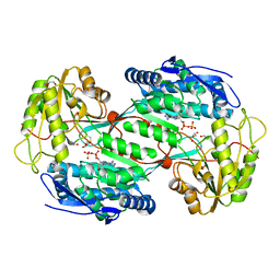
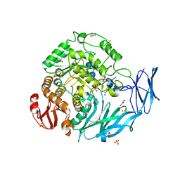
4QFS
 | | Structure of AMPK in complex with Br2-A769662core activator and STAUROSPORINE inhibitor | | 分子名称: | 2-bromo-3-(4-bromophenyl)-4-hydroxy-6-oxo-6,7-dihydrothieno[2,3-b]pyridine-5-carbonitrile, 5'-AMP-activated protein kinase catalytic subunit alpha-1, 5'-AMP-activated protein kinase subunit beta-1, ... | | 著者 | Calabrese, M.F, Kurumbail, R.G. | | 登録日 | 2014-05-21 | | 公開日 | 2014-08-06 | | 最終更新日 | 2024-10-09 | | 実験手法 | X-RAY DIFFRACTION (3.55 Å) | | 主引用文献 | Structural Basis for AMPK Activation: Natural and Synthetic Ligands Regulate Kinase Activity from Opposite Poles by Different Molecular Mechanisms.
Structure, 22, 2014
|
|
3DKU
 | | Crystal structure of Nudix hydrolase Orf153, ymfB, from Escherichia coli K-1 | | 分子名称: | Putative phosphohydrolase | | 著者 | Hong, M.K, Kim, J.K, Jung, J.H, Jung, J.W, Choi, J.Y, Kang, L.W. | | 登録日 | 2008-06-26 | | 公開日 | 2009-06-30 | | 最終更新日 | 2023-08-30 | | 実験手法 | X-RAY DIFFRACTION (2.69 Å) | | 主引用文献 | Crystal structure of Nudix hydrolase Orf153, ymfB, from Escherichia coli K-1.
To be Published
|
|
7T1X
 | |
2Y8C
 | | Plasmodium falciparum dUTPase in complex with a trityl ligand | | 分子名称: | (2S)-2-[(2,4-dioxopyrimidin-1-yl)methyl]-N-(2-hydroxyethyl)-4-trityloxy-butanamide, DEOXYURIDINE 5'-TRIPHOSPHATE NUCLEOTIDOHYDROLASE, SULFATE ION | | 著者 | Baragana, B, McCarthy, O, Sanchez, P, Bosch, C, Kaiser, M, Brun, R, Whittingham, J.L, Roberts, S, Zhou, X.-X, Wilson, K.S, Johansson, N.G, Gonzalez-Pacanowska, D, Gilbert, I.H. | | 登録日 | 2011-02-04 | | 公開日 | 2012-03-07 | | 最終更新日 | 2023-12-20 | | 実験手法 | X-RAY DIFFRACTION (2.1 Å) | | 主引用文献 | Beta-Branched Acyclic Nucleoside Analogues as Inhibitors of Plasmodium Falciparum Dutpase
Bioorg.Med.Chem., 19, 2011
|
|
5SIF
 | | CRYSTAL STRUCTURE OF HUMAN PHOSPHODIESTERASE 10 IN COMPLEX WITH C1=CN(N=C(C1=O)c2ccnn2c3cc(ccc3F)F)c4cc(ccc4)[S](=O)(=O)C, micromolar IC50=0.049318 | | 分子名称: | 3-[1-(2,5-difluorophenyl)-1H-pyrazol-5-yl]-1-[3-(methanesulfonyl)phenyl]pyridazin-4(1H)-one, CHLORIDE ION, GLYCEROL, ... | | 著者 | Joseph, C, Benz, J, Flohr, A, Rogers-Evans, M, Rudolph, M.G. | | 登録日 | 2022-02-01 | | 公開日 | 2022-10-12 | | 最終更新日 | 2024-10-16 | | 実験手法 | X-RAY DIFFRACTION (2.2 Å) | | 主引用文献 | Crystal Structure of a human phosphodiesterase 10 complex
To be published
|
|
2RJ2
 | | Crystal Structure of the Sugar Recognizing SCF Ubiquitin Ligase at 1.7 Resolution | | 分子名称: | CHLORIDE ION, F-box only protein 2, NICKEL (II) ION | | 著者 | Vaijayanthimala, S, Velmurugan, D, Mizushima, T, Yamane, T, Yoshida, Y, Tanaka, K. | | 登録日 | 2007-10-14 | | 公開日 | 2008-10-14 | | 最終更新日 | 2023-11-08 | | 実験手法 | X-RAY DIFFRACTION (1.7 Å) | | 主引用文献 | Crystal Structure of the Sugar Recognizing SCF Ubiquitin Ligase at 1.7 Resolution
To be Published
|
|
9HNO
 | | Structure of the (6-4) photolyase of Caulobacter crescentus in its iron sulfur cluster oxidized state determined by serial femtosecond crystallography | | 分子名称: | 1-deoxy-1-(6,7-dimethyl-2,4-dioxo-3,4-dihydropteridin-8(2H)-yl)-D-ribitol, Cryptochrome/photolyase family protein, FLAVIN-ADENINE DINUCLEOTIDE, ... | | 著者 | Po Hsun, W, Maestre-Reyna, M, Essen, L.-O. | | 登録日 | 2024-12-11 | | 公開日 | 2025-05-07 | | 最終更新日 | 2025-05-21 | | 実験手法 | X-RAY DIFFRACTION (2.3 Å) | | 主引用文献 | Redox-State-Dependent Structural Changes within a Prokaryotic 6-4 Photolyase.
J.Am.Chem.Soc., 147, 2025
|
|
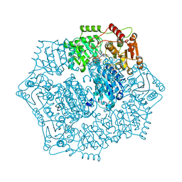
5X3J
 | | Kfla1895 D451A mutant in complex with cyclobis-(1->6)-alpha-nigerosyl | | 分子名称: | Cyclic alpha-D-glucopyranose-(1-3)-alpha-D-glucopyranose-(1-6)-alpha-D-glucopyranose-(1-3)-alpha-D-glucopyranose, GLYCEROL, Glycoside hydrolase family 31, ... | | 著者 | Tanaka, Y, Chen, M, Tagami, T, Yao, M, Kimura, A. | | 登録日 | 2017-02-06 | | 公開日 | 2018-02-07 | | 最終更新日 | 2024-03-27 | | 実験手法 | X-RAY DIFFRACTION (2.3 Å) | | 主引用文献 | Glycoside hydrolase mutant in complex with substrate
To Be Published
|
|
6DEQ
 | | Crystal structure of Candida albicans acetohydroxyacid synthase in complex with the herbicide penoxsulam | | 分子名称: | (3Z)-4-{[(4-AMINO-2-METHYLPYRIMIDIN-5-YL)METHYL]AMINO}-3-MERCAPTOPENT-3-EN-1-YL TRIHYDROGEN DIPHOSPHATE, 2-(2,2-difluoroethoxy)-N-(5,8-dimethoxy[1,2,4]triazolo[1,5-c]pyrimidin-2-yl)-6-(trifluoromethyl)benzenesulfonamide, Acetolactate synthase, ... | | 著者 | Garcia, M.D, Guddat, L.W. | | 登録日 | 2018-05-12 | | 公開日 | 2018-09-26 | | 最終更新日 | 2023-10-11 | | 実験手法 | X-RAY DIFFRACTION (2.127 Å) | | 主引用文献 | Commercial AHAS-inhibiting herbicides are promising drug leads for the treatment of human fungal pathogenic infections.
Proc. Natl. Acad. Sci. U.S.A., 115, 2018
|
|


