5Z6Y
 
 | |
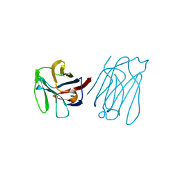
5YR2
 | |
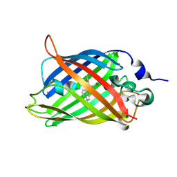
5Y8R
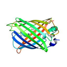
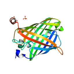
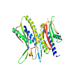
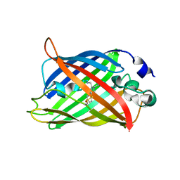
 | | ZsYellow at pH 3.5 | | 分子名称: | GFP-like fluorescent chromoprotein FP538 | | 著者 | Bae, J.E, Kim, I.J, Nam, K.H. | | 登録日 | 2017-08-21 | | 公開日 | 2017-09-13 | | 最終更新日 | 2023-11-22 | | 実験手法 | X-RAY DIFFRACTION (2.3 Å) | | 主引用文献 | Disruption of the hydrogen bonding network determines the pH-induced non-fluorescent state of the fluorescent protein ZsYellow by protonation of Glu221.
Biochem. Biophys. Res. Commun., 493, 2017
|
|
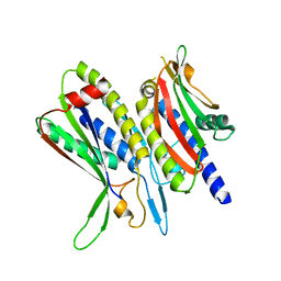
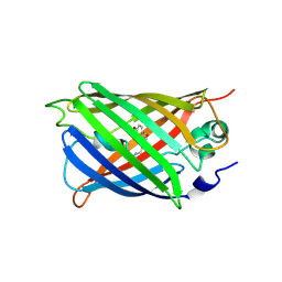
5Y8Q
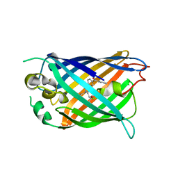
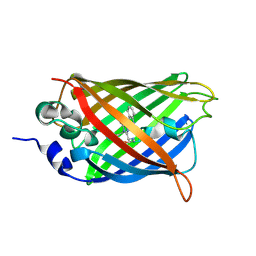
 | | ZsYellow at pH 8.0 | | 分子名称: | GFP-like fluorescent chromoprotein FP538 | | 著者 | Bae, J.E, Kim, I.J, Nam, K.H. | | 登録日 | 2017-08-21 | | 公開日 | 2017-09-13 | | 最終更新日 | 2023-11-22 | | 実験手法 | X-RAY DIFFRACTION (2.9 Å) | | 主引用文献 | Disruption of the hydrogen bonding network determines the pH-induced non-fluorescent state of the fluorescent protein ZsYellow by protonation of Glu221.
Biochem. Biophys. Res. Commun., 493, 2017
|
|
5Y03
 | |
5Y01
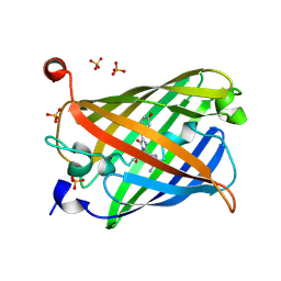 | | Acid-tolerant monomeric GFP, Gamillus, non-fluorescence (OFF) state | | 分子名称: | Green fluorescent protein, PHOSPHATE ION | | 著者 | Nakashima, R, Sakurai, K, Shinoda, H, Matsuda, T, Nagai, T. | | 登録日 | 2017-07-14 | | 公開日 | 2018-01-17 | | 最終更新日 | 2023-11-15 | | 実験手法 | X-RAY DIFFRACTION (2.65 Å) | | 主引用文献 | Acid-Tolerant Monomeric GFP from Olindias formosa.
Cell Chem Biol, 25, 2018
|
|
5Y00
 | | Acid-tolerant monomeric GFP, Gamillus, fluorescence (ON) state | | 分子名称: | CHLORIDE ION, GLYCEROL, Green fluorescent protein, ... | | 著者 | Nakashima, R, Sakurai, K, Shinoda, H, Matsuda, T, Nagai, T. | | 登録日 | 2017-07-14 | | 公開日 | 2018-01-17 | | 最終更新日 | 2023-11-15 | | 実験手法 | X-RAY DIFFRACTION (1.6 Å) | | 主引用文献 | Acid-Tolerant Monomeric GFP from Olindias formosa.
Cell Chem Biol, 25, 2018
|
|
5XV6
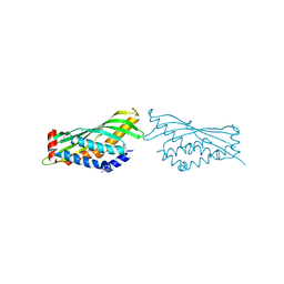
 | | Crystal structure of ATG101-ATG13HORMA | | 分子名称: | Autophagy-related protein 101, Autophagy-related protein 13 | | 著者 | Kim, B.-W, Song, H.K. | | 登録日 | 2017-06-26 | | 公開日 | 2018-07-04 | | 最終更新日 | 2023-11-22 | | 実験手法 | X-RAY DIFFRACTION (2.455 Å) | | 主引用文献 | The C-terminal region of ATG101 bridges ULK1 and PtdIns3K complex in autophagy initiation.
Autophagy, 14, 2018
|
|
5XV4
 | | Crystal structure of ATG101-ATG13HORMA | | 分子名称: | Autophagy-related protein 101, Autophagy-related protein 13 | | 著者 | Kim, B.-W, Song, H.K. | | 登録日 | 2017-06-26 | | 公開日 | 2018-07-04 | | 最終更新日 | 2023-11-22 | | 実験手法 | X-RAY DIFFRACTION (2.95 Å) | | 主引用文献 | The C-terminal region of ATG101 bridges ULK1 and PtdIns3K complex in autophagy initiation.
Autophagy, 14, 2018
|
|
5XV3
 | | Crystal structure of ATG101-ATG13HORMA | | 分子名称: | Autophagy-related protein 101, Autophagy-related protein 13, DI(HYDROXYETHYL)ETHER | | 著者 | Kim, B.-W, Song, H.K. | | 登録日 | 2017-06-26 | | 公開日 | 2018-07-04 | | 最終更新日 | 2023-11-22 | | 実験手法 | X-RAY DIFFRACTION (2.57 Å) | | 主引用文献 | The C-terminal region of ATG101 bridges ULK1 and PtdIns3K complex in autophagy initiation.
Autophagy, 14, 2018
|
|
5XUY
 | | Crystal structure of ATG101-ATG13HORMA | | 分子名称: | Autophagy-related protein 101, Autophagy-related protein 13 | | 著者 | Kim, B.-W, Song, H.K. | | 登録日 | 2017-06-26 | | 公開日 | 2018-07-04 | | 最終更新日 | 2024-03-27 | | 実験手法 | X-RAY DIFFRACTION (2.2 Å) | | 主引用文献 | The C-terminal region of ATG101 bridges ULK1 and PtdIns3K complex in autophagy initiation.
Autophagy, 14, 2018
|
|
5XG8
 | |
5XG7
 | | Galectin-13/Placental Protein 13 crystal structure | | 分子名称: | Galactoside-binding soluble lectin 13 | | 著者 | Su, J.Y, Wang, Y. | | 登録日 | 2017-04-12 | | 公開日 | 2018-01-31 | | 最終更新日 | 2024-04-17 | | 実験手法 | X-RAY DIFFRACTION (1.55 Å) | | 主引用文献 | Galectin-13, a different prototype galectin, does not bind beta-galacto-sides and forms dimers via intermolecular disulfide bridges between Cys-136 and Cys-138
Sci Rep, 8, 2018
|
|
5WWK
 | | Highly stable green fluorescent protein | | 分子名称: | Green fluorescent protein | | 著者 | Sriram, R, George, A, Kesavan, M, Jaimohan, S.M, Kamini, N.R, Easwaramoorthi, S, Ganesh, S, Gunasekaran, K, Ayyadurai, N. | | 登録日 | 2017-01-02 | | 公開日 | 2017-12-13 | | 最終更新日 | 2023-11-22 | | 実験手法 | X-RAY DIFFRACTION (3.199 Å) | | 主引用文献 | Excited State Electronic Interconversion and Structural Transformation of Engineered Red-Emitting Green Fluorescent Protein Mutant.
J.Phys.Chem.B, 123, 2019
|
|
5WUA
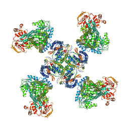 | | Structure of a Pancreatic ATP-sensitive Potassium Channel | | 分子名称: | ATP-sensitive inward rectifier potassium channel 11,superfolder GFP, SUR1 | | 著者 | Li, N, Wu, J.-X, Chen, L, Gao, N. | | 登録日 | 2016-12-16 | | 公開日 | 2017-01-25 | | 実験手法 | ELECTRON MICROSCOPY (5.6 Å) | | 主引用文献 | Structure of a Pancreatic ATP-Sensitive Potassium Channel
Cell, 168, 2017
|
|
5WTS
 | |
5WJ4
 | |
5WJ3
 | |
5WJ2
 | |
5WD6
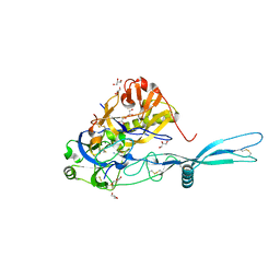
 | | bovine salivary protein form 30b | | 分子名称: | CALCIUM ION, Short palate, lung and nasal epithelium carcinoma-associated protein 2B | | 著者 | Zhang, H, Arcus, V.L. | | 登録日 | 2017-07-04 | | 公開日 | 2018-07-11 | | 最終更新日 | 2019-07-24 | | 実験手法 | X-RAY DIFFRACTION (2 Å) | | 主引用文献 | The three dimensional structure of Bovine Salivary Protein 30b (BSP30b) and its interaction with specific rumen bacteria.
Plos One, 14, 2019
|
|
5T3I
 | |
5OYL
 | | VSV G CR2 | | 分子名称: | 2-acetamido-2-deoxy-beta-D-glucopyranose, CALCIUM ION, GLYCEROL, ... | | 著者 | Albertini, A.A, Belot, L, Legrand, P, Gaudin, Y. | | 登録日 | 2017-09-11 | | 公開日 | 2018-03-21 | | 最終更新日 | 2024-01-17 | | 実験手法 | X-RAY DIFFRACTION (2.25 Å) | | 主引用文献 | Structural basis for the recognition of LDL-receptor family members by VSV glycoprotein.
Nat Commun, 9, 2018
|
|
5OXC
 | | Structure of Cerulean Fluorescent Protein at 1.02 Angstrom resolution | | 分子名称: | Green fluorescent protein | | 著者 | Gotthard, G, von Stetten, D, Clavel, D, Noirclerc-Savoye, M, Royant, A. | | 登録日 | 2017-09-06 | | 公開日 | 2017-11-29 | | 最終更新日 | 2024-01-17 | | 実験手法 | X-RAY DIFFRACTION (1.02 Å) | | 主引用文献 | Chromophore Isomer Stabilization Is Critical to the Efficient Fluorescence of Cyan Fluorescent Proteins.
Biochemistry, 56, 2017
|
|
5OXB
 | | Structure of blue-light irradiated Cerulean | | 分子名称: | Green fluorescent protein | | 著者 | Gotthard, G, von Stetten, D, Clavel, D, Noirclerc-Savoye, M, Royant, A. | | 登録日 | 2017-09-06 | | 公開日 | 2017-11-29 | | 最終更新日 | 2024-01-17 | | 実験手法 | X-RAY DIFFRACTION (1.38 Å) | | 主引用文献 | Chromophore Isomer Stabilization Is Critical to the Efficient Fluorescence of Cyan Fluorescent Proteins.
Biochemistry, 56, 2017
|
|
5OXA
 | | Structure of the S205A mutant of the Cyan Fluorescent Protein Cerulean at pH 7.0 | | 分子名称: | Green fluorescent protein | | 著者 | Gotthard, G, von Stetten, D, Clavel, D, Noirclerc-Savoye, M, Royant, A. | | 登録日 | 2017-09-06 | | 公開日 | 2017-11-29 | | 最終更新日 | 2024-01-17 | | 実験手法 | X-RAY DIFFRACTION (1.16 Å) | | 主引用文献 | Chromophore Isomer Stabilization Is Critical to the Efficient Fluorescence of Cyan Fluorescent Proteins.
Biochemistry, 56, 2017
|
|


