8GJV
 
 | |
1F1F
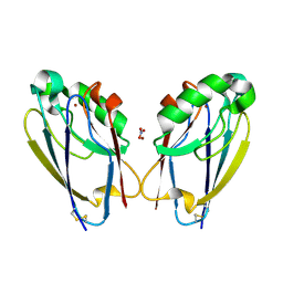
 | | CRYSTAL STRUCTURE OF CYTOCHROME C6 FROM ARTHROSPIRA MAXIMA | | 分子名称: | CYTOCHROME C6, HEME C | | 著者 | Kerfeld, C.A, Serag, A.A, Sawaya, M.R, Krogmann, D.W, Yeates, T.O. | | 登録日 | 2000-05-18 | | 公開日 | 2001-08-08 | | 最終更新日 | 2021-03-03 | | 実験手法 | X-RAY DIFFRACTION (2.7 Å) | | 主引用文献 | Structures of cytochrome c-549 and cytochrome c6 from the cyanobacterium Arthrospira maxima.
Biochemistry, 40, 2001
|
|
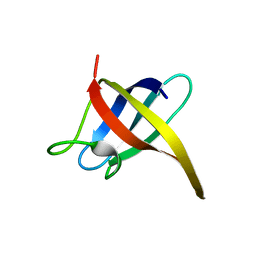
1HYM
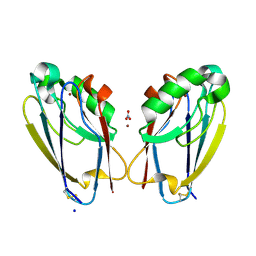
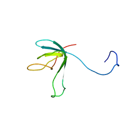
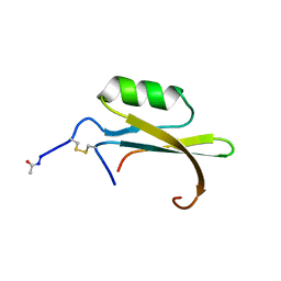 | | HYDROLYZED TRYPSIN INHIBITOR (CMTI-V, MINIMIZED AVERAGE NMR STRUCTURE) | | 分子名称: | HYDROLYZED CUCURBITA MAXIMA TRYPSIN INHIBITOR V | | 著者 | Cai, M, Gong, Y, Prakash, O, Krishnamoorthi, R. | | 登録日 | 1995-06-12 | | 公開日 | 1995-09-15 | | 最終更新日 | 2017-11-29 | | 実験手法 | SOLUTION NMR | | 主引用文献 | Reactive-site hydrolyzed Cucurbita maxima trypsin inhibitor-V: function, thermodynamic stability, and NMR solution structure.
Biochemistry, 34, 1995
|
|
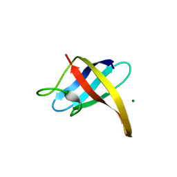
1KIB
 | | cytochrome c6 from Arthrospira maxima: an assembly of 24 subunits in the form of an oblate shell | | 分子名称: | HEME C, cytochrome c6 | | 著者 | Kerfeld, C.A, Sawaya, M.R, Krogmann, D, Yeates, T.O. | | 登録日 | 2001-12-03 | | 公開日 | 2002-07-03 | | 最終更新日 | 2023-08-16 | | 実験手法 | X-RAY DIFFRACTION (3.5 Å) | | 主引用文献 | Structure of cytochrome c6 from Arthrospira maxima: an assembly of 24 subunits in a nearly symmetric shell.
Acta Crystallogr.,Sect.D, 58, 2002
|
|
1TIN
 | |
8OTT
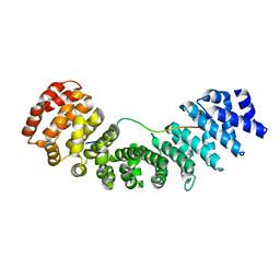
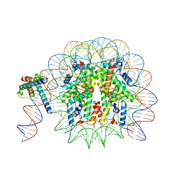 | | MYC-MAX bound to a nucleosome at SHL+5.8 | | 分子名称: | DNA (144-MER), Histone H2A type 1-B/E, Histone H2A type 1-K, ... | | 著者 | Stoos, L, Michael, A.K, Kempf, G, Kater, L, Cavadini, S, Thoma, N. | | 登録日 | 2023-04-21 | | 公開日 | 2023-05-24 | | 最終更新日 | 2023-09-06 | | 実験手法 | ELECTRON MICROSCOPY (3.3 Å) | | 主引用文献 | Cooperation between bHLH transcription factors and histones for DNA access.
Nature, 619, 2023
|
|
2I5L
 | |
2I5M
 | |
2HAX
 | |
2ES2
 | |
7TC5
 | | All Phe-Azurin variant - F15Y | | 分子名称: | Azurin, COPPER (II) ION, NITRATE ION, ... | | 著者 | Fedoretz-Maxwell, B.P, Worrall, L.J, Strynadka, N.C.J, Warren, J.J. | | 登録日 | 2021-12-22 | | 公開日 | 2022-06-22 | | 最終更新日 | 2023-10-18 | | 実験手法 | X-RAY DIFFRACTION (1.45 Å) | | 主引用文献 | The Impact of Second Coordination Sphere Methionine-Aromatic Interactions in Copper Proteins.
Inorg.Chem., 61, 2022
|
|
8FZM
 | |
8GAI
 | |
7TC6
 | | All Phe-Azurin variant - F15W | | 分子名称: | Azurin, COPPER (II) ION, NITRATE ION | | 著者 | Fedoretz-Maxwell, B.P, Worrall, L.J, Strynadka, N.C.J, Warren, J.J. | | 登録日 | 2021-12-22 | | 公開日 | 2022-06-22 | | 最終更新日 | 2023-10-18 | | 実験手法 | X-RAY DIFFRACTION (1.85 Å) | | 主引用文献 | The Impact of Second Coordination Sphere Methionine-Aromatic Interactions in Copper Proteins.
Inorg.Chem., 61, 2022
|
|
7N8J
 | |
1FCG
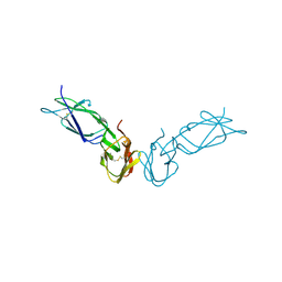 | | ECTODOMAIN OF HUMAN FC GAMMA RECEPTOR, FCGRIIA | | 分子名称: | PROTEIN (FC RECEPTOR FC(GAMMA)RIIA) | | 著者 | Maxwell, K.F, Powell, M.S, Garrett, T.P, Hogarth, P.M. | | 登録日 | 1999-04-07 | | 公開日 | 2000-04-12 | | 最終更新日 | 2023-12-27 | | 実験手法 | X-RAY DIFFRACTION (2 Å) | | 主引用文献 | Crystal structure of the human leukocyte Fc receptor, Fc gammaRIIa.
Nat.Struct.Biol., 6, 1999
|
|
7RZ3
 | |
1HYW
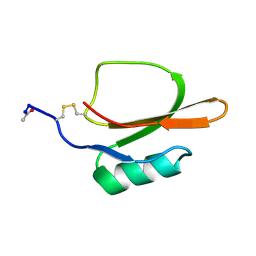
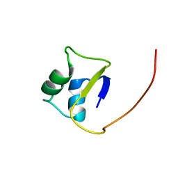
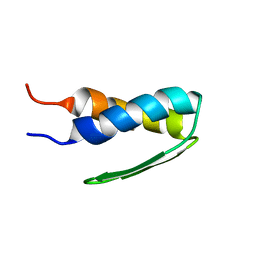 | | SOLUTION STRUCTURE OF BACTERIOPHAGE LAMBDA GPW | | 分子名称: | HEAD-TO-TAIL JOINING PROTEIN W | | 著者 | Maxwell, K.L, Yee, A.A, Booth, V, Arrowsmith, C.H, Gold, M, Davidson, A.R. | | 登録日 | 2001-01-22 | | 公開日 | 2001-04-25 | | 最終更新日 | 2024-05-29 | | 実験手法 | SOLUTION NMR | | 主引用文献 | The solution structure of bacteriophage lambda protein W, a small morphogenetic protein possessing a novel fold.
J.Mol.Biol., 308, 2001
|
|
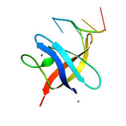
1K0H
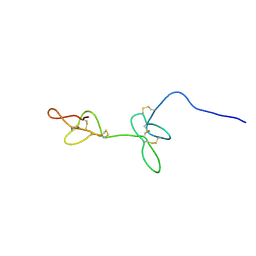
 | | Solution structure of bacteriophage lambda gpFII | | 分子名称: | gpFII | | 著者 | Maxwell, K.L, Yee, A.A, Arrowsmith, C.H, Gold, M, Davidson, A.R. | | 登録日 | 2001-09-19 | | 公開日 | 2002-07-17 | | 最終更新日 | 2024-05-29 | | 実験手法 | SOLUTION NMR | | 主引用文献 | The solution structure of the bacteriophage lambda head-tail joining protein, gpFII.
J.Mol.Biol., 318, 2002
|
|
2LTF
 | |
2BCX
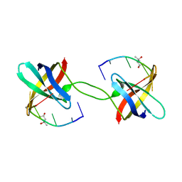
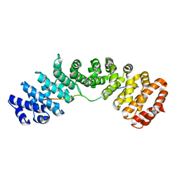
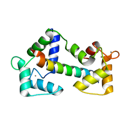 | | Crystal structure of calmodulin in complex with a ryanodine receptor peptide | | 分子名称: | CALCIUM ION, Calmodulin, Ryanodine receptor 1 | | 著者 | Maximciuc, A.A, Shamoo, Y, MacKenzie, K.R. | | 登録日 | 2005-10-19 | | 公開日 | 2006-10-31 | | 最終更新日 | 2024-02-14 | | 実験手法 | X-RAY DIFFRACTION (2 Å) | | 主引用文献 | Complex of calmodulin with a ryanodine receptor target reveals a novel, flexible binding mode.
Structure, 14, 2006
|
|
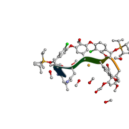
8JRD
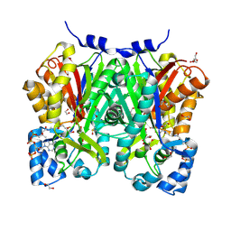 | | Chalcone synthase from Glycine max (L.) Merr (soybean) complexed with naringenin and coenzyme A | | 分子名称: | 1,2-ETHANEDIOL, COENZYME A, DI(HYDROXYETHYL)ETHER, ... | | 著者 | Waki, T, Imaizumi, R, Nakata, S, Yanai, T, Takeshita, K, Sakai, N, Kataoka, K, Yamamoto, M, Nakayama, T, Yamashita, S. | | 登録日 | 2023-06-16 | | 公開日 | 2024-06-19 | | 実験手法 | X-RAY DIFFRACTION (1.83 Å) | | 主引用文献 | Chalcone synthase from Glycine max (L.) Merr (soybean) complexed with naringenin and coenzyme A
To Be Published
|
|
8OTS
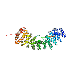
 | | OCT4 and MYC-MAX co-bound to a nucleosome | | 分子名称: | DNA (127-MER), Green fluorescent protein,POU domain, class 5, ... | | 著者 | Michael, A.K, Stoos, L, Kempf, G, Cavadini, S, Thoma, N. | | 登録日 | 2023-04-21 | | 公開日 | 2023-05-24 | | 最終更新日 | 2023-07-26 | | 実験手法 | ELECTRON MICROSCOPY (3.3 Å) | | 主引用文献 | Cooperation between bHLH transcription factors and histones for DNA access.
Nature, 619, 2023
|
|
5ROJ
 | | PanDDA analysis group deposition -- Proteinase K crystal structure Apo59 | | 分子名称: | Proteinase K, SULFATE ION | | 著者 | Lima, G.M.A, Talibov, V, Benz, L.S, Jagudin, E, Mueller, U. | | 登録日 | 2020-09-23 | | 公開日 | 2021-05-26 | | 最終更新日 | 2021-06-23 | | 実験手法 | X-RAY DIFFRACTION (1.02 Å) | | 主引用文献 | FragMAXapp: crystallographic fragment-screening data-analysis and project-management system.
Acta Crystallogr D Struct Biol, 77, 2021
|
|
5ROE
 | | PanDDA analysis group deposition -- Proteinase K crystal structure Apo6 | | 分子名称: | Proteinase K, SULFATE ION | | 著者 | Lima, G.M.A, Talibov, V, Benz, L.S, Jagudin, E, Mueller, U. | | 登録日 | 2020-09-23 | | 公開日 | 2021-05-26 | | 最終更新日 | 2021-06-23 | | 実験手法 | X-RAY DIFFRACTION (1.5 Å) | | 主引用文献 | FragMAXapp: crystallographic fragment-screening data-analysis and project-management system.
Acta Crystallogr D Struct Biol, 77, 2021
|
|


