Loading
PDBj
メニューPDBj@FacebookPDBj@X(formerly Twitter)PDBj@BlueSkyPDBj@YouTubewwPDB FoundationwwPDBDonate
RCSB PDBPDBeBMRBAdv. SearchSearch help

7A3B
DownloadVisualize
BU of 7a3b by Molmil
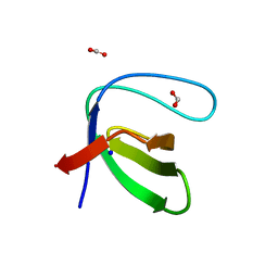
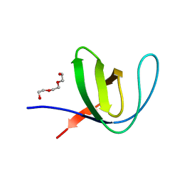
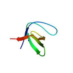
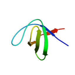
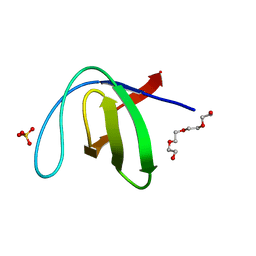
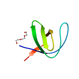
Intertwined dimer of the c-Src SH3 domain mutant E106D
分子名称: GLYCEROL, Proto-oncogene tyrosine-protein kinase Src, TRIETHYLENE GLYCOL
著者Camara-Artigas, A, Plaza-Garrido, M, Salinas-Garcia, M.C.
登録日2020-08-18
公開日2021-08-25
最終更新日2024-01-31
実験手法X-RAY DIFFRACTION (1.91 Å)
主引用文献Intertwined dimer of the c-Src SH3 domain mutant E106D
To be published
5LMH
DownloadVisualize
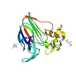
BU of 5lmh by Molmil
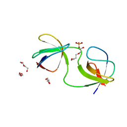
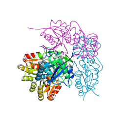
High dose Thaumatin - 160-200 ms.
分子名称: L(+)-TARTARIC ACID, Thaumatin-1
著者Schubert, R, Kapis, S, Heymann, M, Giquel, Y, Bourenkov, G, Schneider, T, Betzel, C, Perbandt, M.
登録日2016-07-30
公開日2016-11-09
最終更新日2024-10-16
実験手法X-RAY DIFFRACTION (1.96 Å)
主引用文献A multicrystal diffraction data-collection approach for studying structural dynamics with millisecond temporal resolution.
IUCrJ, 3, 2016
5LNT
DownloadVisualize
BU of 5lnt by Molmil
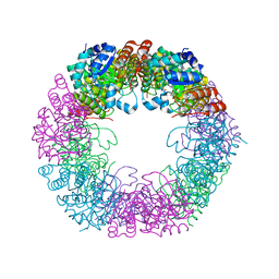
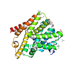
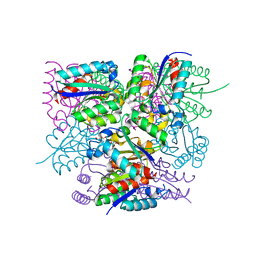
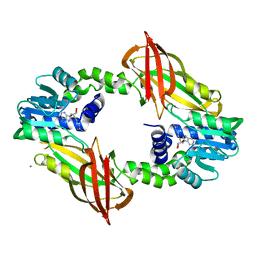
Crystal structure of Arabidopsis thaliana Pdx1K166R-preI320 complex
分子名称: PHOSPHATE ION, Pyridoxal 5'-phosphate synthase subunit PDX1.1, [(~{E},4~{S})-4-azanyl-3-oxidanylidene-pent-1-enyl] dihydrogen phosphate
著者Rodrigues, M.J, Windeisen, V, Zhang, Y, Guedez, G, Weber, S, Strohmeier, M, Hanes, J.W, Royant, A, Evans, G, Sinning, I, Ealick, S.E, Begley, T.P, Tews, I.
登録日2016-08-06
公開日2017-01-18
最終更新日2024-10-16
実験手法X-RAY DIFFRACTION (2.32 Å)
主引用文献Lysine relay mechanism coordinates intermediate transfer in vitamin B6 biosynthesis.
Nat. Chem. Biol., 13, 2017
7A2L
DownloadVisualize
BU of 7a2l by Molmil
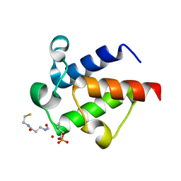
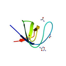
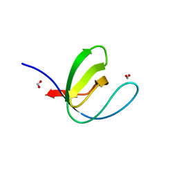
Crystal structure of the Fyn SH3 domain mutant E129Q in space group C21 at pH 4.0
分子名称: FORMIC ACID, SODIUM ION, Tyrosine-protein kinase Fyn
著者Camara-Artigas, A, Plaza-Garrido, M, Salinas-Garcia, M.C.
登録日2020-08-18
公開日2021-08-25
最終更新日2024-01-31
実験手法X-RAY DIFFRACTION (1.9 Å)
主引用文献Crystal structure of the Fyn SH3 domain mutant E129Q in space group C21 at pH 4.0
To be published
6SL4
DownloadVisualize
BU of 6sl4 by Molmil
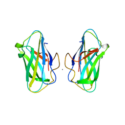
The unique CBM-Cthe_0271 of Ruminiclostridium thermocellum
分子名称: CALCIUM ION, Type 3a cellulose-binding domain protein
著者Milana, M.V, Almog, R, Yaniv, O, Oded, L, Inna, R.G, Felix, F, Edward, A.B, Raphael, L.
登録日2019-08-18
公開日2020-03-18
最終更新日2024-05-15
実験手法X-RAY DIFFRACTION (1.8 Å)
主引用文献The unique CBM-Cthe_0271 of Ruminiclostridium thermocellum
To Be Published
7A2U
DownloadVisualize
BU of 7a2u by Molmil
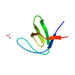
Crystal structure of the Fyn SH3 domain L112V-S114N-S115T at pH 4.5
分子名称: ACETATE ION, Tyrosine-protein kinase Fyn
著者Camara-Artigas, A, Plaza-Garrido, M, Salinas-Garcia, M.C.
登録日2020-08-18
公開日2021-08-25
最終更新日2024-01-31
実験手法X-RAY DIFFRACTION (1.7 Å)
主引用文献Crystal structure of the Fyn SH3 domain L112V-S114N-S115T at pH 4.5
To be published
7A2O
DownloadVisualize
BU of 7a2o by Molmil
Crystal structure of the Fyn SH3 domain L112V-S114N-S115T-E121L-R123H mutant at pH 4.5
分子名称: TRIETHYLENE GLYCOL, Tyrosine-protein kinase Fyn
著者Camara-Artigas, A, Plaza-Garrido, M, Salinas-Garcia, M.C.
登録日2020-08-18
公開日2021-08-25
最終更新日2024-01-31
実験手法X-RAY DIFFRACTION (0.94 Å)
主引用文献Crystal structure of the Fyn SH3 domain L112V-S114N-S115T-E121L-R123H mutant at pH 4.5
To be published
7A3D
DownloadVisualize
BU of 7a3d by Molmil
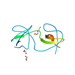
Intertwined dimer of the c-Src SH3 domain mutant T125S
分子名称: DI(HYDROXYETHYL)ETHER, GLYCEROL, Proto-oncogene tyrosine-protein kinase Src, ...
著者Camara-Artigas, A, Plaza-Garrido, M, Salinas-Garcia, M.C.
登録日2020-08-18
公開日2021-08-25
最終更新日2024-01-31
実験手法X-RAY DIFFRACTION (2.2 Å)
主引用文献Intertwined dimer of the c-Src SH3 domain mutant T125S
To be published
7A2F
DownloadVisualize
BU of 7a2f by Molmil
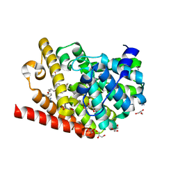
Crystal structure of T. brucei PDE-B1 catalytic domain with inhibitor NPD-656
分子名称: 1-cycloheptyl-3-{4-methoxy-3-[2-(4-methoxyphenyl)ethoxy]phenyl}-4,4-dimethyl-4,5-dihydro-1H-pyrazol-5-one, FORMIC ACID, GLYCEROL, ...
著者Singh, A.K, Blaazer, A.R, Zara, L, de Esch, I.J.P, Leurs, R, Brown, D.G.
登録日2020-08-17
公開日2021-08-25
最終更新日2024-01-31
実験手法X-RAY DIFFRACTION (2 Å)
主引用文献Crystal structure of T. brucei PDE-B1 catalytic domain with inhibitor NPD-656
To be published
6SM6
DownloadVisualize
BU of 6sm6 by Molmil
AntF (holo): type II PKS acyl-carrier protein
分子名称: Acyl carrier protein
著者Braeuer, A, Zhou, Q, Grammbitter, G.L.C, Schmalhofer, M, Ruehl, M, Kaila, V.R.I, Bode, H, Groll, M.
登録日2019-08-21
公開日2020-05-27
最終更新日2024-01-24
実験手法X-RAY DIFFRACTION (2.4 Å)
主引用文献Structural snapshots of the minimal PKS system responsible for octaketide biosynthesis.
Nat.Chem., 12, 2020
5M1A
DownloadVisualize
BU of 5m1a by Molmil
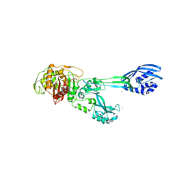
Crystal structure of PBP2a from MRSA in the presence of Ceftazidime ligand
分子名称: CADMIUM ION, Penicillin-binding protein 2, beta-muramic acid
著者Molina, R, Batuecas, M.T, Hermoso, J.A.
登録日2016-10-07
公開日2017-02-08
最終更新日2024-01-17
実験手法X-RAY DIFFRACTION (2 Å)
主引用文献Conformational Dynamics in Penicillin-Binding Protein 2a of Methicillin-Resistant Staphylococcus aureus, Allosteric Communication Network and Enablement of Catalysis.
J. Am. Chem. Soc., 139, 2017
6SME
DownloadVisualize
BU of 6sme by Molmil
THE CRYSTAL STRUCTURE OF TYPE II DEHYDROQUINASE FROM PROPIONIBACTERIUM ACNES
分子名称: 3-dehydroquinate dehydratase, GLYCEROL, SULFATE ION
著者Lapthorn, A.J, Roszak, A.W.
登録日2019-08-21
公開日2020-07-08
最終更新日2024-01-24
実験手法X-RAY DIFFRACTION (1.7 Å)
主引用文献Biomacromolecular charge chirality detected using chiral plasmonic nanostructures
Nanoscale Horiz., 2020
6SOP
DownloadVisualize
BU of 6sop by Molmil
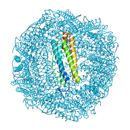
Metal free structure of SynFtn variant E62A
分子名称: CHLORIDE ION, Ferritin
著者Hemmings, A.M, Bradley, J.M.
登録日2019-08-29
公開日2020-01-29
最終更新日2024-01-24
実験手法X-RAY DIFFRACTION (1.93 Å)
主引用文献Routes of iron entry into, and exit from, the catalytic ferroxidase sites of the prokaryotic ferritin SynFtn.
Dalton Trans, 49, 2020
7A2J
DownloadVisualize
BU of 7a2j by Molmil
Crystal structure of the Fyn SH3 domain in space group C21 at pH 7.5
分子名称: SODIUM ION, Tyrosine-protein kinase Fyn
著者Camara-Artigas, A, Plaza-Garrido, M, Salinas-Garcia, M.C.
登録日2020-08-18
公開日2021-08-25
最終更新日2024-01-31
実験手法X-RAY DIFFRACTION (1.5 Å)
主引用文献Crystal structure of the Fyn SH3 domain in space group C21 at pH 7.5
To be published
7A2V
DownloadVisualize
BU of 7a2v by Molmil
Crystal structure of the Fyn SH3 domain L112V-S114N-S115T-E129Q at pH 4.0
分子名称: Tyrosine-protein kinase Fyn
著者Camara-Artigas, A, Plaza-Garrido, M, Salinas-Garcia, M.C.
登録日2020-08-18
公開日2021-08-25
最終更新日2024-01-31
実験手法X-RAY DIFFRACTION (1.81 Å)
主引用文献Crystal structure of the Fyn SH3 domain L112V-S114N-S115T-E129Q at pH 4.0
To be published
7A37
DownloadVisualize
BU of 7a37 by Molmil
Crystal structure of the c-Src SH3 domain mutant V111L-N113S-T114S in 3 M urea
分子名称: Proto-oncogene tyrosine-protein kinase Src, UREA
著者Camara-Artigas, A, Plaza-Garrido, M, Salinas-Garcia, M.C.
登録日2020-08-18
公開日2021-08-25
最終更新日2024-01-31
実験手法X-RAY DIFFRACTION (1.52 Å)
主引用文献Crystal structure of the c-Src SH3 domain mutant V111L-N113S-T114S in 3 M urea
To be published
7A28
DownloadVisualize
BU of 7a28 by Molmil
Crystal structure of T. brucei PDE-B1 catalytic domain with inhibitor NPD-617
分子名称: 1-cycloheptyl-3-(4-methoxy-3-{4-[4-(1H-1,2,3,4-tetrazol-5-yl)phenoxy]butoxy}phenyl)-4,4-dimethyl-4,5-dihydro-1H-pyrazol-5-one, DI(HYDROXYETHYL)ETHER, GLYCEROL, ...
著者Singh, A.K, Blaazer, A.R, Zara, L, de Esch, I.J.P, Leurs, R, Brown, D.G.
登録日2020-08-16
公開日2021-08-25
最終更新日2024-01-31
実験手法X-RAY DIFFRACTION (1.89 Å)
主引用文献Crystal structure of T. brucei PDE-B1 catalytic domain with inhibitor NPD-617
To be published
6SPH
DownloadVisualize
BU of 6sph by Molmil
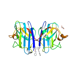
A4V MUTANT OF HUMAN SUPEROXIDE DISMUTASE 1 WITH EBSELEN BOND IN C2 SPACE GROUP
分子名称: DIMETHYL SULFOXIDE, GLYCEROL, N-phenyl-2-selanylbenzamide, ...
著者Shahid, M, Chantadul, V, Amporndanai, K, Wright, G, Antonyuk, S, Hasnain, S.
登録日2019-09-01
公開日2020-03-18
最終更新日2024-11-13
実験手法X-RAY DIFFRACTION (2.25 Å)
主引用文献Ebselen as template for stabilization of A4V mutant dimer for motor neuron disease therapy.
Commun Biol, 3, 2020
7A2Q
DownloadVisualize
BU of 7a2q by Molmil
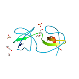
Crystal structure of the Fyn SH3 domain L112V-S114N-S115T-E121L-R123H mutant at pH 3.0 with PEG
分子名称: SULFATE ION, TETRAETHYLENE GLYCOL, Tyrosine-protein kinase Fyn
著者Camara-Artigas, A, Plaza-Garrido, M, Salinas-Garcia, M.C.
登録日2020-08-18
公開日2021-08-25
最終更新日2024-01-31
実験手法X-RAY DIFFRACTION (0.94 Å)
主引用文献Crystal structure of the Fyn SH3 domain L112V-S114N-S115T-E121L-R123H mutant at pH 3.0 with PEG
To be published
7A33
DownloadVisualize
BU of 7a33 by Molmil
Crystal structure of the c-Src SH3 domain mutant S94A-T98D-V111L-N113S-T114S at pH 3.0
分子名称: FORMIC ACID, Proto-oncogene tyrosine-protein kinase Src, SODIUM ION
著者Camara-Artigas, A, Plaza-Garrido, M, Salinas-Garcia, M.C.
登録日2020-08-18
公開日2021-08-25
最終更新日2024-01-31
実験手法X-RAY DIFFRACTION (0.96 Å)
主引用文献Crystal structure of the c-Src SH3 domain mutant S94A-T98D-V111L-N113S-T114S at pH 3.0
To be published
7A2W
DownloadVisualize
BU of 7a2w by Molmil
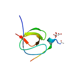
Crystal structure of the Fyn SH3 domain L112V-S114N-S115T-E121L-R123H mutant in complex with VSL12 at pH 3.0
分子名称: CITRIC ACID, Tyrosine-protein kinase Fyn, VSL12
著者Camara-Artigas, A, Plaza-Garrido, M, Salinas-Garcia, M.C.
登録日2020-08-18
公開日2021-08-25
最終更新日2024-10-23
実験手法X-RAY DIFFRACTION (0.99 Å)
主引用文献Crystal structure of the Fyn SH3 domain L112V-S114N-S115T-E121L-R123H mutant in complex with VSL12 at pH 3.0
To be published
7A3A
DownloadVisualize
BU of 7a3a by Molmil
Intertwined dimer of the c-Src SH3 domain mutant V111L-N113S-T114S-Q128E at pH 6.0
分子名称: 1,2-ETHANEDIOL, DI(HYDROXYETHYL)ETHER, Proto-oncogene tyrosine-protein kinase Src, ...
著者Camara-Artigas, A, Plaza-Garrido, M, Salinas-Garcia, M.C.
登録日2020-08-18
公開日2021-08-25
最終更新日2024-01-31
実験手法X-RAY DIFFRACTION (1.8 Å)
主引用文献Intertwined dimer of the c-Src SH3 domain mutant V111L-N113S-T114S-Q128E at pH 6.0
To be published
6SQ4
DownloadVisualize
BU of 6sq4 by Molmil
Crystal structure of mouse PRMT6 in complex with inhibitor U2
分子名称: 9-(7-{[amino(iminio)methyl]amino}-5,6,7-trideoxy-beta-D-ribo-heptofuranosyl)-9H-purin-6-amine, IODIDE ION, Protein arginine N-methyltransferase 6
著者Bonnefond, L, Cavarelli, J.
登録日2019-09-03
公開日2020-09-30
最終更新日2024-05-15
実験手法X-RAY DIFFRACTION (1.7 Å)
主引用文献Crystal structure of mouse PRMT6 in complex with inhibitors
To be published
7A0U
DownloadVisualize
BU of 7a0u by Molmil
LIGAND FREE TYPE II E. COLI ASPARAGINASE T12S/T89S DOUBLE MUTANT
分子名称: L-asparaginase 2
著者Maggi, M, Scotti, C.
登録日2020-08-10
公開日2021-08-18
最終更新日2024-11-06
実験手法X-RAY DIFFRACTION (2.260487 Å)
主引用文献LIGAND FREE TYPE II E. COLI ASPARAGINASE T12S/T89S DOUBLE MUTANT
To Be Published
7A2P
DownloadVisualize
BU of 7a2p by Molmil
Crystal structure of the Fyn SH3 domain L112V-S114N-S115T-E121L-R123H mutant at pH 5.0 with PEG
分子名称: TRIETHYLENE GLYCOL, Tyrosine-protein kinase Fyn
著者Camara-Artigas, A, Plaza-Garrido, M, Salinas-Garcia, M.C.
登録日2020-08-18
公開日2021-08-25
最終更新日2024-01-31
実験手法X-RAY DIFFRACTION (0.9 Å)
主引用文献Crystal structure of the Fyn SH3 domain L112V-S114N-S115T-E121L-R123H mutant at pH 5.0 with PEG
To be published

243911

件を2025-10-29に公開中

PDB statisticsPDBj update infoContact PDBjnumon