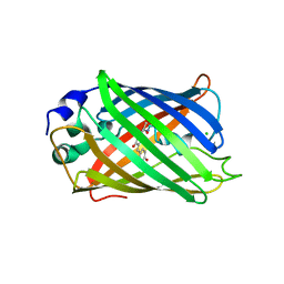
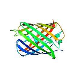
5MA6
 
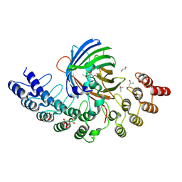
 | | GFP-binding DARPin 3G124nc | | 分子名称: | 1,2-ETHANEDIOL, 3G124nc, Green fluorescent protein, ... | | 著者 | Hansen, S, Stueber, J, Ernst, P, Koch, A, Bojar, D, Batyuk, A, Plueckthun, A. | | 登録日 | 2016-11-03 | | 公開日 | 2017-12-06 | | 実験手法 | X-RAY DIFFRACTION (2.3 Å) | | 主引用文献 | Design and applications of a clamp for Green Fluorescent Protein with picomolar affinity.
Sci Rep, 7, 2017
|
|
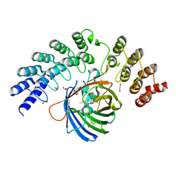
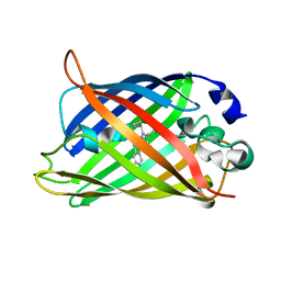
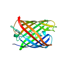
5MA5
 | | GFP-binding DARPin fusion gc_K11 | | 分子名称: | 1,2-ETHANEDIOL, CITRIC ACID, Green fluorescent protein, ... | | 著者 | Hansen, S, Stueber, J, Ernst, P, Koch, A, Bojar, D, Batyuk, A, Plueckthun, A. | | 登録日 | 2016-11-03 | | 公開日 | 2017-11-08 | | 最終更新日 | 2023-11-15 | | 実験手法 | X-RAY DIFFRACTION (1.85 Å) | | 主引用文献 | Design and applications of a clamp for Green Fluorescent Protein with picomolar affinity.
Sci Rep, 7, 2017
|
|
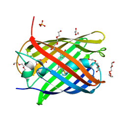
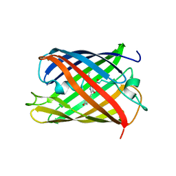
5MA4
 | | GFP-binding DARPin fusion gc_K7 | | 分子名称: | Green fluorescent protein, K7 | | 著者 | Hansen, S, Stueber, J, Ernst, P, Koch, A, Bojar, D, Batyuk, A, Plueckthun, A. | | 登録日 | 2016-11-03 | | 公開日 | 2017-11-08 | | 最終更新日 | 2023-11-15 | | 実験手法 | X-RAY DIFFRACTION (1.4 Å) | | 主引用文献 | Design and applications of a clamp for Green Fluorescent Protein with picomolar affinity.
Sci Rep, 7, 2017
|
|
5MA3
 | | GFP-binding DARPin fusion gc_R11 | | 分子名称: | 1,2-ETHANEDIOL, Green fluorescent protein, R11 | | 著者 | Hansen, S, Stueber, J, Ernst, P, Bojar, D, Batyuk, A, Plueckthun, A. | | 登録日 | 2016-11-03 | | 公開日 | 2017-11-08 | | 最終更新日 | 2023-11-15 | | 実験手法 | X-RAY DIFFRACTION (1.7 Å) | | 主引用文献 | Design and applications of a clamp for Green Fluorescent Protein with picomolar affinity.
Sci Rep, 7, 2017
|
|
5LTR
 | |
5LTQ
 | |
5LTP
 | |
5LK4
 | | Structure of the Red Fluorescent Protein mScarlet at pH 7.8 | | 分子名称: | DI(HYDROXYETHYL)ETHER, PHOSPHATE ION, TETRAETHYLENE GLYCOL, ... | | 著者 | Aumonier, S, Gotthard, G, Royant, A. | | 登録日 | 2016-07-20 | | 公開日 | 2016-12-07 | | 最終更新日 | 2024-01-31 | | 実験手法 | X-RAY DIFFRACTION (1.47 Å) | | 主引用文献 | mScarlet: a bright monomeric red fluorescent protein for cellular imaging.
Nat. Methods, 14, 2017
|
|
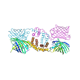
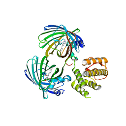
5LEM
 | | Crystal structure of DARPin-DARPin rigid fusion, variant DD_Off7_11_3G124 in complex with Maltose-binding Protein and Green Fluorescent Protein | | 分子名称: | DD_Off7_11_3G124, Green fluorescent protein, Maltose-binding periplasmic protein | | 著者 | Batyuk, A, Wu, Y, Mittl, P.R, Plueckthun, A. | | 登録日 | 2016-06-30 | | 公開日 | 2017-08-02 | | 最終更新日 | 2019-10-16 | | 実験手法 | X-RAY DIFFRACTION (2.98 Å) | | 主引用文献 | Rigidly connected multispecific artificial binders with adjustable geometries.
Sci Rep, 7, 2017
|
|
5LEL
 | | Crystal structure of DARPin-DARPin rigid fusion, variant DD_Off7_10_3G124 in complex with Maltose-binding Protein and Green Fluorescent Protein | | 分子名称: | DD_Off7_10_3G124, Green fluorescent protein, Maltose-binding periplasmic protein | | 著者 | Batyuk, A, Wu, Y, Mittl, P.R, Plueckthun, A. | | 登録日 | 2016-06-30 | | 公開日 | 2017-11-15 | | 最終更新日 | 2019-10-16 | | 実験手法 | X-RAY DIFFRACTION (3.1 Å) | | 主引用文献 | Rigidly connected multispecific artificial binders with adjustable geometries.
Sci Rep, 7, 2017
|
|
5KTG
 | |
5JZL
 | | The Structure of Monomeric Ultra Stable Green Fluorescent Protein | | 分子名称: | CHLORIDE ION, Green fluorescent protein, SODIUM ION | | 著者 | Gunn, N.J, Yong, K.J, Scott, D.J, Griffin, M.D.W. | | 登録日 | 2016-05-17 | | 公開日 | 2017-12-06 | | 最終更新日 | 2023-11-15 | | 実験手法 | X-RAY DIFFRACTION (1.8 Å) | | 主引用文献 | A Novel Ultra-Stable, Monomeric Green Fluorescent Protein For Direct Volumetric Imaging of Whole Organs Using CLARITY.
Sci Rep, 8, 2018
|
|
5JZK
 | | The Structure of Ultra Stable Green Fluorescent Protein | | 分子名称: | 1,2-ETHANEDIOL, CHLORIDE ION, GLYCEROL, ... | | 著者 | Yong, K.J, Gunn, N.J, Scott, D.J, Griffin, M.D.W. | | 登録日 | 2016-05-17 | | 公開日 | 2017-12-06 | | 最終更新日 | 2023-11-15 | | 実験手法 | X-RAY DIFFRACTION (1.9 Å) | | 主引用文献 | A Novel Ultra-Stable, Monomeric Green Fluorescent Protein For Direct Volumetric Imaging of Whole Organs Using CLARITY.
Sci Rep, 8, 2018
|
|
5J3N
 | | C-terminal domain of EcoR124I HsdR subunit fused with the pH-sensitive GFP variant ratiometric pHluorin | | 分子名称: | Green fluorescent protein,HsdR | | 著者 | Grinkevich, P, Iermak, I, Luedtke, N, Mesters, J.R, Ettrich, R, Ludwig, J. | | 登録日 | 2016-03-31 | | 公開日 | 2017-04-12 | | 最終更新日 | 2024-01-10 | | 実験手法 | X-RAY DIFFRACTION (2.45 Å) | | 主引用文献 | Crystal structure of a novel domain of the motor subunit of the Type I restriction enzyme EcoR124 involved in complex assembly and DNA binding.
J. Biol. Chem., 293, 2018
|
|
5J2O
 | |
5HZU
 | | Crystal structure of Dronpa-Ni2+ | | 分子名称: | Fluorescent protein Dronpa, NICKEL (II) ION | | 著者 | Hwang, K.Y, Nam, K.H. | | 登録日 | 2016-02-03 | | 公開日 | 2017-03-15 | | 最終更新日 | 2023-11-15 | | 実験手法 | X-RAY DIFFRACTION (1.89 Å) | | 主引用文献 | Crystal structures of Dronpa complexed with quenchable metal ions provide insight into metal biosensor development
FEBS Lett., 590, 2016
|
|
5HZT
 | | Crystal structure of Dronpa-Cu2+ | | 分子名称: | COPPER (II) ION, Fluorescent protein Dronpa | | 著者 | Hwang, K.Y, Nam, K.H. | | 登録日 | 2016-02-03 | | 公開日 | 2017-03-15 | | 最終更新日 | 2023-11-15 | | 実験手法 | X-RAY DIFFRACTION (2.84 Å) | | 主引用文献 | Crystal structures of Dronpa complexed with quenchable metal ions provide insight into metal biosensor development
FEBS Lett., 590, 2016
|
|
5HZS
 | | Crystal structure of Dronpa-Co2+ | | 分子名称: | COBALT (II) ION, Fluorescent protein Dronpa | | 著者 | Hwang, K.Y, Nam, K.H. | | 登録日 | 2016-02-03 | | 公開日 | 2017-03-15 | | 最終更新日 | 2023-11-15 | | 実験手法 | X-RAY DIFFRACTION (2.17 Å) | | 主引用文献 | Crystal structures of Dronpa complexed with quenchable metal ions provide insight into metal biosensor development
FEBS Lett., 590, 2016
|
|
5HZO
 | | GFP mutant S205G | | 分子名称: | D-MALATE, Green fluorescent protein, UNDECYL-MALTOSIDE | | 著者 | Remington, S.J, Trujillo, K. | | 登録日 | 2016-02-02 | | 公開日 | 2016-02-17 | | 最終更新日 | 2023-11-15 | | 実験手法 | X-RAY DIFFRACTION (2.49 Å) | | 主引用文献 | Ultrafast Dynamics and Mechanisms of Proton Transfer in the GFP S205G Mutant
To Be Published
|
|
5HW9
 | | Filamentous Assembly of Green Fluorescent Protein Supported by a C-terminal fusion of 18-residues, viewed in space group P21 | | 分子名称: | Green fluorescent protein | | 著者 | Sawaya, M.R, Heller, D.M, McPartland, L, Hochschild, A, Eisenberg, D.S. | | 登録日 | 2016-01-29 | | 公開日 | 2017-02-01 | | 最終更新日 | 2023-11-15 | | 実験手法 | X-RAY DIFFRACTION (3 Å) | | 主引用文献 | Green Fluorescent Protein Fusion that Self Assembles as Polar Filaments
to be published
|
|
5HGE
 | | Filamentous Assembly of Green Fluorescent Protein Supported by a C-terminal fusion of 18-residues, viewed in space group P212121 | | 分子名称: | (4S)-2-METHYL-2,4-PENTANEDIOL, Green fluorescent protein | | 著者 | Sawaya, M.R, Heller, D.M, McPartland, L, Hochschild, A, Eisenberg, D.S. | | 登録日 | 2016-01-08 | | 公開日 | 2017-01-11 | | 最終更新日 | 2023-11-15 | | 実験手法 | X-RAY DIFFRACTION (1.863 Å) | | 主引用文献 | Green Fluorescent Protein Fusion that Self Assembles as Polar Filaments
to be published
|
|
5HBD
 | | Filamentous Assembly of Green Fluorescent Protein Supported by a C-terminal fusion of 18-residues, viewed in space group C2 | | 分子名称: | Green fluorescent protein | | 著者 | Sawaya, M.R, Hochschild, A, Heller, D.M, McPartland, L, Eisenberg, D.S. | | 登録日 | 2015-12-31 | | 公開日 | 2017-01-04 | | 最終更新日 | 2024-03-06 | | 実験手法 | X-RAY DIFFRACTION (1.65 Å) | | 主引用文献 | Green Fluorescent Protein Fusion that Self Assembles as Polar Filaments
to be published
|
|
5H89
 | | Crystal structure of mRojoA mutant - T16V - P63Y - W143G - L163V | | 分子名称: | mRojoA fluorescent protein | | 著者 | Pandelieva, A.T, Tremblay, V, Sarvan, S, Chica, R.A, Couture, J.-F. | | 登録日 | 2015-12-23 | | 公開日 | 2016-01-27 | | 最終更新日 | 2016-03-02 | | 実験手法 | X-RAY DIFFRACTION (1.76 Å) | | 主引用文献 | Brighter Red Fluorescent Proteins by Rational Design of Triple-Decker Motif.
Acs Chem.Biol., 11, 2016
|
|
5H88
 | | Crystal structure of mRojoA mutant - T16V -P63F - W143A - L163V | | 分子名称: | mRojoA fluorescent protein | | 著者 | Pandelieva, A.T, Tremblay, V, Sarvan, S, Chica, R.A, Couture, J.-F. | | 登録日 | 2015-12-23 | | 公開日 | 2016-01-27 | | 最終更新日 | 2016-03-02 | | 実験手法 | X-RAY DIFFRACTION (2.06 Å) | | 主引用文献 | Brighter Red Fluorescent Proteins by Rational Design of Triple-Decker Motif.
Acs Chem.Biol., 11, 2016
|
|
5H87
 | | Crystal structure of mRojoA mutant - P63H - W143S | | 分子名称: | mRojoA fluorescent protein | | 著者 | Pandelieva, A.T, Tremblay, V, Sarvan, S, Chica, R.A, Couture, J.-F. | | 登録日 | 2015-12-23 | | 公開日 | 2016-01-27 | | 最終更新日 | 2016-03-02 | | 実験手法 | X-RAY DIFFRACTION (2.24 Å) | | 主引用文献 | Brighter Red Fluorescent Proteins by Rational Design of Triple-Decker Motif.
Acs Chem.Biol., 11, 2016
|
|


