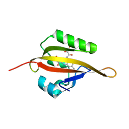
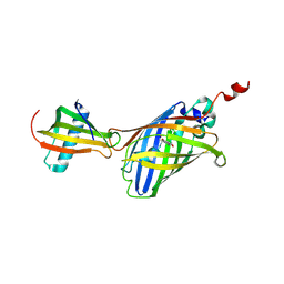
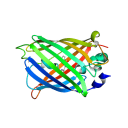
3FZ9
 
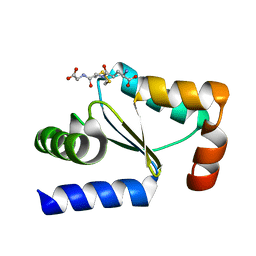 | | Crystal structure of poplar glutaredoxin S12 in complex with glutathione | | 分子名称: | GLUTATHIONE, Glutaredoxin | | 著者 | Didierjean, C, Corbier, C, Koh, C.S, Rouhier, N, Jacquot, J.P. | | 登録日 | 2009-01-24 | | 公開日 | 2009-02-24 | | 最終更新日 | 2023-09-06 | | 実験手法 | X-RAY DIFFRACTION (1.7 Å) | | 主引用文献 | Structure-function relationship of the chloroplastic glutaredoxin S12 with an atypical WCSYS active site.
J.Biol.Chem., 284, 2009
|
|
1QF9
 | |
2A52
 | | fluorescent protein asFP595, S158V, on-state | | 分子名称: | GFP-like non-fluorescent chromoprotein FP595 chain 1, GFP-like non-fluorescent chromoprotein FP595 chain 2 | | 著者 | Andresen, M, Wahl, M.C, Stiel, A.C, Graeter, F, Schaefer, L, Trowitzsch, S, Weber, G, Eggeling, C, Grubmueller, H, Hell, S.W, Jakobs, S. | | 登録日 | 2005-06-30 | | 公開日 | 2005-08-16 | | 最終更新日 | 2021-11-10 | | 実験手法 | X-RAY DIFFRACTION (1.7 Å) | | 主引用文献 | Structure and mechanism of the reversible photoswitch of a fluorescent protein
Proc.Natl.Acad.Sci.Usa, 102, 2005
|
|
4IR5
 | | Crystal Structure of the bromodomain of human BAZ2B in complex with 1-{1-[2-(hydroxymethyl)phenyl]-7-phenoxyindolizin-3-yl}ethanone (GSK2847449A) | | 分子名称: | 1,2-ETHANEDIOL, 1-{1-[2-(hydroxymethyl)phenyl]-7-phenoxyindolizin-3-yl}ethanone, Bromodomain adjacent to zinc finger domain protein 2B | | 著者 | Chaikuad, A, Felletar, I, Chung, C.W, Drewry, D, Chen, P, Filippakopoulos, P, Fedorov, O, Krojer, T, von Delft, F, Arrowsmith, C.H, Edwards, A.M, Bountra, C, Knapp, S, Structural Genomics Consortium (SGC) | | 登録日 | 2013-01-14 | | 公開日 | 2013-01-23 | | 最終更新日 | 2023-09-20 | | 実験手法 | X-RAY DIFFRACTION (1.7 Å) | | 主引用文献 | Discovery and Characterization of GSK2801, a Selective Chemical Probe for the Bromodomains BAZ2A and BAZ2B.
J.Med.Chem., 59, 2016
|
|
4EEP
 | | Crystal structure of LOV2 domain of Arabidopsis thaliana phototropin 2 | | 分子名称: | FLAVIN MONONUCLEOTIDE, Phototropin-2 | | 著者 | Hitomi, K, Christie, J.M, Arvai, A.S, Hartfield, K.A, Pratt, A.J, Tainer, J.A, Getzoff, E.D. | | 登録日 | 2012-03-28 | | 公開日 | 2012-05-16 | | 最終更新日 | 2023-09-13 | | 実験手法 | X-RAY DIFFRACTION (1.7 Å) | | 主引用文献 | Structural Tuning of the Fluorescent Protein iLOV for Improved Photostability.
J.Biol.Chem., 287, 2012
|
|
3SG6
 | | Crystal Structure of Dimeric GCaMP2-LIA(linker 1) | | 分子名称: | CALCIUM ION, Myosin light chain kinase, Green fluorescent protein, ... | | 著者 | Schreiter, E.R, Akerboom, J, Looger, L.L. | | 登録日 | 2011-06-14 | | 公開日 | 2012-06-20 | | 最終更新日 | 2023-12-06 | | 実験手法 | X-RAY DIFFRACTION (1.7 Å) | | 主引用文献 | Optimization of a GCaMP calcium indicator for neural activity imaging.
J.Neurosci., 32, 2012
|
|
2QU1
 | | Crystal Structure of a Cyclized GFP Variant | | 分子名称: | CALCIUM ION, Green fluorescent protein | | 著者 | Bailey, L.J, McCoy, J.G, Bitto, E, Bingman, C.A, Fox, B.G, Wesenberg, G.E, Phillips Jr, G.N, Center for Eukaryotic Structural Genomics (CESG) | | 登録日 | 2007-08-03 | | 公開日 | 2007-09-11 | | 最終更新日 | 2023-11-15 | | 実験手法 | X-RAY DIFFRACTION (1.7 Å) | | 主引用文献 | Crystal Structure of a Cyclized GFP Variant.
To be Published
|
|
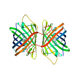
4W6J
 | | Crystal Structure of Full-Length Split GFP Mutant D117C Disulfide Dimer, P 31 2 1 Space Group | | 分子名称: | (4S)-2-METHYL-2,4-PENTANEDIOL, ACETATE ION, fluorescent protein D117C | | 著者 | Leibly, D.J, Waldo, G.S, Yeates, T.O. | | 登録日 | 2014-08-20 | | 公開日 | 2015-02-18 | | 最終更新日 | 2023-11-15 | | 実験手法 | X-RAY DIFFRACTION (1.702 Å) | | 主引用文献 | A Suite of Engineered GFP Molecules for Oligomeric Scaffolding.
Structure, 23, 2015
|
|
3ST3
 | | Dreiklang - off state | | 分子名称: | Dreiklang, PHOSPHATE ION | | 著者 | Brakemann, T, Weber, G, Andresen, M, Stiel, A.C, Jakobs, S, Wahl, M.C. | | 登録日 | 2011-07-08 | | 公開日 | 2011-09-14 | | 最終更新日 | 2017-11-08 | | 実験手法 | X-RAY DIFFRACTION (1.702 Å) | | 主引用文献 | A reversibly photoswitchable GFP-like protein with fluorescence excitation decoupled from switching.
Nat.Biotechnol., 29, 2011
|
|
7CD7
 | | GFP-40/GFPuv complex, Form I | | 分子名称: | GFP-40, Green fluorescent protein | | 著者 | Yasui, N, Yamashita, A. | | 登録日 | 2020-06-18 | | 公開日 | 2021-01-13 | | 最終更新日 | 2023-11-29 | | 実験手法 | X-RAY DIFFRACTION (1.704 Å) | | 主引用文献 | A sweet protein monellin as a non-antibody scaffold for synthetic binding proteins.
J.Biochem., 169, 2021
|
|
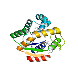
4ORN
 | | Blue Fluorescent Protein mKalama1 | | 分子名称: | 2-(N-MORPHOLINO)-ETHANESULFONIC ACID, CHLORIDE ION, SULFATE ION, ... | | 著者 | Vegh, R.B, Bloch, D.A, Bommarius, A.S, Verkhovsky, M, Pletnev, S, Iwai, H, Bochenkova, A.V, Solntsev, K.M. | | 登録日 | 2014-02-11 | | 公開日 | 2015-02-11 | | 最終更新日 | 2023-11-15 | | 実験手法 | X-RAY DIFFRACTION (1.71 Å) | | 主引用文献 | Hidden photoinduced reactivity of the blue fluorescent protein mKalama1.
Phys Chem Chem Phys, 17, 2015
|
|
3E5W
 | | Crystal Structure Analysis of FP611 | | 分子名称: | Red fluorescent protein eqFP611 | | 著者 | Nienhaus, K, Nar, H, Heilker, R, Wiedenmann, J, Nienhaus, G.U. | | 登録日 | 2008-08-14 | | 公開日 | 2008-09-23 | | 最終更新日 | 2023-11-15 | | 実験手法 | X-RAY DIFFRACTION (1.71 Å) | | 主引用文献 | Trans-cis isomerization is responsible for the red-shifted fluorescence in variants of the red fluorescent protein eqFP611.
J.Am.Chem.Soc., 130, 2008
|
|
2A47
 | | Crystal structure of amFP486 H199T | | 分子名称: | BETA-MERCAPTOETHANOL, GFP-like fluorescent chromoprotein amFP486 | | 著者 | Henderson, J.N, Remington, S.J. | | 登録日 | 2005-06-28 | | 公開日 | 2005-08-16 | | 最終更新日 | 2023-11-15 | | 実験手法 | X-RAY DIFFRACTION (1.72 Å) | | 主引用文献 | Crystal structures and mutational analysis of amFP486, a cyan fluorescent protein from Anemonia majano
Proc.Natl.Acad.Sci.Usa, 102, 2005
|
|
7A7S
 | |
7A83
 | |
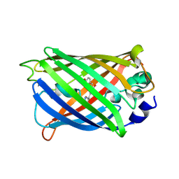
6B7R
 | | Truncated strand 11-less green fluorescent protein | | 分子名称: | 2-[N-CYCLOHEXYLAMINO]ETHANE SULFONIC ACID, Green fluorescent protein | | 著者 | Deng, A, Boxer, S.G. | | 登録日 | 2017-10-05 | | 公開日 | 2017-12-27 | | 最終更新日 | 2023-11-15 | | 実験手法 | X-RAY DIFFRACTION (1.73 Å) | | 主引用文献 | Structural Insight into the Photochemistry of Split Green Fluorescent Proteins: A Unique Role for a His-Tag.
J. Am. Chem. Soc., 140, 2018
|
|
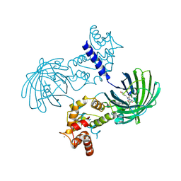
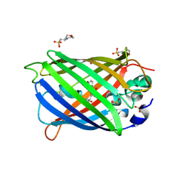
5G5Y
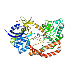 | | S.pneumoniae ABC-transporter substrate binding protein FusA apo structure | | 分子名称: | ABC TRANSPORTER, SUBSTRATE-BINDING PROTEIN, CALCIUM ION, ... | | 著者 | Culurgioni, S, Harris, G, Singh, A.K, King, S.J, Walsh, M.A. | | 登録日 | 2016-06-10 | | 公開日 | 2017-01-18 | | 最終更新日 | 2024-05-08 | | 実験手法 | X-RAY DIFFRACTION (1.73 Å) | | 主引用文献 | Structural Basis for Regulation and Specificity of Fructooligosaccharide Import in Streptococcus pneumoniae.
Structure, 25, 2017
|
|
4JLO
 | |
7UGR
 | | Crystal structure of hyperfolder YFP | | 分子名称: | 1,2-ETHANEDIOL, DI(HYDROXYETHYL)ETHER, Hyperfolder yellow fluorescent protein, ... | | 著者 | Campbell, B.C, Liu, C.F, Petsko, G.A. | | 登録日 | 2022-03-25 | | 公開日 | 2022-10-26 | | 最終更新日 | 2023-11-15 | | 実験手法 | X-RAY DIFFRACTION (1.74 Å) | | 主引用文献 | Chemically stable fluorescent proteins for advanced microscopy.
Nat.Methods, 19, 2022
|
|
4KW4
 | |
7RRH
 | | Crystal structure of fast switching R66M/M159T mutant of fluorescent protein Dronpa (Dronpa2) | | 分子名称: | Fluorescent protein Dronpa | | 著者 | Lin, C.-Y, Romei, M.G, Mathews, I.I, Boxer, S.G. | | 登録日 | 2021-08-09 | | 公開日 | 2021-10-13 | | 最終更新日 | 2023-11-15 | | 実験手法 | X-RAY DIFFRACTION (1.747 Å) | | 主引用文献 | Energetic Basis and Design of Enzyme Function Demonstrated Using GFP, an Excited-State Enzyme.
J.Am.Chem.Soc., 144, 2022
|
|
6ZWB
 | | Z-SBTub3 photoswitch bound to tubulin-DARPin D1 complex | | 分子名称: | 5-[2-(1,3-benzothiazol-2-yl)ethyl]-2-methoxy-phenol, Designed Ankyrin Repeat Protein (DARPIN) D1, GUANOSINE-5'-DIPHOSPHATE, ... | | 著者 | Wranik, M, Weinert, T, Olieric, N, Gao, L, Kraus, Y.C.M, Bingham, R, Ntouliou, E, Ahlfeld, J, Thorn-Seshold, O, Steinmetz, M.O, Standfuss, J. | | 登録日 | 2020-07-28 | | 公開日 | 2020-12-23 | | 最終更新日 | 2024-01-31 | | 実験手法 | X-RAY DIFFRACTION (1.747 Å) | | 主引用文献 | A Robust, GFP-Orthogonal Photoswitchable Inhibitor Scaffold Extends Optical Control over the Microtubule Cytoskeleton.
Cell Chem Biol, 28, 2021
|
|
7TSU
 | |
7TSS
 | |
7TSV
 | |


