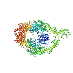
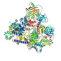
6UZD
 
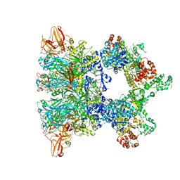
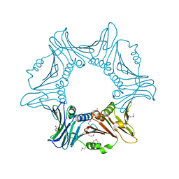
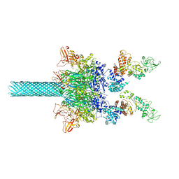 | | Anthrax toxin protective antigen channels bound to edema factor | | 分子名称: | CALCIUM ION, Calmodulin-sensitive adenylate cyclase, Protective antigen | | 著者 | Hardenbrook, N.J, Liu, S, Zhou, K, Zhou, Z.H, Krantz, B.A. | | 登録日 | 2019-11-14 | | 公開日 | 2020-03-04 | | 最終更新日 | 2024-03-06 | | 実験手法 | ELECTRON MICROSCOPY (3.4 Å) | | 主引用文献 | Atomic structures of anthrax toxin protective antigen channels bound to partially unfolded lethal and edema factors.
Nat Commun, 11, 2020
|
|
6DM6
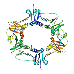 | |
3HI8
 | | Crystal structure of proliferating cell nuclear antigen (PCNA) from Haloferax volcanii | | 分子名称: | Proliferating cell nuclear antigen PcnA | | 著者 | Morgunova, E, Gray, F.C, MacNeill, S.A, Ladenstein, R. | | 登録日 | 2009-05-19 | | 公開日 | 2009-09-29 | | 最終更新日 | 2023-09-06 | | 実験手法 | X-RAY DIFFRACTION (3.202 Å) | | 主引用文献 | Structural insights into the adaptation of proliferating cell nuclear antigen (PCNA) from Haloferax volcanii to a high-salt environment.
Acta Crystallogr.,Sect.D, 65, 2009
|
|
6ZXK
 | | Fully-loaded anthrax lethal toxin in its heptameric pre-pore state and PA7LF(2+1B) arrangement | | 分子名称: | Lethal factor, Protective antigen | | 著者 | Quentin, D, Antoni, C, Gatsogiannis, C, Raunser, S. | | 登録日 | 2020-07-29 | | 公開日 | 2020-09-02 | | 最終更新日 | 2024-05-01 | | 実験手法 | ELECTRON MICROSCOPY (3.8 Å) | | 主引用文献 | Cryo-EM structure of the fully-loaded asymmetric anthrax lethal toxin in its heptameric pre-pore state.
Plos Pathog., 16, 2020
|
|
6ZXL
 | | Fully-loaded anthrax lethal toxin in its heptameric pre-pore state and PA7LF(2+1A) arrangement | | 分子名称: | Lethal factor, Protective antigen | | 著者 | Quentin, D, Antoni, C, Gatsogiannis, C, Raunser, S. | | 登録日 | 2020-07-29 | | 公開日 | 2020-09-02 | | 最終更新日 | 2024-05-01 | | 実験手法 | ELECTRON MICROSCOPY (4.2 Å) | | 主引用文献 | Cryo-EM structure of the fully-loaded asymmetric anthrax lethal toxin in its heptameric pre-pore state.
Plos Pathog., 16, 2020
|
|
6ZXJ
 | | Fully-loaded anthrax lethal toxin in its heptameric pre-pore state, in which the third lethal factor is masked out (PA7LF3-masked) | | 分子名称: | Lethal factor, Protective antigen | | 著者 | Quentin, D, Antoni, C, Gatsogiannis, C, Raunser, S. | | 登録日 | 2020-07-29 | | 公開日 | 2020-09-02 | | 最終更新日 | 2024-05-01 | | 実験手法 | ELECTRON MICROSCOPY (3.5 Å) | | 主引用文献 | Cryo-EM structure of the fully-loaded asymmetric anthrax lethal toxin in its heptameric pre-pore state.
Plos Pathog., 16, 2020
|
|
5OKI
 | |
5OKC
 | |
7YPE
 | |
7YPF
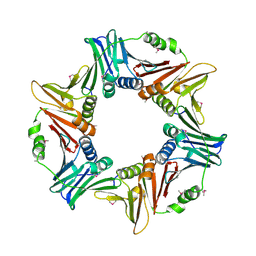 | |
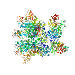
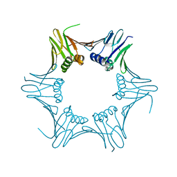
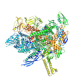
6VRA
 | | Anthrax octamer prechannel bound to full-length edema factor | | 分子名称: | CALCIUM ION, Calmodulin-sensitive adenylate cyclase, Protective antigen | | 著者 | Zhou, K, Hardenbrook, N.J, Liu, S, Cui, Y.X, Krantz, B.A, Zhou, Z.H. | | 登録日 | 2020-02-07 | | 公開日 | 2020-12-16 | | 最終更新日 | 2024-03-06 | | 実験手法 | ELECTRON MICROSCOPY (3.3 Å) | | 主引用文献 | Atomic Structures of Anthrax Prechannel Bound with Full-Length Lethal and Edema Factors.
Structure, 28, 2020
|
|
8E84
 | |
6I5F
 | | Crystal structure of DNA-free E.coli MutS P839E dimer mutant | | 分子名称: | ADENOSINE-5'-DIPHOSPHATE, DNA mismatch repair protein MutS, GLYCEROL, ... | | 著者 | Bhairosing-Kok, D, Groothuizen, F.S, Fish, A, Dharadhar, S, Winterwerp, H.H.K, Sixma, T.K. | | 登録日 | 2018-11-13 | | 公開日 | 2019-08-14 | | 最終更新日 | 2024-01-24 | | 実験手法 | X-RAY DIFFRACTION (2.6 Å) | | 主引用文献 | Sharp kinking of a coiled-coil in MutS allows DNA binding and release.
Nucleic Acids Res., 47, 2019
|
|
6T7X
 | | Crystal structure of PCNA from P. abyssi | | 分子名称: | DNA polymerase sliding clamp | | 著者 | Madru, C, Raia, P, Hugonneau Beaufet, I, Delarue, M, Carroni, M, Sauguet, L. | | 登録日 | 2019-10-23 | | 公開日 | 2020-03-04 | | 最終更新日 | 2024-01-24 | | 実験手法 | X-RAY DIFFRACTION (2.3 Å) | | 主引用文献 | Structural basis for the increased processivity of D-family DNA polymerases in complex with PCNA.
Nat Commun, 11, 2020
|
|
5AUJ
 | |
3A2F
 | |
1PLQ
 | | CRYSTAL STRUCTURE OF THE EUKARYOTIC DNA POLYMERASE PROCESSIVITY FACTOR PCNA | | 分子名称: | MERCURY (II) ION, PROLIFERATING CELL NUCLEAR ANTIGEN (PCNA) | | 著者 | Krishna, T.S.R, Kong, X.-P, Gary, S, Burgers, P.M, Kuriyan, J. | | 登録日 | 1995-01-02 | | 公開日 | 1995-03-31 | | 最終更新日 | 2024-02-14 | | 実験手法 | X-RAY DIFFRACTION (2.3 Å) | | 主引用文献 | Crystal structure of the eukaryotic DNA polymerase processivity factor PCNA.
Cell(Cambridge,Mass.), 79, 1994
|
|
8DT6
 | |
1PLR
 | | CRYSTAL STRUCTURE OF THE EUKARYOTIC DNA POLYMERASE PROCESSIVITY FACTOR PCNA | | 分子名称: | PROLIFERATING CELL NUCLEAR ANTIGEN (PCNA) | | 著者 | Krishna, T.S.R, Kong, X.-P, Gary, S, Burgers, P.M, Kuriyan, J. | | 登録日 | 1995-01-02 | | 公開日 | 1995-03-31 | | 最終更新日 | 2024-06-05 | | 実験手法 | X-RAY DIFFRACTION (3 Å) | | 主引用文献 | Crystal structure of the eukaryotic DNA polymerase processivity factor PCNA.
Cell(Cambridge,Mass.), 79, 1994
|
|
6GWS
 | | Crystal structure of human PCNA in complex with three p15 peptides | | 分子名称: | PCNA-associated factor, Proliferating cell nuclear antigen | | 著者 | De March, M, Merino, N, Gonzalez-Magana, A, Romano-Moreno, M, Onesti, S, Blanco, F.J, De Biasio, A. | | 登録日 | 2018-06-25 | | 公開日 | 2018-08-22 | | 最終更新日 | 2024-01-17 | | 実験手法 | X-RAY DIFFRACTION (2.9 Å) | | 主引用文献 | p15PAF binding to PCNA modulates the DNA sliding surface.
Nucleic Acids Res., 46, 2018
|
|
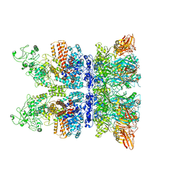
6T8H
 | | Cryo-EM structure of the DNA-bound PolD-PCNA processive complex from P. abyssi | | 分子名称: | DNA polymerase II small subunit, DNA polymerase sliding clamp, DNA primer, ... | | 著者 | Madru, C, Raia, P, Hugonneau Beaufet, I, Pehau-Arnaudet, G, England, P, Lindhal, E, Delarue, M, Carroni, M, Sauguet, L. | | 登録日 | 2019-10-24 | | 公開日 | 2020-03-04 | | 最終更新日 | 2020-04-08 | | 実験手法 | ELECTRON MICROSCOPY (3.77 Å) | | 主引用文献 | Structural basis for the increased processivity of D-family DNA polymerases in complex with PCNA.
Nat Commun, 11, 2020
|
|
1M2Z
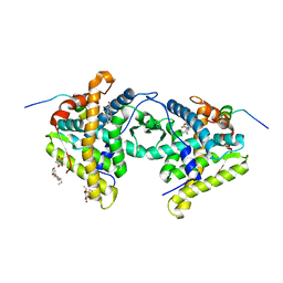 | | Crystal structure of a dimer complex of the human glucocorticoid receptor ligand-binding domain bound to dexamethasone and a TIF2 coactivator motif | | 分子名称: | DEXAMETHASONE, glucocorticoid receptor, nuclear receptor coactivator 2, ... | | 著者 | Bledsoe, R.B, Montana, V.G, Stanley, T.B, Delves, C.J, Apolito, C.J, Mckee, D.D, Consler, T.G, Parks, D.J, Stewart, E.L, Willson, T.M, Lambert, M.H, Moore, J.T, Pearce, K.H, Xu, H.E. | | 登録日 | 2002-06-26 | | 公開日 | 2003-07-15 | | 最終更新日 | 2024-04-03 | | 実験手法 | X-RAY DIFFRACTION (2.5 Å) | | 主引用文献 | Crystal Structure of the Glucocorticoid Receptor Ligand Binding Domain Reveals a Novel Mode of Receptor Dimerization and Coactivator Recognition
Cell(Cambridge,Mass.), 110, 2002
|
|
8ABY
 | |
8AD1
 | |
8AC0
 | |


