6I0Z
 
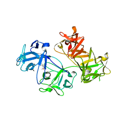 | | CRYSTAL STRUCTURE OF FASCIN IN COMPLEX WITH COMPOUND 1 | | 分子名称: | Fascin, ~{N}-(2,4-dichlorophenyl)-~{N}-methyl-ethanamide | | 著者 | Schuettelkopf, A.W. | | 登録日 | 2018-10-27 | | 公開日 | 2019-02-27 | | 最終更新日 | 2024-01-24 | | 実験手法 | X-RAY DIFFRACTION (1.77 Å) | | 主引用文献 | Structure-based design, synthesis and biological evaluation of a novel series of isoquinolone and pyrazolo[4,3-c]pyridine inhibitors of fascin 1 as potential anti-metastatic agents.
Bioorg.Med.Chem.Lett., 29, 2019
|
|
6I13
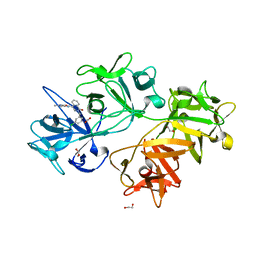 | | CRYSTAL STRUCTURE OF FASCIN IN COMPLEX WITH COMPOUND 7 | | 分子名称: | 1,2-ETHANEDIOL, 2-[(3-chlorophenyl)methyl]-~{N}-(1-methylpyrazol-4-yl)-1-oxidanylidene-isoquinoline-4-carboxamide, ACETATE ION, ... | | 著者 | Schuettelkopf, A.W. | | 登録日 | 2018-10-27 | | 公開日 | 2019-02-27 | | 最終更新日 | 2024-01-24 | | 実験手法 | X-RAY DIFFRACTION (1.79 Å) | | 主引用文献 | Structure-based design, synthesis and biological evaluation of a novel series of isoquinolone and pyrazolo[4,3-c]pyridine inhibitors of fascin 1 as potential anti-metastatic agents.
Bioorg.Med.Chem.Lett., 29, 2019
|
|
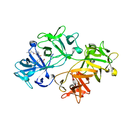
6I17
 | | CRYSTAL STRUCTURE OF FASCIN IN COMPLEX WITH COMPOUND 24 | | 分子名称: | 1,2-ETHANEDIOL, 2-[(3,4-dichlorophenyl)methyl]-~{N}-(1-methylpyrazol-4-yl)-1-oxidanylidene-6-piperidin-4-yl-2,7-naphthyridine-4-carboxamide, ACETATE ION, ... | | 著者 | Schuettelkopf, A.W. | | 登録日 | 2018-10-27 | | 公開日 | 2019-02-27 | | 最終更新日 | 2024-01-24 | | 実験手法 | X-RAY DIFFRACTION (1.56 Å) | | 主引用文献 | Structure-based design, synthesis and biological evaluation of a novel series of isoquinolone and pyrazolo[4,3-c]pyridine inhibitors of fascin 1 as potential anti-metastatic agents.
Bioorg.Med.Chem.Lett., 29, 2019
|
|
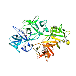
6I16
 | | CRYSTAL STRUCTURE OF FASCIN IN COMPLEX WITH COMPOUND 15 | | 分子名称: | 6-[(3,4-dichlorophenyl)methyl]-~{N}-(1-methylpyrazol-4-yl)-5-oxidanylidene-1,6-naphthyridine-8-carboxamide, ACETATE ION, Fascin | | 著者 | Schuettelkopf, A.W. | | 登録日 | 2018-10-27 | | 公開日 | 2019-02-27 | | 最終更新日 | 2024-01-24 | | 実験手法 | X-RAY DIFFRACTION (2 Å) | | 主引用文献 | Structure-based design, synthesis and biological evaluation of a novel series of isoquinolone and pyrazolo[4,3-c]pyridine inhibitors of fascin 1 as potential anti-metastatic agents.
Bioorg.Med.Chem.Lett., 29, 2019
|
|
2RJY
 | |
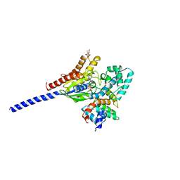
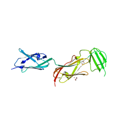
1Z2C
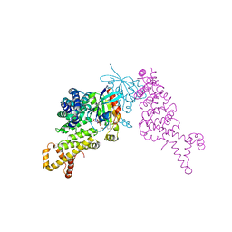 | | Crystal structure of mDIA1 GBD-FH3 in complex with RhoC-GMPPNP | | 分子名称: | Diaphanous protein homolog 1, MAGNESIUM ION, PHOSPHOAMINOPHOSPHONIC ACID-GUANYLATE ESTER, ... | | 著者 | Rose, R, Weyand, M, Lammers, M, Ishizaki, T, Ahmadian, M.R, Wittinghofer, A. | | 登録日 | 2005-03-08 | | 公開日 | 2005-05-10 | | 最終更新日 | 2024-02-14 | | 実験手法 | X-RAY DIFFRACTION (3 Å) | | 主引用文献 | Structural and mechanistic insights into the interaction between Rho and mammalian Dia.
Nature, 435, 2005
|
|
6ZYM
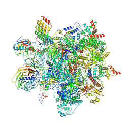 | | Human C Complex Spliceosome - High-resolution CORE | | 分子名称: | 116 kDa U5 small nuclear ribonucleoprotein component, Cell division cycle 5-like protein, Corepressor interacting with RBPJ 1, ... | | 著者 | Bertram, K, Kastner, B. | | 登録日 | 2020-08-02 | | 公開日 | 2020-10-14 | | 実験手法 | ELECTRON MICROSCOPY (3.4 Å) | | 主引用文献 | Structural Insights into the Roles of Metazoan-Specific Splicing Factors in the Human Step 1 Spliceosome.
Mol.Cell, 80, 2020
|
|
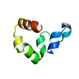
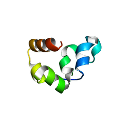
1Y2O
 | | Structure of N-terminal domain IRSp53/BAIAP2 | | 分子名称: | BAI1-associated protein 2 isoform 1 | | 著者 | Millard, T.H, Bompard, G, Heung, M.-Y, Dafforn, T.R, Scott, D.J, Machesky, L.M, Futterer, K. | | 登録日 | 2004-11-23 | | 公開日 | 2005-02-15 | | 最終更新日 | 2011-07-13 | | 実験手法 | X-RAY DIFFRACTION (2.2 Å) | | 主引用文献 | Structural basis of filopodia formation induced by the IRSp53/MIM homology domain of human IRSp53
Embo J., 24, 2005
|
|
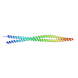
2QU4
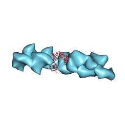 | | Model for Bacterial ParM Filament | | 分子名称: | Plasmid segregation protein parM | | 著者 | Orlova, A, Garner, E.C, Galkin, V.E, Heuser, J, Mullins, R.D, Egelman, E.H. | | 登録日 | 2007-08-03 | | 公開日 | 2007-09-18 | | 最終更新日 | 2024-02-21 | | 実験手法 | ELECTRON MICROSCOPY (16 Å) | | 主引用文献 | The structure of bacterial ParM filaments.
Nat.Struct.Mol.Biol., 14, 2007
|
|
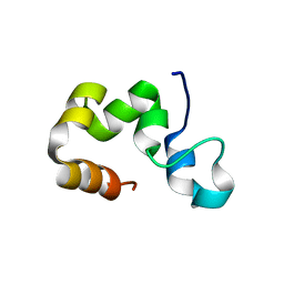
1EJ5
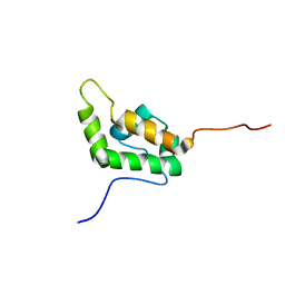 | | SOLUTION STRUCTURE OF THE AUTOINHIBITED CONFORMATION OF WASP | | 分子名称: | WISKOTT-ALDRICH SYNDROME PROTEIN | | 著者 | Kim, A.S, Kakalis, L.T, Abdul-Manan, N, Liu, G.A, Rosen, M.K. | | 登録日 | 2000-02-29 | | 公開日 | 2000-04-05 | | 最終更新日 | 2024-05-22 | | 実験手法 | SOLUTION NMR | | 主引用文献 | Autoinhibition and activation mechanisms of the Wiskott-Aldrich syndrome protein.
Nature, 404, 2000
|
|
1FMV
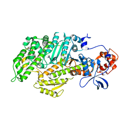 | | CRYSTAL STRUCTURE OF THE APO MOTOR DOMAIN OF DICTYOSTELLIUM MYOSIN II | | 分子名称: | CHLORIDE ION, MYOSIN II HEAVY CHAIN | | 著者 | Bauer, C.B, Holden, H.M, Thoden, J.B, Smith, R, Rayment, I. | | 登録日 | 2000-08-18 | | 公開日 | 2000-11-22 | | 最終更新日 | 2024-02-07 | | 実験手法 | X-RAY DIFFRACTION (2.1 Å) | | 主引用文献 | X-ray structures of the apo and MgATP-bound states of Dictyostelium discoideum myosin motor domain.
J.Biol.Chem., 275, 2000
|
|
1FMW
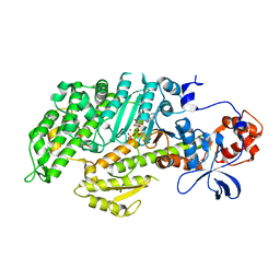 | | CRYSTAL STRUCTURE OF THE MGATP COMPLEX FOR THE MOTOR DOMAIN OF DICTYOSTELIUM MYOSIN II | | 分子名称: | ADENOSINE-5'-TRIPHOSPHATE, MAGNESIUM ION, MYOSIN II HEAVY CHAIN | | 著者 | Bauer, C.B, Holden, H.M, Thoden, J.B, Smith, R, Rayment, I. | | 登録日 | 2000-08-18 | | 公開日 | 2000-11-22 | | 最終更新日 | 2024-02-07 | | 実験手法 | X-RAY DIFFRACTION (2.15 Å) | | 主引用文献 | X-ray structures of the apo and MgATP-bound states of Dictyostelium discoideum myosin motor domain.
J.Biol.Chem., 275, 2000
|
|
1AGR
 | | COMPLEX OF ALF4-ACTIVATED GI-ALPHA-1 WITH RGS4 | | 分子名称: | CITRIC ACID, GUANINE NUCLEOTIDE-BINDING PROTEIN G(I), GUANOSINE-5'-DIPHOSPHATE, ... | | 著者 | Tesmer, J.J.G, Sprang, S.R. | | 登録日 | 1997-03-25 | | 公開日 | 1997-06-16 | | 最終更新日 | 2024-05-22 | | 実験手法 | X-RAY DIFFRACTION (2.8 Å) | | 主引用文献 | Structure of RGS4 bound to AlF4--activated G(i alpha1): stabilization of the transition state for GTP hydrolysis.
Cell(Cambridge,Mass.), 89, 1997
|
|
2RJX
 | |
2RJW
 | |
4J7O
 | |
7MF3
 | | Structure of the autoinhibited state of smooth muscle myosin-2 | | 分子名称: | ADENOSINE-5'-DIPHOSPHATE, MAGNESIUM ION, Myosin light polypeptide 6, ... | | 著者 | Heissler, S.M, Arora, A.S, Billington, N, Sellers, J.R, Chinthalapudi, K. | | 登録日 | 2021-04-08 | | 公開日 | 2022-01-05 | | 最終更新日 | 2024-05-29 | | 実験手法 | ELECTRON MICROSCOPY (3.4 Å) | | 主引用文献 | Cryo-EM structure of the autoinhibited state of myosin-2.
Sci Adv, 7, 2021
|
|
2FGH
 | | ATP bound gelsolin | | 分子名称: | ADENOSINE-5'-TRIPHOSPHATE, gelsolin | | 著者 | Ma, Q, Robinson, R.C, Burtnick, L.D, Urosev, D. | | 登録日 | 2005-12-22 | | 公開日 | 2006-04-18 | | 最終更新日 | 2017-12-20 | | 実験手法 | X-RAY DIFFRACTION (2.8 Å) | | 主引用文献 | The structure of gelsolin bound to ATP
J.Mol.Biol., 357, 2006
|
|
6BFI
 | |
6H3A
 | | Crystal structure of the KAP1 RBCC domain in complex with the SMARCAD1 CUE1 domain. | | 分子名称: | SWI/SNF-related matrix-associated actin-dependent regulator of chromatin subfamily A containing DEAD/H box 1, Transcription intermediary factor 1-beta, ZINC ION | | 著者 | Newman, J.A, Aitkenhead, H, Lim, M, Williams, H.L, Svejstrup, J.Q, von Delft, F, Arrowsmith, C.H, Edwards, A, Bountra, C, Gileadi, O. | | 登録日 | 2018-07-17 | | 公開日 | 2019-06-26 | | 最終更新日 | 2024-05-15 | | 実験手法 | X-RAY DIFFRACTION (5.505 Å) | | 主引用文献 | A Ubiquitin-Binding Domain that Binds a Structural Fold Distinct from that of Ubiquitin.
Structure, 27, 2019
|
|
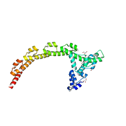
3U1A
 | |
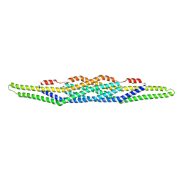
1WLH
 | | Molecular structure of the rod domain of Dictyostelium filamin | | 分子名称: | Gelation factor | | 著者 | Popowicz, G.M, Mueller, R, Noegel, A.A, Schleicher, M, Huber, R, Holak, T.A. | | 登録日 | 2004-06-27 | | 公開日 | 2004-10-05 | | 最終更新日 | 2023-10-25 | | 実験手法 | X-RAY DIFFRACTION (2.8 Å) | | 主引用文献 | Molecular structure of the rod domain of dictyostelium filamin
J.Mol.Biol., 342, 2004
|
|
3U59
 | |
1HUX
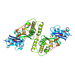 | | CRYSTAL STRUCTURE OF THE ACIDAMINOCOCCUS FERMENTANS (R)-2-HYDROXYGLUTARYL-COA DEHYDRATASE COMPONENT A | | 分子名称: | ACTIVATOR OF (R)-2-HYDROXYGLUTARYL-COA DEHYDRATASE, ADENOSINE-5'-DIPHOSPHATE, IRON/SULFUR CLUSTER | | 著者 | Locher, K.P, Hans, M, Yeh, A.P, Schmid, B, Buckel, W, Rees, D.C. | | 登録日 | 2001-01-04 | | 公開日 | 2001-03-21 | | 最終更新日 | 2024-02-07 | | 実験手法 | X-RAY DIFFRACTION (3 Å) | | 主引用文献 | Crystal structure of the Acidaminococcus fermentans 2-hydroxyglutaryl-CoA dehydratase component A.
J.Mol.Biol., 307, 2001
|
|
2J3S
 | |


