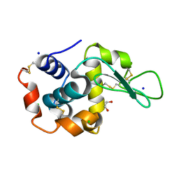
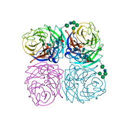
6QRX
 
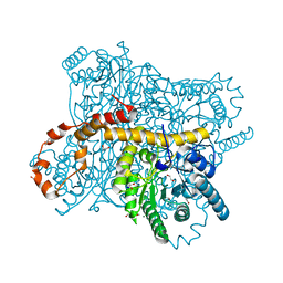 | | X-ray radiation dose series on xylose isomerase - 3.88 MGy | | 分子名称: | 1,2-ETHANEDIOL, ISOPROPYL ALCOHOL, MAGNESIUM ION, ... | | 著者 | Taberman, H, Bury, C.S, van der Woerd, M.J, Snell, E.H, Garman, E.F. | | 登録日 | 2019-02-19 | | 公開日 | 2019-07-17 | | 最終更新日 | 2024-01-24 | | 実験手法 | X-RAY DIFFRACTION (1.17 Å) | | 主引用文献 | Structural knowledge or X-ray damage? A case study on xylose isomerase illustrating both.
J.Synchrotron Radiat., 26, 2019
|
|
5ZPD
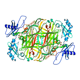 | | Copper amine oxidase from Arthrobacter globiformis anaerobically reduced by ethylamine at pH 6 at 288 K (1) | | 分子名称: | COPPER (II) ION, Phenylethylamine oxidase, SODIUM ION | | 著者 | Murakawa, T, Baba, S, Kawano, Y, Hayashi, H, Yano, T, Tanizawa, K, Kumasaka, T, Yamamoto, M, Okajima, T. | | 登録日 | 2018-04-16 | | 公開日 | 2018-12-19 | | 最終更新日 | 2023-11-22 | | 実験手法 | X-RAY DIFFRACTION (1.665 Å) | | 主引用文献 | In crystallothermodynamic analysis of conformational change of the topaquinone cofactor in bacterial copper amine oxidase
Proc. Natl. Acad. Sci. U.S.A., 116, 2019
|
|
5ZPF
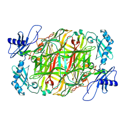 | | Copper amine oxidase from Arthrobacter globiformis anaerobically reduced by ethylamine at pH 6 at 288 K (3) | | 分子名称: | COPPER (II) ION, Phenylethylamine oxidase, SODIUM ION | | 著者 | Murakawa, T, Baba, S, Kawano, Y, Hayashi, H, Yano, T, Tanizawa, K, Kumasaka, T, Yamamoto, M, Okajima, T. | | 登録日 | 2018-04-16 | | 公開日 | 2018-12-19 | | 最終更新日 | 2023-11-22 | | 実験手法 | X-RAY DIFFRACTION (1.759 Å) | | 主引用文献 | In crystallothermodynamic analysis of conformational change of the topaquinone cofactor in bacterial copper amine oxidase
Proc. Natl. Acad. Sci. U.S.A., 116, 2019
|
|
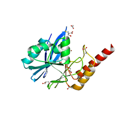
5DPG
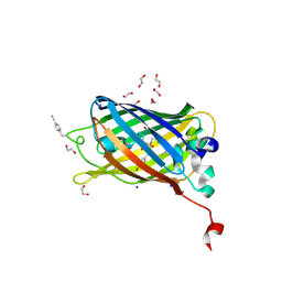 | | sfGFP mutant - 133 p-cyano-L-phenylalanine | | 分子名称: | 1,2-ETHANEDIOL, Green fluorescent protein, SODIUM ION | | 著者 | Dippel, A.B, Olenginski, G.M, Maurici, N, Liskov, M.T, Brewer, S.H, Phillips-Piro, C.M. | | 登録日 | 2015-09-12 | | 公開日 | 2016-01-13 | | 最終更新日 | 2019-11-27 | | 実験手法 | X-RAY DIFFRACTION (1.85 Å) | | 主引用文献 | Probing the effectiveness of spectroscopic reporter unnatural amino acids: a structural study.
Acta Crystallogr D Struct Biol, 72, 2016
|
|
4JDO
 | | Secreted chlamydial protein pgp3, coiled-coil deletion | | 分子名称: | SODIUM ION, Virulence plasmid protein pGP3-D | | 著者 | Galaleldeen, A, Taylor, A.B, Chen, D, Holloway, S.P, Zhong, G, Hart, P.J. | | 登録日 | 2013-02-25 | | 公開日 | 2013-06-05 | | 最終更新日 | 2023-12-06 | | 実験手法 | X-RAY DIFFRACTION (2.3 Å) | | 主引用文献 | Structure of the Chlamydia trachomatis immunodominant antigen Pgp3.
J.Biol.Chem., 288, 2013
|
|
5X5P
 | | Human serum transferrin bound to ruthenium NTA | | 分子名称: | FE (III) ION, MALONATE ION, NITRILOTRIACETIC ACID, ... | | 著者 | Sun, H, Wang, M. | | 登録日 | 2017-02-17 | | 公開日 | 2018-02-21 | | 最終更新日 | 2023-11-22 | | 実験手法 | X-RAY DIFFRACTION (2.7 Å) | | 主引用文献 | Binding of ruthenium and osmium at non‐iron sites of transferrin accounts for their iron-independent cellular uptake.
J.Inorg.Biochem., 234, 2022
|
|
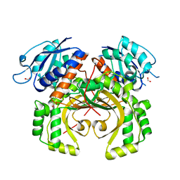
5DKP
 | | Crystal Structure of N. meningitidis ClpP in complex with agonist ADEP A54556. | | 分子名称: | ATP-dependent Clp protease proteolytic subunit, POTASSIUM ION, SODIUM ION, ... | | 著者 | Goodreid, J.D, Janetzko, J, Santa Maria Jr, J.P, Wong, K, Leung, E, Eger, B.T, Bryson, S, Pai, E.F, Gray-Owen, S.D, Walker, S, Houry, W.A, Batey, R.A. | | 登録日 | 2015-09-03 | | 公開日 | 2016-01-27 | | 最終更新日 | 2023-11-15 | | 実験手法 | X-RAY DIFFRACTION (2.381 Å) | | 主引用文献 | Development and Characterization of Potent Cyclic Acyldepsipeptide Analogues with Increased Antimicrobial Activity.
J.Med.Chem., 59, 2016
|
|
5X2P
 | | Crystal structure of the medaka fish taste receptor T1r2a-T1r3 ligand binding domains in complex with L-glutamate | | 分子名称: | 2-acetamido-2-deoxy-beta-D-glucopyranose, 2-acetamido-2-deoxy-beta-D-glucopyranose-(1-4)-2-acetamido-2-deoxy-beta-D-glucopyranose, CALCIUM ION, ... | | 著者 | Nuemket, N, Yasui, N, Atsumi, N, Yamashita, A. | | 登録日 | 2017-02-02 | | 公開日 | 2017-05-24 | | 最終更新日 | 2023-11-22 | | 実験手法 | X-RAY DIFFRACTION (2.608 Å) | | 主引用文献 | Structural basis for perception of diverse chemical substances by T1r taste receptors
Nat Commun, 8, 2017
|
|
4JQG
 | | Crystal structure of an inactive mutant of MMP-9 catalytic domain in complex with a fluorogenic synthetic peptidic substrate with a fluorine atom. | | 分子名称: | 1,2-ETHANEDIOL, AZIDE ION, CALCIUM ION, ... | | 著者 | Stura, E.A, Vera, L, Cassar-Lajeunesse, E, Tranchant, I, Amoura, M, Dive, V. | | 登録日 | 2013-03-20 | | 公開日 | 2014-03-12 | | 最終更新日 | 2023-12-06 | | 実験手法 | X-RAY DIFFRACTION (1.849 Å) | | 主引用文献 | Halogen Bonding Controls Selectivity of FRET Substrate Probes for MMP-9.
Chem.Biol., 21, 2014
|
|
6QY1
 | | Pink beam serial crystallography: Lysozyme, 5 us exposure, 1500 patterns merged | | 分子名称: | 1,2-ETHANEDIOL, CHLORIDE ION, Lysozyme C, ... | | 著者 | Lieske, J, Tolstikova, A, Meents, A. | | 登録日 | 2019-03-08 | | 公開日 | 2019-09-25 | | 最終更新日 | 2024-01-24 | | 実験手法 | X-RAY DIFFRACTION (1.7 Å) | | 主引用文献 | 1 kHz fixed-target serial crystallography using a multilayer monochromator and an integrating pixel detector.
Iucrj, 6, 2019
|
|
6A0R
 | | Homoserine dehydrogenase from Thermus thermophilus HB8 unliganded form | | 分子名称: | 3-CYCLOHEXYL-1-PROPYLSULFONIC ACID, FORMIC ACID, GLYCEROL, ... | | 著者 | Akai, S, Ikushiro, H, Sawai, T, Yano, T, Kamiya, N, Miyahara, I. | | 登録日 | 2018-06-06 | | 公開日 | 2018-11-28 | | 最終更新日 | 2023-11-22 | | 実験手法 | X-RAY DIFFRACTION (1.83 Å) | | 主引用文献 | The crystal structure of homoserine dehydrogenase complexed with l-homoserine and NADPH in a closed form
J. Biochem., 165, 2019
|
|
6QXX
 | | Pink beam serial crystallography: Lysozyme, 5 us exposure, 14793 patterns merged | | 分子名称: | 1,2-ETHANEDIOL, CHLORIDE ION, Lysozyme C, ... | | 著者 | Lieske, J, Tolstikova, A, Meents, A. | | 登録日 | 2019-03-08 | | 公開日 | 2019-10-16 | | 最終更新日 | 2024-01-24 | | 実験手法 | X-RAY DIFFRACTION (1.7 Å) | | 主引用文献 | 1 kHz fixed-target serial crystallography using a multilayer monochromator and an integrating pixel detector.
Iucrj, 6, 2019
|
|
6A0S
 | | Homoserine dehydrogenase from Thermus thermophilus HB8 complexed with HSE and NADPH | | 分子名称: | 3-CYCLOHEXYL-1-PROPYLSULFONIC ACID, FORMIC ACID, GLYCEROL, ... | | 著者 | Akai, S, Ikushiro, H, Sawai, T, Yano, T, Kamiya, N, Miyahara, I. | | 登録日 | 2018-06-06 | | 公開日 | 2018-11-28 | | 最終更新日 | 2023-11-22 | | 実験手法 | X-RAY DIFFRACTION (2 Å) | | 主引用文献 | The crystal structure of homoserine dehydrogenase complexed with l-homoserine and NADPH in a closed form
J. Biochem., 165, 2019
|
|
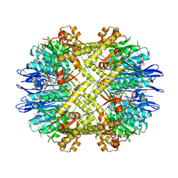
5ZS3
 | | Small heat shock protein from M. marinum:Form-1 | | 分子名称: | CHLORIDE ION, GLY-ARG-LEU-LEU-PRO, Molecular chaperone (Small heat shock protein), ... | | 著者 | Bhandari, S, Suguna, K. | | 登録日 | 2018-04-27 | | 公開日 | 2019-01-30 | | 最終更新日 | 2019-04-17 | | 実験手法 | X-RAY DIFFRACTION (2.001 Å) | | 主引用文献 | Dodecameric structure of a small heat shock protein from Mycobacterium marinum M.
Proteins, 87, 2019
|
|
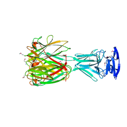
6R1J
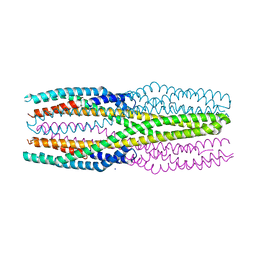 | | Structure of the soluble AhlC triple head mutant of the tripartite alpha-pore forming toxin, AHL, from Aeromonas hydrophila. | | 分子名称: | SODIUM ION, Uncharacterized protein | | 著者 | Churchill-Angus, A.M, Wilson, J.S, Baker, P.J. | | 登録日 | 2019-03-14 | | 公開日 | 2019-07-10 | | 最終更新日 | 2024-01-24 | | 実験手法 | X-RAY DIFFRACTION (1.92 Å) | | 主引用文献 | Identification and structural analysis of the tripartite alpha-pore forming toxin of Aeromonas hydrophila.
Nat Commun, 10, 2019
|
|
2FTX
 | |
5X2N
 | | Crystal structure of the medaka fish taste receptor T1r2a-T1r3 ligand binding domains in complex with L-alanine | | 分子名称: | 2-acetamido-2-deoxy-beta-D-glucopyranose, ALANINE, CHLORIDE ION, ... | | 著者 | Nuemket, N, Yasui, N, Atsumi, N, Yamashita, A. | | 登録日 | 2017-02-02 | | 公開日 | 2017-05-24 | | 最終更新日 | 2023-11-22 | | 実験手法 | X-RAY DIFFRACTION (2.2 Å) | | 主引用文献 | Structural basis for perception of diverse chemical substances by T1r taste receptors
Nat Commun, 8, 2017
|
|
6R6D
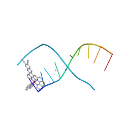 | | [Ru(TAP)2(11,12-CN2-dppz)]2+ bound to d(TCGGCGCCGA)2 | | 分子名称: | BARIUM ION, DNA (5'-D(*TP*CP*GP*GP*CP*GP*CP*CP*GP*A)-3'), Ruthenium (bis-(tetraazaphenanthrene)) (11,12-dicyano-dipyridophenazine), ... | | 著者 | McQuaid, K.T, Hall, J.P, Cardin, C.J. | | 登録日 | 2019-03-27 | | 公開日 | 2020-05-06 | | 最終更新日 | 2024-05-15 | | 実験手法 | X-RAY DIFFRACTION (1.838 Å) | | 主引用文献 | X-ray Crystal Structures Show DNA Stacking Advantage of Terminal Nitrile Substitution in Ru-dppz Complexes.
Chemistry, 24, 2018
|
|
1IVG
 | |
6R79
 | | Structure of IMP-13 metallo-beta-lactamase in apo form (loop open) | | 分子名称: | BETA-MERCAPTOETHANOL, Beta-lactamase, GLYCEROL, ... | | 著者 | Zak, K.M, Softley, C, Kolonko, M, Sattler, M, Popowicz, G.M. | | 登録日 | 2019-03-28 | | 公開日 | 2020-04-01 | | 最終更新日 | 2024-01-24 | | 実験手法 | X-RAY DIFFRACTION (1.9 Å) | | 主引用文献 | Structure and Molecular Recognition Mechanism of IMP-13 Metallo-beta-Lactamase.
Antimicrob.Agents Chemother., 64, 2020
|
|
5XDF
 | |
5XDZ
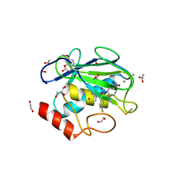
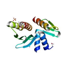
 | | Crystal structure of zebrafish SNX25 PX domain | | 分子名称: | CHLORIDE ION, Cellular trafficking protein, SODIUM ION | | 著者 | Su, K, Zhang, Y, Xu, J, Liu, J. | | 登録日 | 2017-03-30 | | 公開日 | 2017-06-14 | | 最終更新日 | 2023-11-22 | | 実験手法 | X-RAY DIFFRACTION (1.7 Å) | | 主引用文献 | Structure of the PX domain of SNX25 reveals a novel phospholipid recognition model by dimerization in the PX domain
FEBS Lett., 591, 2017
|
|
4KLS
 | | DNA polymerase beta mismatched reactant complex with Mn2+, 10 min | | 分子名称: | 2'-DEOXYADENOSINE 5'-TRIPHOSPHATE, 5'-D(*CP*CP*GP*AP*CP*GP*GP*CP*GP*CP*AP*TP*CP*AP*GP*C)-3', 5'-D(*GP*CP*TP*GP*AP*TP*GP*CP*GP*CP*A)-3', ... | | 著者 | Freudenthal, B.D, Beard, W.A, Shock, D.D, Wilson, S.H. | | 登録日 | 2013-05-07 | | 公開日 | 2013-07-17 | | 最終更新日 | 2023-09-20 | | 実験手法 | X-RAY DIFFRACTION (1.978 Å) | | 主引用文献 | Observing a DNA polymerase choose right from wrong.
Cell(Cambridge,Mass.), 154, 2013
|
|
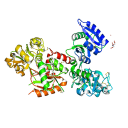
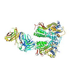
6A8Z
 | | Crystal structure of M1 zinc metallopeptidase from Deinococcus radiodurans | | 分子名称: | SODIUM ION, TYROSINE, ZINC ION, ... | | 著者 | Agrawal, R, Kumar, A, Makde, R.D. | | 登録日 | 2018-07-11 | | 公開日 | 2019-07-17 | | 最終更新日 | 2023-11-22 | | 実験手法 | X-RAY DIFFRACTION (2.045 Å) | | 主引用文献 | Two-domain aminopeptidase of M1 family: Structural features for substrate binding and gating in absence of C-terminal domain.
J.Struct.Biol., 208, 2019
|
|
5X2Q
 | | Crystal structure of the medaka fish taste receptor T1r2a-T1r3 ligand binding domains in complex with glycine | | 分子名称: | 2-acetamido-2-deoxy-beta-D-glucopyranose, 2-acetamido-2-deoxy-beta-D-glucopyranose-(1-4)-2-acetamido-2-deoxy-beta-D-glucopyranose, CALCIUM ION, ... | | 著者 | Nuemket, N, Yasui, N, Atsumi, N, Yamashita, A. | | 登録日 | 2017-02-02 | | 公開日 | 2017-05-24 | | 最終更新日 | 2023-11-22 | | 実験手法 | X-RAY DIFFRACTION (2.6 Å) | | 主引用文献 | Structural basis for perception of diverse chemical substances by T1r taste receptors
Nat Commun, 8, 2017
|
|


