6MSO
 
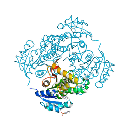
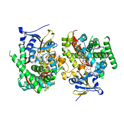
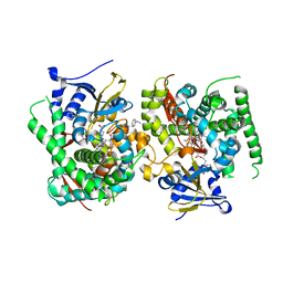
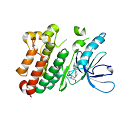
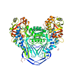 | | Crystal structure of mitochondrial fumarate hydratase from Leishmania major in a complex with inhibitor thiomalate | | 分子名称: | (2S)-2-sulfanylbutanedioic acid, GLYCEROL, IRON/SULFUR CLUSTER, ... | | 著者 | Feliciano, P.R, Drennan, C.L, Nonato, M.C. | | 登録日 | 2018-10-17 | | 公開日 | 2019-01-30 | | 最終更新日 | 2023-10-11 | | 実験手法 | X-RAY DIFFRACTION (2.053 Å) | | 主引用文献 | Crystal Structures of Fumarate Hydratases from Leishmania major in a Complex with Inhibitor 2-Thiomalate.
ACS Chem. Biol., 14, 2019
|
|
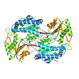
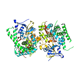
6MYI
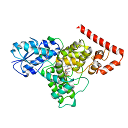
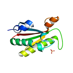
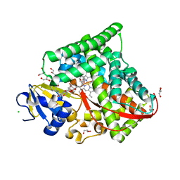
 | | Pleurotus ostreatus OstreolysinA | | 分子名称: | 1,2-ETHANEDIOL, Ostreolysin A6, SODIUM ION | | 著者 | Tomchick, D.R, Radhakrishnan, A, Endapally, S. | | 登録日 | 2018-11-01 | | 公開日 | 2019-02-13 | | 最終更新日 | 2024-03-13 | | 実験手法 | X-RAY DIFFRACTION (1.15 Å) | | 主引用文献 | Molecular Discrimination between Two Conformations of Sphingomyelin in Plasma Membranes.
Cell, 176, 2019
|
|
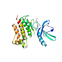
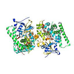
5U0L
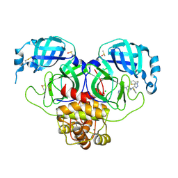
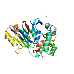
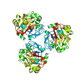
 | | X-ray crystal structure of fatty aldehyde dehydrogenase enzymes from Marinobacter aquaeolei VT8 complexed with a substrate | | 分子名称: | (4S)-2-METHYL-2,4-PENTANEDIOL, 1,2-ETHANEDIOL, CHLORIDE ION, ... | | 著者 | Shi, K, Mulliner, K, Barney, B.M, Aihara, H. | | 登録日 | 2016-11-24 | | 公開日 | 2017-04-26 | | 最終更新日 | 2023-10-04 | | 実験手法 | X-RAY DIFFRACTION (2.285 Å) | | 主引用文献 | Five Fatty Aldehyde Dehydrogenase Enzymes from Marinobacter and Acinetobacter spp. and Structural Insights into the Aldehyde Binding Pocket.
Appl. Environ. Microbiol., 83, 2017
|
|
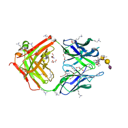
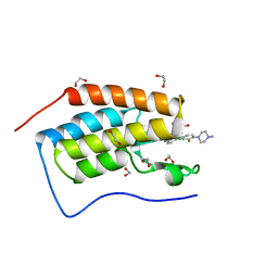
1PET
 | | NMR SOLUTION STRUCTURE OF THE TETRAMERIC MINIMUM TRANSFORMING DOMAIN OF P53 | | 分子名称: | TUMOR SUPPRESSOR P53 | | 著者 | Lee, W, Harvey, T.S, Yin, Y, Yau, P, Litchfield, D, Arrowsmith, C.H. | | 登録日 | 1994-11-24 | | 公開日 | 1995-02-07 | | 最終更新日 | 2024-05-22 | | 実験手法 | SOLUTION NMR | | 主引用文献 | Solution structure of the tetrameric minimum transforming domain of p53.
Nat.Struct.Biol., 1, 1994
|
|
7M5Z
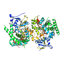
 | | Crystal Structure of the MerTK Kinase Domain in Complex with Inhibitor MIPS15692 | | 分子名称: | 2-(butylamino)-N-[1-(3-fluoropropyl)piperidin-4-yl]-4-{[(1r,4r)-4-hydroxycyclohexyl]amino}pyrimidine-5-carboxamide, Tyrosine-protein kinase Mer | | 著者 | Hermans, S.J, Hancock, N.C, Baell, J.B, Parker, M.W. | | 登録日 | 2021-03-25 | | 公開日 | 2021-10-06 | | 最終更新日 | 2023-10-18 | | 実験手法 | X-RAY DIFFRACTION (3.06 Å) | | 主引用文献 | Development of [ 18 F]MIPS15692, a radiotracer with in vitro proof-of-concept for the imaging of MER tyrosine kinase (MERTK) in neuroinflammatory disease.
Eur.J.Med.Chem., 226, 2021
|
|
5ZQQ
 | | Tankyrase-2 in complex with compound 52 | | 分子名称: | 1-methyl-1'-(4-oxo-3,4,5,6,7,8-hexahydroquinazolin-2-yl)spiro[indole-3,4'-piperidin]-2(1H)-one, GLYCEROL, PHOSPHATE ION, ... | | 著者 | Niwa, H, Shirai, F, Sato, S, Yoshimoto, N, Tsumura, T, Okue, M, Shirouzu, M, Seimiya, H, Umehara, T. | | 登録日 | 2018-04-19 | | 公開日 | 2019-04-03 | | 最終更新日 | 2023-11-22 | | 実験手法 | X-RAY DIFFRACTION (2.29 Å) | | 主引用文献 | Discovery of Novel Spiroindoline Derivatives as Selective Tankyrase Inhibitors.
J. Med. Chem., 62, 2019
|
|
9G0I
 | | Crystal structure of SARS-CoV-2 main protease (MPro) in complex with the noncovalently bound inhibitor C5N17B | | 分子名称: | 3C-like proteinase nsp5, DIMETHYL SULFOXIDE, IMIDAZOLE, ... | | 著者 | Yang, C.-C, Strater, N, Sylvester, K, Muller, C.E, Yang, M, Lee, M.K, Gao, S, Song, L, Liu, X, Kim, M, Zhan, P. | | 登録日 | 2024-07-08 | | 公開日 | 2025-07-02 | | 実験手法 | X-RAY DIFFRACTION (1.67 Å) | | 主引用文献 | Miniaturized Modular Click Chemistry-enabled Rapid Discovery of Unique SARS-CoV-2 M pro Inhibitors With Robust Potency and Drug-like Profile.
Adv Sci, 11, 2024
|
|
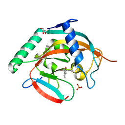
6SQ9
 | | Crystal structure of M. tuberculosis InhA in complex with NAD+ and 3-hydroxynaphthalene-2-carboxylic acid | | 分子名称: | 3-hydroxynaphthalene-2-carboxylic acid, 4-(2-HYDROXYETHYL)-1-PIPERAZINE ETHANESULFONIC ACID, Enoyl-[acyl-carrier-protein] reductase [NADH], ... | | 著者 | Mendes, V, Sabbah, M, Coyne, A.G, Abell, C, Blundell, T.L. | | 登録日 | 2019-09-03 | | 公開日 | 2020-04-22 | | 最終更新日 | 2024-01-24 | | 実験手法 | X-RAY DIFFRACTION (1.75 Å) | | 主引用文献 | Fragment-Based Design ofMycobacterium tuberculosisInhA Inhibitors.
J.Med.Chem., 63, 2020
|
|
6DD6
 | |
9FKJ
 | | The structure of glycosynthase IXT6 (E241G mutant), the intracellular xylanase of G.proteiniphilus T-6 in complex with two xylobiose-F molecules | | 分子名称: | Beta-xylanase, GLYCEROL, beta-D-xylopyranose-(1-4)-1-fluoro-D-xylopyranose | | 著者 | Hadad, N, Chmelnik, O, Dessau, M, Shoham, Y, Shoham, G. | | 登録日 | 2024-06-03 | | 公開日 | 2025-06-18 | | 実験手法 | X-RAY DIFFRACTION (2 Å) | | 主引用文献 | The structure of glycosynthase IXT6 (E241G mutant), the intracellular xylanase of G.proteiniphilus T-6 in complex with two xylobiose-F molecules
To Be Published
|
|
5B2W
 | | Crystal Structure of P450BM3 with N-perfluorododecanoyl-L-tryptophan | | 分子名称: | (2~{S})-3-(1~{H}-indol-3-yl)-2-[2,2,3,3,4,4,5,5,6,6,7,7,8,8,9,9,10,10,11,11,12,12,12-tricosakis(fluoranyl)dodecanoylamino]propanoic acid, Bifunctional cytochrome P450/NADPH--P450 reductase, PROTOPORPHYRIN IX CONTAINING FE | | 著者 | Cong, Z, Shoji, O, Kasai, C, Sugimoto, H, Shiro, Y, Watanabe, Y. | | 登録日 | 2016-02-03 | | 公開日 | 2017-02-08 | | 最終更新日 | 2023-11-08 | | 実験手法 | X-RAY DIFFRACTION (1.65 Å) | | 主引用文献 | Crystal Structure of P450BM3 with decoy molecules
to be published
|
|
9FKI
 | | The structure of glycosynthase IXT6 (E241G mutant), the intracellular xylanase of G.proteiniphilus T-6 in complex with xylobiose-F molecule | | 分子名称: | Beta-xylanase, beta-D-xylopyranose-(1-4)-1-fluoro-D-xylopyranose | | 著者 | Hadad, N, Chmelnik, O, Dessau, M, Shoham, Y, Shoham, G. | | 登録日 | 2024-06-03 | | 公開日 | 2025-06-18 | | 実験手法 | X-RAY DIFFRACTION (2.3 Å) | | 主引用文献 | The structure of glycosynthase IXT6 (E241G mutant), the intracellular xylanase of G.proteiniphilus T-6 in complex with xylobiose-F molecule
To Be Published
|
|
9G0H
 | | Crystal structure of SARS-CoV-2 main protease (MPro) in complex with the noncovalently bound inhibitor C5N17A | | 分子名称: | 3C-like proteinase nsp5, CHLORIDE ION, DIMETHYL SULFOXIDE, ... | | 著者 | Yang, C.-C, Strater, N, Sylvester, K, Muller, C.E, Yang, M, Lee, M.K, Gao, S, Song, L, Liu, X, Kim, M, Zhan, P. | | 登録日 | 2024-07-08 | | 公開日 | 2025-07-02 | | 実験手法 | X-RAY DIFFRACTION (1.65 Å) | | 主引用文献 | Miniaturized Modular Click Chemistry-enabled Rapid Discovery of Unique SARS-CoV-2 M pro Inhibitors With Robust Potency and Drug-like Profile.
Adv Sci, 11, 2024
|
|
8HRH
 | | SN-131/1B2 anti-MUC1 antibody with a glycopeptide | | 分子名称: | 1-ACETYL-L-PROLINE, 2-AMINO-2-HYDROXYMETHYL-PROPANE-1,3-DIOL, ALANINE, ... | | 著者 | Wakui, H, Horidome, C, Yao, M, Ose, T, Nishimura, S.-I. | | 登録日 | 2022-12-15 | | 公開日 | 2023-08-30 | | 実験手法 | X-RAY DIFFRACTION (2.07 Å) | | 主引用文献 | Structural and molecular insight into antibody recognition of dynamic neoepitopes in membrane tethered MUC1 of pancreatic cancer cells and secreted exosomes.
Rsc Chem Biol, 4, 2023
|
|
7WDI
 | | Crystal structure of the P450 BM3 heme domain mutant F87K in complex with N-imidazolyl-hexanoyl-L-phenylalanine and hydroxylamine | | 分子名称: | (2S)-2-(6-imidazol-1-ylhexanoylamino)-3-phenyl-propanoic acid, Bifunctional cytochrome P450/NADPH--P450 reductase, HYDROXYAMINE, ... | | 著者 | Jiang, Y, Dong, S, Feng, Y, Cong, Z. | | 登録日 | 2021-12-21 | | 公開日 | 2022-12-28 | | 最終更新日 | 2023-11-29 | | 実験手法 | X-RAY DIFFRACTION (2.1 Å) | | 主引用文献 | Crystal structure of the P450 BM3 heme domain mutant F87A in complex with N-imidazolyl-hexanoyl-L-phenylalanine and hydroxylamine
To Be Published
|
|
7WDH
 | | Crystal structure of the P450 BM3 heme domain mutant F87A in complex with N-imidazolyl-hexanoyl-L-phenylalanine, phenol and hydroxylamine | | 分子名称: | (2S)-2-(6-imidazol-1-ylhexanoylamino)-3-phenyl-propanoic acid, Bifunctional cytochrome P450/NADPH--P450 reductase, HYDROXYAMINE, ... | | 著者 | Jiang, Y, Dong, S, Feng, Y, Cong, Z. | | 登録日 | 2021-12-21 | | 公開日 | 2022-12-28 | | 最終更新日 | 2023-11-29 | | 実験手法 | X-RAY DIFFRACTION (1.68 Å) | | 主引用文献 | Engineering Cytochrome P450BM3 Enzymes for Direct Nitration of Unsaturated Hydrocarbons.
Angew.Chem.Int.Ed.Engl., 62, 2023
|
|
7WDG
 | | Crystal structure of the P450 BM3 heme domain mutant F87L in complex with N-imidazolyl-hexanoyl-L-phenylalanine, phenol and hydroxylamine | | 分子名称: | (2S)-2-(6-imidazol-1-ylhexanoylamino)-3-phenyl-propanoic acid, Bifunctional cytochrome P450/NADPH--P450 reductase, HYDROXYAMINE, ... | | 著者 | Jiang, Y, Dong, S, Feng, Y, Cong, Z. | | 登録日 | 2021-12-21 | | 公開日 | 2022-12-28 | | 最終更新日 | 2023-11-29 | | 実験手法 | X-RAY DIFFRACTION (2.07 Å) | | 主引用文献 | Engineering Cytochrome P450BM3 Enzymes for Direct Nitration of Unsaturated Hydrocarbons.
Angew.Chem.Int.Ed.Engl., 62, 2023
|
|
5F62
 | | Crystal structure of the first bromodomain of human BRD4 in complex with MA4-022-2 | | 分子名称: | 1,2-ETHANEDIOL, Bromodomain-containing protein 4, ~{N}-[2-chloranyl-5-[[2-[[3-fluoranyl-4-(4-methylpiperazin-1-yl)phenyl]amino]-5-methyl-pyrimidin-4-yl]amino]phenyl]-2-methyl-propane-2-sulfonamide | | 著者 | Ember, S.W, Zhu, J.-Y, Schonbrunn, E. | | 登録日 | 2015-12-04 | | 公開日 | 2017-02-08 | | 最終更新日 | 2023-09-27 | | 実験手法 | X-RAY DIFFRACTION (1.35 Å) | | 主引用文献 | Potent Dual BET Bromodomain-Kinase Inhibitors as Value-Added Multitargeted Chemical Probes and Cancer Therapeutics.
Mol. Cancer Ther., 16, 2017
|
|
7WDE
 | | Crystal structure of the P450 BM3 heme domain mutant F87L in complex with N-imidazolyl-hexanoyl-L-phenylalanine, styrene and hydroxylamine | | 分子名称: | (2S)-2-(6-imidazol-1-ylhexanoylamino)-3-phenyl-propanoic acid, Bifunctional cytochrome P450/NADPH--P450 reductase, GLYCEROL, ... | | 著者 | Jiang, Y, Dong, S, Feng, Y, Cong, Z. | | 登録日 | 2021-12-21 | | 公開日 | 2022-12-28 | | 最終更新日 | 2023-11-29 | | 実験手法 | X-RAY DIFFRACTION (2.11 Å) | | 主引用文献 | Crystal structure of the P450 BM3 heme domain mutant F87A in complex with N-imidazolyl-hexanoyl-L-phenylalanine and hydroxylamine
To Be Published
|
|
4B62
 | | The structure of the cell wall anchor of the T6SS from Pseudomonas aeruginosa | | 分子名称: | 1,2-ETHANEDIOL, PHOSPHATE ION, TSSL1 | | 著者 | Robb, C.S, Carlson, M, Nano, F.E, Boraston, A.B. | | 登録日 | 2012-08-08 | | 公開日 | 2013-10-02 | | 最終更新日 | 2023-12-20 | | 実験手法 | X-RAY DIFFRACTION (1.45 Å) | | 主引用文献 | Crystal Structure of the Periplasmic Peptidoglycan Binding Anchor of a T6Ss from Pseudomonas Aeruginosa.
To be Published
|
|
6DY0
 | | Rabbit N-acylethanolamine-hydrolyzing acid amidase (NAAA) covalently bound to beta-lactam inhibitor ARN726, in presence of Triton X-100 | | 分子名称: | (2S)-3-amino-2-{[(4-cyclohexylbutoxy)carbonyl]amino}propanethioic S-acid, 2-acetamido-2-deoxy-beta-D-glucopyranose, 2-{2-[4-(1,1,3,3-TETRAMETHYLBUTYL)PHENOXY]ETHOXY}ETHANOL, ... | | 著者 | Gorelik, A, Gebai, A, Illes, K, Piomelli, D, Nagar, B. | | 登録日 | 2018-07-01 | | 公開日 | 2018-09-26 | | 最終更新日 | 2024-10-09 | | 実験手法 | X-RAY DIFFRACTION (3.014 Å) | | 主引用文献 | Molecular mechanism of activation of the immunoregulatory amidase NAAA.
Proc. Natl. Acad. Sci. U.S.A., 115, 2018
|
|
7WDD
 | | Crystal structure of the P450 BM3 heme domain mutant F87K in complex with N-imidazolyl-hexanoyl-L-phenylalanine, styrene and hydroxylamine | | 分子名称: | (2S)-2-(6-imidazol-1-ylhexanoylamino)-3-phenyl-propanoic acid, Bifunctional cytochrome P450/NADPH--P450 reductase, HYDROXYAMINE, ... | | 著者 | Jiang, Y, Dong, S, Feng, Y, Cong, Z. | | 登録日 | 2021-12-21 | | 公開日 | 2022-12-28 | | 最終更新日 | 2023-11-29 | | 実験手法 | X-RAY DIFFRACTION (2.21 Å) | | 主引用文献 | Crystal structure of the P450 BM3 heme domain mutant F87A in complex with N-imidazolyl-hexanoyl-L-phenylalanine and hydroxylamine
To Be Published
|
|
6JXT
 | |
7WY4
 | | Structure of the CYP102A1 F87A Haem Domain with N-Enanthyl-L-Prolyl-L-Phenylalanine in complex with Styrene | | 分子名称: | (2S)-2-[[(2S)-1-heptylpyrrolidin-2-yl]carbonylamino]-3-phenyl-propanoic acid, Bifunctional cytochrome P450/NADPH--P450 reductase, CHLORIDE ION, ... | | 著者 | Suzuki, K, Stanfield, J.K, Shisaka, Y, Omura, K, Kasai, C, Sugimoto, H, Shoji, O. | | 登録日 | 2022-02-15 | | 公開日 | 2023-01-04 | | 最終更新日 | 2023-11-29 | | 実験手法 | X-RAY DIFFRACTION (1.45 Å) | | 主引用文献 | A Compound I Mimic Reveals the Transient Active Species of a Cytochrome P450 Enzyme: Insight into the Stereoselectivity of P450-Catalysed Oxidations.
Angew.Chem.Int.Ed.Engl., 62, 2023
|
|
5ZC0
 | |


