3DQ9
 
 | |
3DQA
 | |
3DQC
 | |
3DQD
 | |
3DQE
 | |
3DQF
 | |
3DQH
 | |
3DQI
 | |
3DQJ
 | |
3DQK
 | |
3DQL
 | |
3DQM
 | |
3DQN
 | |
3DQO
 | |
3DQU
 | |
3E5T
 | |
3E5V
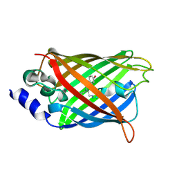
 | | Crystal Structure Analysis of eqFP611 Double Mutant T122R, N143S | | 分子名称: | Red fluorescent protein eqFP611 | | 著者 | Nar, H, Nienhaus, K, Nienhaus, U, Wiedenmann, J. | | 登録日 | 2008-08-14 | | 公開日 | 2008-09-23 | | 最終更新日 | 2023-11-15 | | 実験手法 | X-RAY DIFFRACTION (2.1 Å) | | 主引用文献 | Trans-cis isomerization is responsible for the red-shifted fluorescence in variants of the red fluorescent protein eqFP611.
J.Am.Chem.Soc., 130, 2008
|
|
3E5W
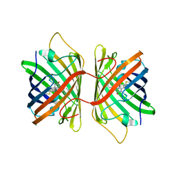 | | Crystal Structure Analysis of FP611 | | 分子名称: | Red fluorescent protein eqFP611 | | 著者 | Nienhaus, K, Nar, H, Heilker, R, Wiedenmann, J, Nienhaus, G.U. | | 登録日 | 2008-08-14 | | 公開日 | 2008-09-23 | | 最終更新日 | 2023-11-15 | | 実験手法 | X-RAY DIFFRACTION (1.71 Å) | | 主引用文献 | Trans-cis isomerization is responsible for the red-shifted fluorescence in variants of the red fluorescent protein eqFP611.
J.Am.Chem.Soc., 130, 2008
|
|
3EEO
 | |
3EK4
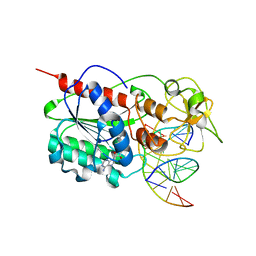
 | | Calcium-saturated GCaMP2 Monomer | | 分子名称: | CALCIUM ION, Myosin light chain kinase, Green fluorescent protein, ... | | 著者 | Akerboom, J, Velez Rivera, J.D, Looger, L.L, Schreiter, E.R. | | 登録日 | 2008-09-18 | | 公開日 | 2008-12-16 | | 最終更新日 | 2023-11-15 | | 実験手法 | X-RAY DIFFRACTION (2.65 Å) | | 主引用文献 | Crystal Structures of the GCaMP Calcium Sensor Reveal the Mechanism of Fluorescence Signal Change and Aid Rational Design
J.Biol.Chem., 284, 2009
|
|
3EK7
 | | Calcium-saturated GCaMP2 dimer | | 分子名称: | CALCIUM ION, Myosin light chain kinase, Green fluorescent protein, ... | | 著者 | Akerboom, J, Velez Rivera, J.D, Looger, L.L, Schreiter, E.R. | | 登録日 | 2008-09-18 | | 公開日 | 2008-12-16 | | 最終更新日 | 2023-11-15 | | 実験手法 | X-RAY DIFFRACTION (1.85 Å) | | 主引用文献 | Crystal Structures of the GCaMP Calcium Sensor Reveal the Mechanism of Fluorescence Signal Change and Aid Rational Design
J.Biol.Chem., 284, 2009
|
|
3EK8
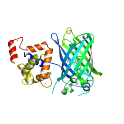
 | | Calcium-saturated GCaMP2 T116V/G87R mutant monomer | | 分子名称: | CALCIUM ION, Myosin light chain kinase, Green fluorescent protein, ... | | 著者 | Akerboom, J, Velez Rivera, J.D, Looger, L.L, Schreiter, E.R. | | 登録日 | 2008-09-19 | | 公開日 | 2008-12-16 | | 最終更新日 | 2023-11-15 | | 実験手法 | X-RAY DIFFRACTION (2.8 Å) | | 主引用文献 | Crystal Structures of the GCaMP Calcium Sensor Reveal the Mechanism of Fluorescence Signal Change and Aid Rational Design
J.Biol.Chem., 284, 2009
|
|
3EKH
 | | Calcium-saturated GCaMP2 T116V/K378W mutant monomer | | 分子名称: | CALCIUM ION, GLYCEROL, Myosin light chain kinase, ... | | 著者 | Akerboom, J, Velez Rivera, J.D, Looger, L.L, Schreiter, E.R. | | 登録日 | 2008-09-19 | | 公開日 | 2008-12-16 | | 最終更新日 | 2023-11-15 | | 実験手法 | X-RAY DIFFRACTION (2 Å) | | 主引用文献 | Crystal Structures of the GCaMP Calcium Sensor Reveal the Mechanism of Fluorescence Signal Change and Aid Rational Design
J.Biol.Chem., 284, 2009
|
|
3EKJ
 | | Calcium-free GCaMP2 (calcium binding deficient mutant) | | 分子名称: | Myosin light chain kinase, Green fluorescent protein, Calmodulin chimera | | 著者 | Akerboom, J, Velez Rivera, J.D, Looger, L.L, Schreiter, E.R. | | 登録日 | 2008-09-19 | | 公開日 | 2008-12-16 | | 最終更新日 | 2023-11-15 | | 実験手法 | X-RAY DIFFRACTION (2.8 Å) | | 主引用文献 | Crystal Structures of the GCaMP Calcium Sensor Reveal the Mechanism of Fluorescence Signal Change and Aid Rational Design
J.Biol.Chem., 284, 2009
|
|
3EOZ
 | | Crystal Structure of Phosphoglycerate Mutase from Plasmodium Falciparum, PFD0660w | | 分子名称: | GLYCEROL, PHOSPHATE ION, putative Phosphoglycerate mutase | | 著者 | Wernimont, A.K, Tempel, W, Lam, A, Zhao, Y, Lew, J, Lin, Y.H, Wasney, G, Vedadi, M, Kozieradzki, I, Cossar, D, Schapira, M, Weigelt, J, Arrowsmith, C.H, Bochkarev, A, Edwards, A.M, Hui, R, Pizarro, J, Hills, T, Structural Genomics Consortium (SGC) | | 登録日 | 2008-09-29 | | 公開日 | 2008-11-25 | | 最終更新日 | 2017-10-25 | | 実験手法 | X-RAY DIFFRACTION (2.4 Å) | | 主引用文献 | Characterization of a new phosphatase from Plasmodium.
Mol.Biochem.Parasitol., 179, 2011
|
|


