1CU2
 
 | | T4 LYSOZYME MUTANT L84M | | 分子名称: | 2-HYDROXYETHYL DISULFIDE, CHLORIDE ION, LYSOZYME | | 著者 | Gassner, N.C, Baase, W.A, Lindstrom, J.D, Lu, J, Matthews, B.W. | | 登録日 | 1999-08-20 | | 公開日 | 1999-11-10 | | 最終更新日 | 2024-02-07 | | 実験手法 | X-RAY DIFFRACTION (1.85 Å) | | 主引用文献 | Methionine and alanine substitutions show that the formation of wild-type-like structure in the carboxy-terminal domain of T4 lysozyme is a rate-limiting step in folding.
Biochemistry, 38, 1999
|
|
1CU3
 | | T4 LYSOZYME MUTANT V87M | | 分子名称: | 2-HYDROXYETHYL DISULFIDE, CHLORIDE ION, LYSOZYME | | 著者 | Gassner, N.C, Baase, W.A, Lindstrom, J.D, Lu, J, Matthews, B.W. | | 登録日 | 1999-08-20 | | 公開日 | 1999-11-10 | | 最終更新日 | 2024-02-07 | | 実験手法 | X-RAY DIFFRACTION (2.12 Å) | | 主引用文献 | Methionine and alanine substitutions show that the formation of wild-type-like structure in the carboxy-terminal domain of T4 lysozyme is a rate-limiting step in folding.
Biochemistry, 38, 1999
|
|
1CU4
 | | CRYSTAL STRUCTURE OF THE ANTI-PRION FAB 3F4 IN COMPLEX WITH ITS PEPTIDE EPITOPE | | 分子名称: | FAB HEAVY CHAIN, FAB LIGHT CHAIN, RECOGNITION PEPTIDE | | 著者 | Kanyo, Z.F, Pan, K.M, Williamson, R.A, Burton, D.R, Prusiner, S.B, Fletterick, R.J, Cohen, F.E. | | 登録日 | 1999-08-20 | | 公開日 | 2000-04-17 | | 最終更新日 | 2018-04-04 | | 実験手法 | X-RAY DIFFRACTION (2.9 Å) | | 主引用文献 | Antibody binding defines a structure for an epitope that participates in the PrPC-->PrPSc conformational change.
J.Mol.Biol., 293, 1999
|
|
1CU5
 | | T4 LYSOZYME MUTANT L91M | | 分子名称: | 2-HYDROXYETHYL DISULFIDE, CHLORIDE ION, LYSOZYME | | 著者 | Gassner, N.C, Baase, W.A, Lindstrom, J.D, Lu, J, Matthews, B.W. | | 登録日 | 1999-08-20 | | 公開日 | 1999-11-10 | | 最終更新日 | 2024-02-14 | | 実験手法 | X-RAY DIFFRACTION (2.05 Å) | | 主引用文献 | Methionine and alanine substitutions show that the formation of wild-type-like structure in the carboxy-terminal domain of T4 lysozyme is a rate-limiting step in folding.
Biochemistry, 38, 1999
|
|
1CU6
 | | T4 LYSOZYME MUTANT L91A | | 分子名称: | 2-HYDROXYETHYL DISULFIDE, CHLORIDE ION, LYSOZYME | | 著者 | Gassner, N.C, Baase, W.A, Lindstrom, J.D, Lu, J, Matthews, B.W. | | 登録日 | 1999-08-20 | | 公開日 | 1999-11-17 | | 最終更新日 | 2024-02-07 | | 実験手法 | X-RAY DIFFRACTION (2.1 Å) | | 主引用文献 | Methionine and alanine substitutions show that the formation of wild-type-like structure in the carboxy-terminal domain of T4 lysozyme is a rate-limiting step in folding.
Biochemistry, 38, 1999
|
|
1CUA
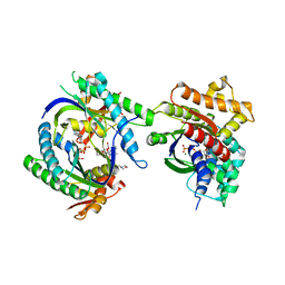
 | | CUTINASE, N172K MUTANT | | 分子名称: | CUTINASE | | 著者 | Longhi, S, Cambillau, C. | | 登録日 | 1995-11-16 | | 公開日 | 1996-07-11 | | 最終更新日 | 2021-11-03 | | 実験手法 | X-RAY DIFFRACTION (1.8 Å) | | 主引用文献 | Dynamics of Fusarium solani cutinase investigated through structural comparison among different crystal forms of its variants.
Proteins, 26, 1996
|
|
1CUB
 | |
1CUC
 | |
1CUD
 | |
1CUE
 | | CUTINASE, Q121L MUTANT | | 分子名称: | CUTINASE | | 著者 | Martinez, C, Cambillau, C. | | 登録日 | 1995-11-16 | | 公開日 | 1996-07-11 | | 最終更新日 | 2021-11-03 | | 実験手法 | X-RAY DIFFRACTION (2.1 Å) | | 主引用文献 | Dynamics of Fusarium solani cutinase investigated through structural comparison among different crystal forms of its variants.
Proteins, 26, 1996
|
|
1CUF
 | | CUTINASE, R156L MUTANT | | 分子名称: | CUTINASE | | 著者 | Longhi, S, Cambillau, C. | | 登録日 | 1995-11-16 | | 公開日 | 1996-07-11 | | 最終更新日 | 2021-11-03 | | 実験手法 | X-RAY DIFFRACTION (1.75 Å) | | 主引用文献 | Dynamics of Fusarium solani cutinase investigated through structural comparison among different crystal forms of its variants.
Proteins, 26, 1996
|
|
1CUG
 | | CUTINASE, R17E, N172K MUTANT | | 分子名称: | CUTINASE | | 著者 | Longhi, S, Cambillau, C. | | 登録日 | 1995-11-16 | | 公開日 | 1996-07-11 | | 最終更新日 | 2021-11-03 | | 実験手法 | X-RAY DIFFRACTION (1.75 Å) | | 主引用文献 | Dynamics of Fusarium solani cutinase investigated through structural comparison among different crystal forms of its variants.
Proteins, 26, 1996
|
|
1CUH
 | | CUTINASE, R196E MUTANT | | 分子名称: | CUTINASE | | 著者 | Longhi, S, Cambillau, C. | | 登録日 | 1995-11-16 | | 公開日 | 1996-07-11 | | 最終更新日 | 2021-11-03 | | 実験手法 | X-RAY DIFFRACTION (1.75 Å) | | 主引用文献 | Dynamics of Fusarium solani cutinase investigated through structural comparison among different crystal forms of its variants.
Proteins, 26, 1996
|
|
1CUI
 | | CUTINASE, S120A MUTANT | | 分子名称: | CUTINASE | | 著者 | Martinez, C, Cambillau, C. | | 登録日 | 1995-11-16 | | 公開日 | 1996-07-11 | | 最終更新日 | 2021-11-03 | | 実験手法 | X-RAY DIFFRACTION (2.5 Å) | | 主引用文献 | Dynamics of Fusarium solani cutinase investigated through structural comparison among different crystal forms of its variants.
Proteins, 26, 1996
|
|
1CUJ
 | | CUTINASE, S120C MUTANT | | 分子名称: | CUTINASE | | 著者 | Martinez, C, Cambillau, C. | | 登録日 | 1995-11-16 | | 公開日 | 1996-07-11 | | 最終更新日 | 2021-11-03 | | 実験手法 | X-RAY DIFFRACTION (1.6 Å) | | 主引用文献 | Dynamics of Fusarium solani cutinase investigated through structural comparison among different crystal forms of its variants.
Proteins, 26, 1996
|
|
1CUK
 | |
1CUL
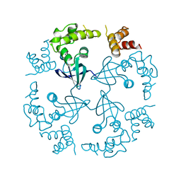
 | | COMPLEX OF GS-ALPHA WITH THE CATALYTIC DOMAINS OF MAMMALIAN ADENYLYL CYCLASE: COMPLEX WITH 2',5'-DIDEOXY-ADENOSINE 3'-TRIPHOSPHATE AND MG | | 分子名称: | 2',5'-DIDEOXY-ADENOSINE 3'-MONOPHOSPHATE, 2-(N-MORPHOLINO)-ETHANESULFONIC ACID, 5'-GUANOSINE-DIPHOSPHATE-MONOTHIOPHOSPHATE, ... | | 著者 | Tesmer, J.J.G, Dessauer, C.A, Sunahara, R.K, Johnson, R.A, Gilman, A.G, Sprang, S.R. | | 登録日 | 1999-08-20 | | 公開日 | 2001-01-10 | | 最終更新日 | 2024-02-07 | | 実験手法 | X-RAY DIFFRACTION (2.4 Å) | | 主引用文献 | Molecular basis for P-site inhibition of adenylyl cyclase.
Biochemistry, 39, 2000
|
|
1CUN
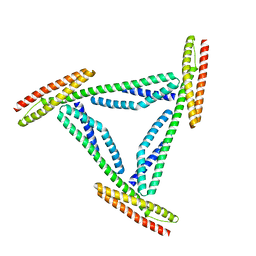 | | CRYSTAL STRUCTURE OF REPEATS 16 AND 17 OF CHICKEN BRAIN ALPHA SPECTRIN | | 分子名称: | PROTEIN (ALPHA SPECTRIN) | | 著者 | Grum, V.L, Li, D, MacDonald, R.I, Mondragon, A. | | 登録日 | 1999-08-20 | | 公開日 | 1999-10-06 | | 最終更新日 | 2024-02-07 | | 実験手法 | X-RAY DIFFRACTION (2 Å) | | 主引用文献 | Structures of two repeats of spectrin suggest models of flexibility.
Cell(Cambridge,Mass.), 98, 1999
|
|
1CUO
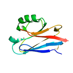 | | CRYSTAL STRUCTURE ANALYSIS OF ISOMER-2 AZURIN FROM METHYLOMONAS J | | 分子名称: | COPPER (II) ION, PROTEIN (AZURIN ISO-2) | | 著者 | Inoue, T, Nishio, N, Kai, Y, Suzuki, S, Kataoka, K. | | 登録日 | 1999-08-21 | | 公開日 | 2000-08-23 | | 最終更新日 | 2011-07-13 | | 実験手法 | X-RAY DIFFRACTION (1.6 Å) | | 主引用文献 | The significance of the flexible loop in the azurin (Az-iso2) from the obligate methylotroph Methylomonas sp. strain J.
J.Mol.Biol., 333, 2003
|
|
1CUP
 | | METHIONINE CORE MUTANT OF T4 LYSOZYME | | 分子名称: | 2-HYDROXYETHYL DISULFIDE, CHLORIDE ION, LYSOZYME | | 著者 | Gassner, N.C, Baase, W.A, Lindstrom, J.D, Lu, J, Matthews, B.W. | | 登録日 | 1999-08-20 | | 公開日 | 1999-11-10 | | 最終更新日 | 2024-02-07 | | 実験手法 | X-RAY DIFFRACTION (1.89 Å) | | 主引用文献 | Methionine and alanine substitutions show that the formation of wild-type-like structure in the carboxy-terminal domain of T4 lysozyme is a rate-limiting step in folding.
Biochemistry, 38, 1999
|
|
1CUQ
 | | T4 LYSOZYME MUTANT V103M | | 分子名称: | 2-HYDROXYETHYL DISULFIDE, CHLORIDE ION, LYSOZYME | | 著者 | Gassner, N.C, Baase, W.A, Lindstrom, J.D, Lu, J, Matthews, B.W. | | 登録日 | 1999-08-20 | | 公開日 | 1999-11-10 | | 最終更新日 | 2024-02-07 | | 実験手法 | X-RAY DIFFRACTION (2.05 Å) | | 主引用文献 | Methionine and alanine substitutions show that the formation of wild-type-like structure in the carboxy-terminal domain of T4 lysozyme is a rate-limiting step in folding.
Biochemistry, 38, 1999
|
|
1CUR
 | | REDUCED RUSTICYANIN, NMR | | 分子名称: | COPPER (II) ION, CU(I) RUSTICYANIN | | 著者 | Botuyan, M.V, Dyson, H.J. | | 登録日 | 1996-04-19 | | 公開日 | 1996-11-08 | | 最終更新日 | 2024-05-22 | | 実験手法 | SOLUTION NMR | | 主引用文献 | NMR solution structure of Cu(I) rusticyanin from Thiobacillus ferrooxidans: structural basis for the extreme acid stability and redox potential.
J.Mol.Biol., 263, 1996
|
|
1CUS
 | |
1CUU
 | | CUTINASE, A199C MUTANT | | 分子名称: | CUTINASE | | 著者 | Longhi, S, Cambillau, C. | | 登録日 | 1995-11-16 | | 公開日 | 1996-07-11 | | 最終更新日 | 2021-11-03 | | 実験手法 | X-RAY DIFFRACTION (1.69 Å) | | 主引用文献 | Dynamics of Fusarium solani cutinase investigated through structural comparison among different crystal forms of its variants.
Proteins, 26, 1996
|
|
1CUV
 | | CUTINASE, A85F MUTANT | | 分子名称: | CUTINASE | | 著者 | Longhi, S, Cambillau, C. | | 登録日 | 1995-11-16 | | 公開日 | 1996-07-11 | | 最終更新日 | 2021-11-03 | | 実験手法 | X-RAY DIFFRACTION (2.01 Å) | | 主引用文献 | Dynamics of Fusarium solani cutinase investigated through structural comparison among different crystal forms of its variants.
Proteins, 26, 1996
|
|


