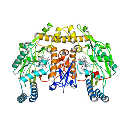
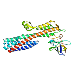
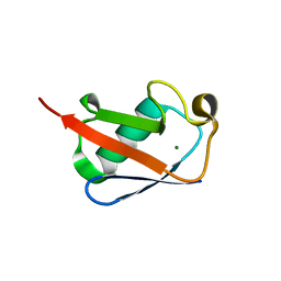
5VUX
 
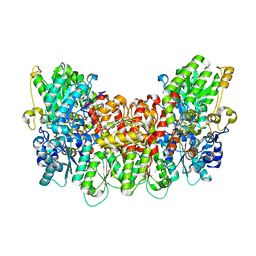
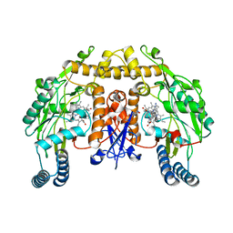 | | Structure of human neuronal nitric oxide synthase heme domain in complex with 7-(((4-(Dimethylamino)benzyl)amino)methyl)quinolin-2-amine | | 分子名称: | 5,6,7,8-TETRAHYDROBIOPTERIN, 7-[({[4-(dimethylamino)phenyl]methyl}amino)methyl]quinolin-2-amine, Nitric oxide synthase, ... | | 著者 | Huiying, L, Thomas, L.P. | | 登録日 | 2017-05-19 | | 公開日 | 2017-08-23 | | 最終更新日 | 2023-10-04 | | 実験手法 | X-RAY DIFFRACTION (2.3 Å) | | 主引用文献 | Hydrophilic, Potent, and Selective 7-Substituted 2-Aminoquinolines as Improved Human Neuronal Nitric Oxide Synthase Inhibitors.
J. Med. Chem., 60, 2017
|
|
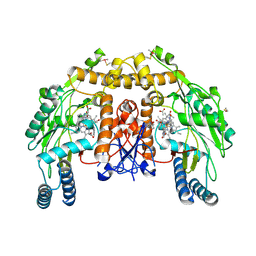
5VV8
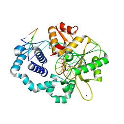
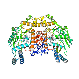 | | Structure of bovine endothelial nitric oxide synthase heme domain in complex with 4-(2-(((2-Aminoquinolin-7-yl)methyl)amino)ethyl)-2-methylbenzonitrile | | 分子名称: | 4-(2-{[(2-aminoquinolin-7-yl)methyl]amino}ethyl)-2-methylbenzonitrile, 5,6,7,8-TETRAHYDROBIOPTERIN, ACETATE ION, ... | | 著者 | Li, H, Poulos, T.L. | | 登録日 | 2017-05-19 | | 公開日 | 2017-08-16 | | 最終更新日 | 2023-10-04 | | 実験手法 | X-RAY DIFFRACTION (2.15 Å) | | 主引用文献 | Hydrophilic, Potent, and Selective 7-Substituted 2-Aminoquinolines as Improved Human Neuronal Nitric Oxide Synthase Inhibitors.
J. Med. Chem., 60, 2017
|
|
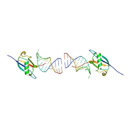
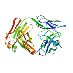
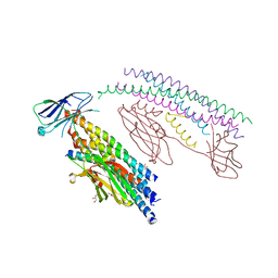
5VVK
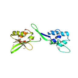
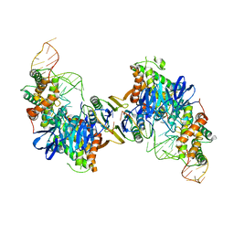 | | Cas1-Cas2 bound to full-site mimic | | 分子名称: | CRISPR-associated endonuclease Cas1, CRISPR-associated endoribonuclease Cas2, DNA (5'-D(*GP*AP*CP*CP*AP*CP*CP*AP*GP*TP*G)-3'), ... | | 著者 | Wright, A.V, Knott, G.J, Doxzen, K.D, Doudna, J.A. | | 登録日 | 2017-05-19 | | 公開日 | 2017-08-02 | | 最終更新日 | 2023-10-04 | | 実験手法 | X-RAY DIFFRACTION (2.9 Å) | | 主引用文献 | Structures of the CRISPR genome integration complex.
Science, 357, 2017
|
|
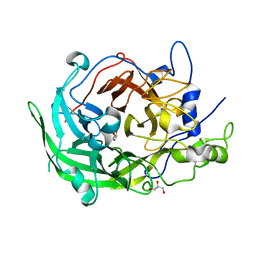
5VUL
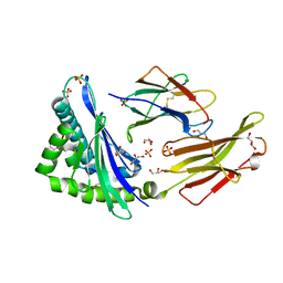
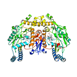 | | Structure of rat neuronal nitric oxide synthase heme domain in complex with 7-(((4-(Dimethylamino)phenethyl)amino)methyl)quinolin-2-amine | | 分子名称: | 5,6,7,8-TETRAHYDROBIOPTERIN, 7-[({2-[4-(dimethylamino)phenyl]ethyl}amino)methyl]quinolin-2-amine, ACETATE ION, ... | | 著者 | Li, H, Poulos, T.L. | | 登録日 | 2017-05-19 | | 公開日 | 2017-08-16 | | 最終更新日 | 2023-10-04 | | 実験手法 | X-RAY DIFFRACTION (2 Å) | | 主引用文献 | Hydrophilic, Potent, and Selective 7-Substituted 2-Aminoquinolines as Improved Human Neuronal Nitric Oxide Synthase Inhibitors.
J. Med. Chem., 60, 2017
|
|
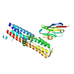
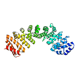
8ID2
 | | Crystal structure of the ubiquitin-like domain in the SF3A1 subunit of human U2 snRNP complexed with the stem-loop 4 of U1 snRNA | | 分子名称: | RNA (5'-R(*GP*GP*GP*GP*AP*CP*UP*GP*CP*GP*UP*UP*CP*GP*CP*GP*CP*UP*UP*UP*CP*CP*CP*C)-3'), Splicing factor 3A subunit 1 | | 著者 | Terawaki, S, Nameki, N, Kuwasako, K. | | 登録日 | 2023-02-12 | | 公開日 | 2023-05-17 | | 最終更新日 | 2024-05-29 | | 実験手法 | X-RAY DIFFRACTION (1.8 Å) | | 主引用文献 | Structural insights into recognition of SL4, the UUCG stem-loop, of human U1 snRNA by the ubiquitin-like domain, including the C-terminal tail in the SF3A1 subunit of U2 snRNP.
J.Biochem., 174, 2023
|
|
5VUW
 | | Structure of human neuronal nitric oxide synthase heme domain in complex with 7-(((3-(Dimethylamino)benzyl)amino)methyl)quinolin-2-amine | | 分子名称: | 5,6,7,8-TETRAHYDROBIOPTERIN, 7-[[[3-(dimethylamino)phenyl]methylamino]methyl]quinolin-2-amine, Nitric oxide synthase, ... | | 著者 | Huiying, L, Thomas, L.P. | | 登録日 | 2017-05-19 | | 公開日 | 2017-08-23 | | 最終更新日 | 2023-10-04 | | 実験手法 | X-RAY DIFFRACTION (2.03 Å) | | 主引用文献 | Hydrophilic, Potent, and Selective 7-Substituted 2-Aminoquinolines as Improved Human Neuronal Nitric Oxide Synthase Inhibitors.
J. Med. Chem., 60, 2017
|
|
5VV6
 | | Structure of bovine endothelial nitric oxide synthase heme domain in complex with 7-(((4-(Dimethylamino)phenethyl)amino)methyl)quinolin-2-amine | | 分子名称: | 5,6,7,8-TETRAHYDROBIOPTERIN, 7-[({2-[4-(dimethylamino)phenyl]ethyl}amino)methyl]quinolin-2-amine, ACETATE ION, ... | | 著者 | Li, H, Poulos, T.L. | | 登録日 | 2017-05-19 | | 公開日 | 2017-08-16 | | 最終更新日 | 2019-12-25 | | 実験手法 | X-RAY DIFFRACTION (2 Å) | | 主引用文献 | Hydrophilic, Potent, and Selective 7-Substituted 2-Aminoquinolines as Improved Human Neuronal Nitric Oxide Synthase Inhibitors.
J. Med. Chem., 60, 2017
|
|
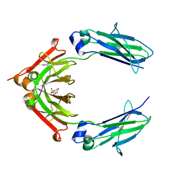
5VXM
 | | 2.05 A resolution structure of IpaD from Shigella flexneri in complex with single-domain antibody 20ipaD | | 分子名称: | Invasin IpaD, Single-domain antibody 20ipaD | | 著者 | Barta, M.L, Lovell, S, Battaile, K.P, Picking, W.D, Picking, W.L. | | 登録日 | 2017-05-23 | | 公開日 | 2017-08-30 | | 最終更新日 | 2023-10-04 | | 実験手法 | X-RAY DIFFRACTION (2.05 Å) | | 主引用文献 | 2.05 A resolution structure of IpaD from Shigella flexneri in complex with single-domain antibody 20ipaD
To Be Published
|
|
5VXK
 | | 2.55 A resolution structure of IpaD from Shigella flexneri in complex with single-domain antibody JMK-H2 | | 分子名称: | Invasin IpaD, single-domain antibody JMK-H2 | | 著者 | Barta, M.L, Lovell, S, Battaile, K.P, Picking, W.D, Picking, W.L. | | 登録日 | 2017-05-23 | | 公開日 | 2017-08-30 | | 最終更新日 | 2023-10-04 | | 実験手法 | X-RAY DIFFRACTION (2.55 Å) | | 主引用文献 | 2.55 A resolution structure of IpaD from Shigella flexneri in complex with single-domain antibody JMK-H2
To Be Published
|
|
5W38
 | | 1.80A resolution structure of human IgG3 Fc (N392K) | | 分子名称: | CITRATE ANION, Immunoglobulin heavy constant gamma 3, beta-D-mannopyranose-(1-4)-2-acetamido-2-deoxy-beta-D-glucopyranose-(1-4)-2-acetamido-2-deoxy-beta-D-glucopyranose | | 著者 | Lovell, S, Mehzabeen, N, Battaile, K.P, Shah, I, Tolbert, T.J. | | 登録日 | 2017-06-07 | | 公開日 | 2017-10-18 | | 最終更新日 | 2023-10-04 | | 実験手法 | X-RAY DIFFRACTION (1.8 Å) | | 主引用文献 | Structural characterization of the Man5 glycoform of human IgG3 Fc.
Mol. Immunol., 92, 2017
|
|
5VYH
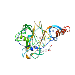 | | Crystal Structure of MERS-CoV S1 N-terminal Domain | | 分子名称: | (4S)-2-METHYL-2,4-PENTANEDIOL, 2-acetamido-2-deoxy-beta-D-glucopyranose, FOLIC ACID, ... | | 著者 | Wang, N, Wrapp, D, Pallesen, J, Ward, A.B, McLellan, J.S. | | 登録日 | 2017-05-25 | | 公開日 | 2017-08-30 | | 最終更新日 | 2024-04-03 | | 実験手法 | X-RAY DIFFRACTION (2 Å) | | 主引用文献 | Immunogenicity and structures of a rationally designed prefusion MERS-CoV spike antigen.
Proc. Natl. Acad. Sci. U.S.A., 114, 2017
|
|
8IN9
 | | The structure of the GfsA KSQ-AT didomain in complex with the GfsA ACP domain | | 分子名称: | N-[2-(acetylamino)ethyl]-N~3~-[(2R)-2-hydroxy-3,3-dimethyl-4-(phosphonooxy)butanoyl]-beta-alaninamide, Polyketide synthase | | 著者 | Chisuga, T, Murakami, S, Miyanaga, A, Kudo, F, Eguchi, T. | | 登録日 | 2023-03-09 | | 公開日 | 2023-05-31 | | 最終更新日 | 2023-06-28 | | 実験手法 | X-RAY DIFFRACTION (3.4 Å) | | 主引用文献 | Structure-Based Analysis of Transient Interactions between Ketosynthase-like Decarboxylase and Acyl Carrier Protein in a Loading Module of Modular Polyketide Synthase.
Acs Chem.Biol., 18, 2023
|
|
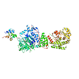
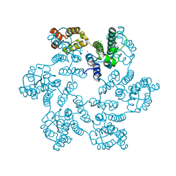
5VQ3
 | | Nitrogenase Cp1 at pH 6.5 | | 分子名称: | 3-HYDROXY-3-CARBOXY-ADIPIC ACID, FE (II) ION, FE(8)-S(7) CLUSTER, ... | | 著者 | Morrison, C.N, Spatzal, T, Rees, D.C. | | 登録日 | 2017-05-07 | | 公開日 | 2017-07-26 | | 最終更新日 | 2023-10-04 | | 実験手法 | X-RAY DIFFRACTION (1.72 Å) | | 主引用文献 | Reversible Protonated Resting State of the Nitrogenase Active Site.
J. Am. Chem. Soc., 139, 2017
|
|
5VS2
 | |
5W2F
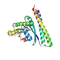
 | | Crystal Structure of the C-terminal Domain of Human eIF2D at 1.4 A resolution | | 分子名称: | Eukaryotic translation initiation factor 2D, FORMIC ACID | | 著者 | Vaidya, A.T, Lomakin, I.B, Joseph, N.N, Dmitriev, S.E, Steitz, T.A. | | 登録日 | 2017-06-06 | | 公開日 | 2017-08-02 | | 最終更新日 | 2024-03-13 | | 実験手法 | X-RAY DIFFRACTION (1.4 Å) | | 主引用文献 | Crystal Structure of the C-terminal Domain of Human eIF2D and Its Implications on Eukaryotic Translation Initiation.
J. Mol. Biol., 429, 2017
|
|
5VZ5
 | | Crystal structure of an anaplastic lymphoma kinase-derived neuroblastoma tumor antigen bound to the Human Major Histocompatibility Complex Class I molecule HLA-B*1501 | | 分子名称: | Beta-2-microglobulin, GLYCEROL, HLA class I histocompatibility antigen, ... | | 著者 | Toor, J, Rao, A.A, Salama, S, Tripathi, S, Haussler, D, Sgourakis, N.G. | | 登録日 | 2017-05-26 | | 公開日 | 2018-03-21 | | 最終更新日 | 2023-10-04 | | 実験手法 | X-RAY DIFFRACTION (2.5901 Å) | | 主引用文献 | A Recurrent Mutation in Anaplastic Lymphoma Kinase with Distinct Neoepitope Conformations.
Front Immunol, 9, 2018
|
|
5W46
 | |
5W24
 | | Crystal Structure of 5C4 Fab | | 分子名称: | (R,R)-2,3-BUTANEDIOL, 5C4 Fab Heavy Chain, 5C4 Fab Light Chain | | 著者 | Battles, M.B, McLellan, J.S, Taher, N.M. | | 登録日 | 2017-06-05 | | 公開日 | 2017-12-06 | | 最終更新日 | 2023-10-04 | | 実験手法 | X-RAY DIFFRACTION (1.5 Å) | | 主引用文献 | Structural basis of respiratory syncytial virus subtype-dependent neutralization by an antibody targeting the fusion glycoprotein.
Nat Commun, 8, 2017
|
|
8IBT
 | | Crystal structure of GH42 beta-galactosidase BiBga42A from Bifidobacterium longum subspecies infantis E318S mutant in complex with lacto-N-tetraose | | 分子名称: | Beta-galactosidase, beta-D-galactopyranose-(1-3)-2-acetamido-2-deoxy-beta-D-glucopyranose-(1-3)-beta-D-galactopyranose-(1-4)-beta-D-glucopyranose | | 著者 | Hidaka, M, Fushinobu, S, Gotoh, A, Katayama, T. | | 登録日 | 2023-02-10 | | 公開日 | 2023-06-07 | | 最終更新日 | 2023-12-20 | | 実験手法 | X-RAY DIFFRACTION (2.2 Å) | | 主引用文献 | Substrate recognition mode of a glycoside hydrolase family 42 beta-galactosidase from Bifidobacterium longum subspecies infantis ( Bi Bga42A) revealed by crystallographic and mutational analyses.
Microbiome Res Rep, 2, 2023
|
|
8IBS
 | | Crystal structure of GH42 beta-galactosidase BiBga42A from Bifidobacterium longum subspecies infantis E160A/E318A mutant in complex with galactose | | 分子名称: | Beta-galactosidase, alpha-D-galactopyranose | | 著者 | Hidaka, M, Fushinobu, S, Gotoh, A, Katayama, T. | | 登録日 | 2023-02-10 | | 公開日 | 2023-06-07 | | 最終更新日 | 2023-12-20 | | 実験手法 | X-RAY DIFFRACTION (1.9 Å) | | 主引用文献 | Substrate recognition mode of a glycoside hydrolase family 42 beta-galactosidase from Bifidobacterium longum subspecies infantis ( Bi Bga42A) revealed by crystallographic and mutational analyses.
Microbiome Res Rep, 2, 2023
|
|
5W41
 | |
8I2Q
 | |
5W4Q
 | |
8IJ9
 | | Crystal structure of the ELKS1/Rab6B complex | | 分子名称: | ELKS/Rab6-interacting/CAST family member 1, GUANOSINE-5'-TRIPHOSPHATE, MAGNESIUM ION, ... | | 著者 | Jin, G, Wei, Z. | | 登録日 | 2023-02-26 | | 公開日 | 2023-06-14 | | 最終更新日 | 2024-05-29 | | 実験手法 | X-RAY DIFFRACTION (2.04 Å) | | 主引用文献 | Structural basis of ELKS/Rab6B interaction and its role in vesicle capturing enhanced by liquid-liquid phase separation.
J.Biol.Chem., 299, 2023
|
|
5W5C
 | |


