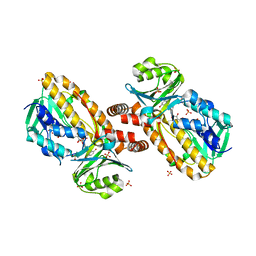
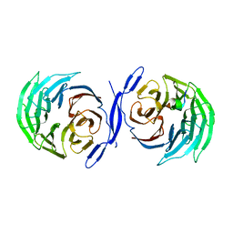
1W14
 
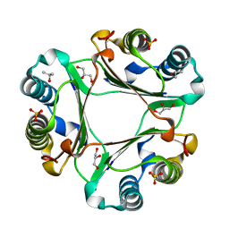
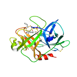 | | UROKINASE TYPE PLASMINOGEN ACTIVATOR | | 分子名称: | N-[(2-PHENYLETHYL)SULFONYL]-D-SERYL-N-[(1S)-4-[(DIAMINOMETHYLENE)AMINO]-1-(HYDROXYMETHYL)BUTYL]-L-ALANINAMIDE, SULFATE ION, UROKINASE-TYPE PLASMINOGEN ACTIVATOR | | 著者 | Jacob, U. | | 登録日 | 2004-06-15 | | 公開日 | 2008-05-20 | | 最終更新日 | 2019-05-22 | | 実験手法 | X-RAY DIFFRACTION (2.2 Å) | | 主引用文献 | Crystals of Urokinase Type Plasminogen Activator Complexes Reveal the Binding Mode of Peptidomimetic Inhibitors.
J.Mol.Biol., 328, 2003
|
|
8T8T
 | |
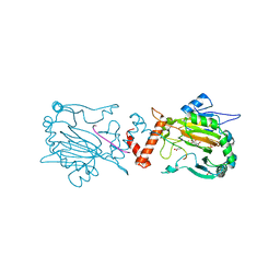
3S9L
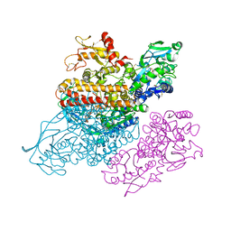 | | Complex between transferrin receptor 1 and transferrin with iron in the N-Lobe, cryocooled 2 | | 分子名称: | 2-acetamido-2-deoxy-beta-D-glucopyranose, CALCIUM ION, CARBONATE ION, ... | | 著者 | Eckenroth, B.E, Steere, A.N, Mason, A.B, Everse, S.J. | | 登録日 | 2011-06-01 | | 公開日 | 2011-08-10 | | 最終更新日 | 2020-07-29 | | 実験手法 | X-RAY DIFFRACTION (3.22 Å) | | 主引用文献 | How the binding of human transferrin primes the transferrin receptor potentiating iron release at endosomal pH.
Proc.Natl.Acad.Sci.USA, 108, 2011
|
|
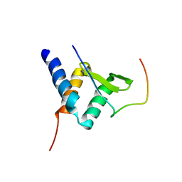
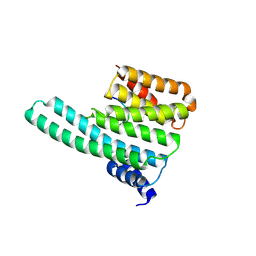
2BIV
 | | Crystal structure of the wild-type MBT domains of Human SCML2 | | 分子名称: | IODIDE ION, SEX COMB ON MIDLEG-LIKE PROTEIN 2, SODIUM ION | | 著者 | Santiveri, C.M, Allen, M.D, Sait, F, Bycroft, M. | | 登録日 | 2005-01-26 | | 公開日 | 2005-02-08 | | 最終更新日 | 2023-12-13 | | 実験手法 | X-RAY DIFFRACTION (1.7 Å) | | 主引用文献 | The Malignant Brain Tumor Repeats of Human Scml2 Bind to Peptides Containing Monomethylated Lysine.
J.Mol.Biol., 382, 2008
|
|
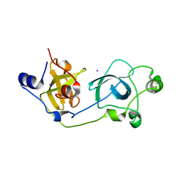
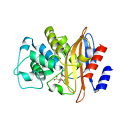
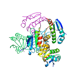
1YKJ
 | | A45G p-hydroxybenzoate hydroxylase with p-hydroxybenzoate bound | | 分子名称: | FLAVIN-ADENINE DINUCLEOTIDE, P-HYDROXYBENZOIC ACID, P-hydroxybenzoate hydroxylase, ... | | 著者 | Cole, L.J, Gatti, D.L, Entsch, B, Ballou, D.P. | | 登録日 | 2005-01-18 | | 公開日 | 2005-07-26 | | 最終更新日 | 2023-08-23 | | 実験手法 | X-RAY DIFFRACTION (2 Å) | | 主引用文献 | Removal of a methyl group causes global changes in p-hydroxybenzoate hydroxylase.
Biochemistry, 44, 2005
|
|
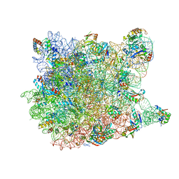
1YI2
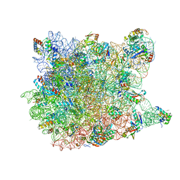 | | Crystal Structure Of Erythromycin Bound To The G2099A Mutant 50S Ribosomal Subunit Of Haloarcula Marismortui | | 分子名称: | 23S Ribosomal RNA, 50S RIBOSOMAL PROTEIN L10E, 50S RIBOSOMAL PROTEIN L11P, ... | | 著者 | Tu, D, Blaha, G, Moore, P.B, Steitz, T.A. | | 登録日 | 2005-01-11 | | 公開日 | 2005-04-26 | | 最終更新日 | 2024-02-14 | | 実験手法 | X-RAY DIFFRACTION (2.65 Å) | | 主引用文献 | Structures of MLSBK antibiotics bound to mutated large ribosomal subunits provide a structural explanation for resistance.
Cell(Cambridge,Mass.), 121, 2005
|
|
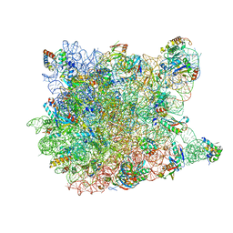
1YHQ
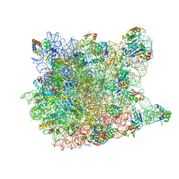 | | Crystal Structure Of Azithromycin Bound To The G2099A Mutant 50S Ribosomal Subunit Of Haloarcula Marismortui | | 分子名称: | 23S Ribosomal RNA, 50S RIBOSOMAL PROTEIN L10E, 50S RIBOSOMAL PROTEIN L11P, ... | | 著者 | Tu, D, Blaha, G, Moore, P.B, Steitz, T.A. | | 登録日 | 2005-01-10 | | 公開日 | 2005-04-26 | | 最終更新日 | 2024-02-14 | | 実験手法 | X-RAY DIFFRACTION (2.4 Å) | | 主引用文献 | Structures of MLSBK antibiotics bound to mutated large ribosomal subunits provide a structural explanation for resistance.
Cell(Cambridge,Mass.), 121, 2005
|
|
1DUQ
 | |
1YIT
 | | Crystal Structure Of Virginiamycin M and S Bound To The 50S Ribosomal Subunit Of Haloarcula Marismortui | | 分子名称: | 23S RIBOSOMAL RNA, 50S RIBOSOMAL PROTEIN L10E, 50S RIBOSOMAL PROTEIN L11P, ... | | 著者 | Tu, D, Blaha, G, Moore, P.B, Steitz, T.A. | | 登録日 | 2005-01-13 | | 公開日 | 2005-04-26 | | 最終更新日 | 2024-07-10 | | 実験手法 | X-RAY DIFFRACTION (2.8 Å) | | 主引用文献 | Structures of Mlsbk Antibiotics Bound to Mutated Large Ribosomal Subunits Provide a Structural Explanation for Resistance.
Cell(Cambridge,Mass.), 121, 2005
|
|
3CR6
 | |
2CE8
 | |
1W7F
 | |
3D8C
 | | Factor inhibiting HIF-1 alpha D201G mutant in complex with ZN(II), alpha-ketoglutarate and HIF-1 alpha 19mer | | 分子名称: | 2-OXOGLUTARIC ACID, GLYCEROL, HYPOXIA-INDUCIBLE FACTOR 1 ALPHA, ... | | 著者 | McDonough, M.A, Chowdhury, R, Schofield, C.J. | | 登録日 | 2008-05-23 | | 公開日 | 2008-08-12 | | 最終更新日 | 2023-08-30 | | 実験手法 | X-RAY DIFFRACTION (2.1 Å) | | 主引用文献 | Evidence that two enzyme-derived histidine ligands are sufficient for iron binding and catalysis by factor inhibiting HIF (FIH).
J.Biol.Chem., 283, 2008
|
|
2MV7
 | |
2MUK
 | |
3D96
 | |
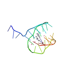
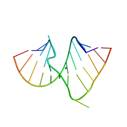
1CSL
 | | CRYSTAL STRUCTURE OF THE RRE HIGH AFFINITY SITE | | 分子名称: | 5'-R(*AP*AP*CP*GP*GP*GP*CP*GP*CP*AP*GP*AP*A)-3', 5'-R(*UP*CP*UP*GP*AP*CP*GP*GP*UP*AP*CP*GP*UP*UP*U)-3' | | 著者 | Ippolito, J.A, Steitz, T.A. | | 登録日 | 1999-08-18 | | 公開日 | 2000-02-14 | | 最終更新日 | 2024-02-07 | | 実験手法 | X-RAY DIFFRACTION (1.6 Å) | | 主引用文献 | The structure of the HIV-1 RRE high affinity rev binding site at 1.6 A resolution.
J.Mol.Biol., 295, 2000
|
|
2CMN
 | | A Proximal Arginine Residue in the Switching Mechanism of the FixL Oxygen Sensor | | 分子名称: | PROTOPORPHYRIN IX CONTAINING FE, SENSOR PROTEIN FIXL | | 著者 | Gilles-Gonzalez, M.-A, Caceres, A.I, Silva Sousa, E.H, Tomchick, D.R, Brautigam, C.A, Gonzalez, C, Machius, M. | | 登録日 | 2006-05-11 | | 公開日 | 2007-05-15 | | 最終更新日 | 2023-12-13 | | 実験手法 | X-RAY DIFFRACTION (2.3 Å) | | 主引用文献 | A Proximal Arginine R206 Participates in Switching of the Bradyrhizobium Japonicum Fixl Oxygen Sensor
J.Mol.Biol., 360, 2006
|
|
3DDK
 | | Coxsackievirus B3 3Dpol RNA Dependent RNA Polymerase | | 分子名称: | RNA polymerase B3 3Dpol, SODIUM ION, SULFATE ION | | 著者 | Campagnola, G, Weygandt, M.H, Scoggin, K.E, Peersen, O.B. | | 登録日 | 2008-06-05 | | 公開日 | 2008-09-23 | | 最終更新日 | 2023-08-30 | | 実験手法 | X-RAY DIFFRACTION (2.25 Å) | | 主引用文献 | Crystal Structure of Coxsackievirus B3 3Dpol Highlights Functional Importance of Residue 5 in Picornaviral Polymerases
J.Virol., 82, 2008
|
|
3DAI
 | | Crystal structure of the bromodomain of the human ATAD2 | | 分子名称: | ATPase family AAA domain-containing protein 2, CHLORIDE ION, SULFATE ION | | 著者 | Filippakopoulos, P, Keates, T, Picaud, S, Fedorov, O, Roos, A.K, von Delft, F, Arrowsmith, C.H, Edwards, A.M, Bountra, C, Knapp, S, Structural Genomics Consortium (SGC) | | 登録日 | 2008-05-29 | | 公開日 | 2008-09-09 | | 最終更新日 | 2023-08-30 | | 実験手法 | X-RAY DIFFRACTION (1.95 Å) | | 主引用文献 | Histone recognition and large-scale structural analysis of the human bromodomain family.
Cell(Cambridge,Mass.), 149, 2012
|
|
1VQ6
 | |
2C1N
 | | Molecular basis for the recognition of phosphorylated and phosphoacetylated histone H3 by 14-3-3 | | 分子名称: | 14-3-3 PROTEIN ZETA/DELTA, HISTONE H3 ACETYLPHOSPHOPEPTIDE | | 著者 | Welburn, J.P.I, Macdonald, N, Noble, M.E.M, Nguyen, A, Yaffe, M.B, Clynes, D, Moggs, J.G, Orphanides, G, Thomson, S, Edmunds, J.W, Clayton, A.L, Endicott, J.A, Mahadevan, L.C. | | 登録日 | 2005-09-16 | | 公開日 | 2005-11-02 | | 最終更新日 | 2023-12-13 | | 実験手法 | X-RAY DIFFRACTION (2 Å) | | 主引用文献 | Molecular Basis for the Recognition of Phosphorylated and Phosphoacetylated Histone H3 by 14-3-3.
Mol.Cell, 20, 2005
|
|
3T6O
 | | The Structure of an Anti-sigma-factor antagonist (STAS) domain protein from Planctomyces limnophilus. | | 分子名称: | CHLORIDE ION, Sulfate transporter/antisigma-factor antagonist STAS | | 著者 | Cuff, M.E, Moser, C, Hatzos-Skintges, C, Bearden, J, Joachimiak, A, Midwest Center for Structural Genomics (MCSG) | | 登録日 | 2011-07-28 | | 公開日 | 2011-09-07 | | 最終更新日 | 2017-11-08 | | 実験手法 | X-RAY DIFFRACTION (2.1 Å) | | 主引用文献 | The Structure of an Anti-sigma-factor antagonist (STAS) domain protein from Planctomyces limnophilus.
TO BE PUBLISHED
|
|
1VQ5
 | |
5EIZ
 | |


