6QUH
 
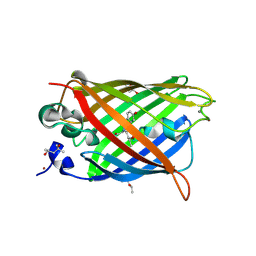 | | GHK tagged GFP variant crystal form II at 1.34A wavelength | | 分子名称: | (4S)-2-METHYL-2,4-PENTANEDIOL, CALCIUM ION, COPPER (II) ION, ... | | 著者 | Huyton, T, Gorlich, D. | | 登録日 | 2019-02-27 | | 公開日 | 2020-05-27 | | 最終更新日 | 2022-12-07 | | 実験手法 | X-RAY DIFFRACTION (1.5 Å) | | 主引用文献 | The copper(II)-binding tripeptide GHK, a valuable crystallization and phasing tag for macromolecular crystallography.
Acta Crystallogr D Struct Biol, 76, 2020
|
|
6QUJ
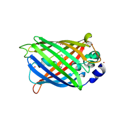 | | GHK tagged GFP variant | | 分子名称: | COPPER (II) ION, GLYCEROL, Green fluorescent protein, ... | | 著者 | Huyton, T, Gorlich, D. | | 登録日 | 2019-02-27 | | 公開日 | 2020-05-27 | | 最終更新日 | 2022-12-07 | | 実験手法 | X-RAY DIFFRACTION (1.68 Å) | | 主引用文献 | The copper(II)-binding tripeptide GHK, a valuable crystallization and phasing tag for macromolecular crystallography.
Acta Crystallogr D Struct Biol, 76, 2020
|
|
6QUI
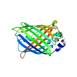 | | GHK tagged GFP variant at 17Kev | | 分子名称: | COPPER (II) ION, GLYCEROL, Green fluorescent protein, ... | | 著者 | Huyton, T, Gorlich, D. | | 登録日 | 2019-02-27 | | 公開日 | 2020-05-27 | | 最終更新日 | 2022-12-07 | | 実験手法 | X-RAY DIFFRACTION (1.94 Å) | | 主引用文献 | The copper(II)-binding tripeptide GHK, a valuable crystallization and phasing tag for macromolecular crystallography.
Acta Crystallogr D Struct Biol, 76, 2020
|
|
6LNP
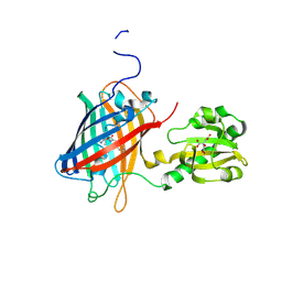 | | Crystal structure of citrate Biosensor | | 分子名称: | CITRIC ACID, Fusion protein of Green fluorescent protein and Sensor histidine kinase CitA | | 著者 | Wen, Y, Campbell, R. | | 登録日 | 2019-12-31 | | 公開日 | 2020-05-06 | | 最終更新日 | 2023-11-22 | | 実験手法 | X-RAY DIFFRACTION (2.993 Å) | | 主引用文献 | High-Performance Intensiometric Direct- and Inverse-Response Genetically Encoded Biosensors for Citrate.
Acs Cent.Sci., 6, 2020
|
|
6YOV
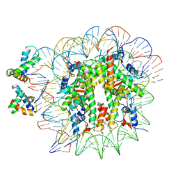 | | OCT4-SOX2-bound nucleosome - SHL+6 | | 分子名称: | DNA (142-MER), Green fluorescent protein,POU domain, class 5, ... | | 著者 | Michael, A.K, Kempf, G, Cavadini, S, Bunker, R.D, Thoma, N.H. | | 登録日 | 2020-04-15 | | 公開日 | 2020-05-06 | | 最終更新日 | 2020-07-08 | | 実験手法 | ELECTRON MICROSCOPY (3.42 Å) | | 主引用文献 | Mechanisms of OCT4-SOX2 motif readout on nucleosomes.
Science, 368, 2020
|
|
6T90
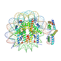 | | OCT4-SOX2-bound nucleosome - SHL-6 | | 分子名称: | DNA (146-MER), Green fluorescent protein,POU domain, class 5, ... | | 著者 | Michael, A.K, Kempf, G, Cavadini, S, Bunker, R.D, Thoma, N.H. | | 登録日 | 2019-10-25 | | 公開日 | 2020-05-06 | | 最終更新日 | 2020-07-08 | | 実験手法 | ELECTRON MICROSCOPY (3.05 Å) | | 主引用文献 | Mechanisms of OCT4-SOX2 motif readout on nucleosomes.
Science, 368, 2020
|
|
6LR7
 | |
6OGA
 | | Crystal structure of Green Fluorescent Protein (GFP); S65T, Y66(3-Br1Y), H148D; circular permutant (50-51) | | 分子名称: | ACETATE ION, Crystal structure of Green Fluorescent Protein (GFP); S65T, Y66(3-Br1Y), ... | | 著者 | Lin, C.-Y, Boxer, S.G. | | 登録日 | 2019-04-02 | | 公開日 | 2020-04-08 | | 最終更新日 | 2023-11-15 | | 実験手法 | X-RAY DIFFRACTION (1.6 Å) | | 主引用文献 | Unusual Spectroscopic and Electric Field Sensitivity of Chromophores with Short Hydrogen Bonds: GFP and PYP as Model Systems.
J.Phys.Chem.B, 124, 2020
|
|
6OGB
 | | Crystal structure of Green Fluorescent Protein (GFP); S65T, Y66(3-I1Y), H148D; circular permutant (50-51) | | 分子名称: | ACETATE ION, Crystal structure of Green Fluorescent Protein (GFP); S65T, Y66(3-I1Y), ... | | 著者 | Lin, C.-Y, Boxer, S.G. | | 登録日 | 2019-04-02 | | 公開日 | 2020-04-08 | | 最終更新日 | 2023-11-15 | | 実験手法 | X-RAY DIFFRACTION (1.65 Å) | | 主引用文献 | Unusual Spectroscopic and Electric Field Sensitivity of Chromophores with Short Hydrogen Bonds: GFP and PYP as Model Systems.
J.Phys.Chem.B, 124, 2020
|
|
6OGC
 | | Crystal structure of Green Fluorescent Protein (GFP); S65T, Y66(2,3-F2Y), H148D; circular permutant (50-51) | | 分子名称: | Crystal structure of Green Fluorescent Protein (GFP); S65T, Y66(2,3-F2Y), H148D; circular permutant (50-51) | | 著者 | Lin, C.-Y, Boxer, S.G. | | 登録日 | 2019-04-02 | | 公開日 | 2020-04-08 | | 最終更新日 | 2023-11-15 | | 実験手法 | X-RAY DIFFRACTION (1.178 Å) | | 主引用文献 | Unusual Spectroscopic and Electric Field Sensitivity of Chromophores with Short Hydrogen Bonds: GFP and PYP as Model Systems.
J.Phys.Chem.B, 124, 2020
|
|
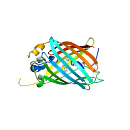
6L26
 | | Neutron crystal structure of the mutant green fluorescent protein (EGFP) | | 分子名称: | Green fluorescent protein, trideuteriooxidanium | | 著者 | Adachi, M, Shimizu, R, Shibazaki, C, Kagotani, Y, Ostermann, A, Schrader, T.E. | | 登録日 | 2019-10-02 | | 公開日 | 2020-04-08 | | 最終更新日 | 2023-11-22 | | 実験手法 | NEUTRON DIFFRACTION (1.444 Å) | | 主引用文献 | Direct Observation of the Protonation States in the Mutant Green Fluorescent Protein.
J Phys Chem Lett, 11, 2020
|
|
6OG9
 | | Crystal structure of Green Fluorescent Protein (GFP); S65T, Y66(3,5-F2Y), H148D; circular permutant (50-51) | | 分子名称: | Crystal structure of Green Fluorescent Protein (GFP); S65T, Y66(3,5-F2Y), H148D; circular permutant (50-51) | | 著者 | Lin, C.-Y, Deng, A, Boxer, S.G. | | 登録日 | 2019-04-02 | | 公開日 | 2020-04-08 | | 最終更新日 | 2023-11-15 | | 実験手法 | X-RAY DIFFRACTION (1.798 Å) | | 主引用文献 | Unusual Spectroscopic and Electric Field Sensitivity of Chromophores with Short Hydrogen Bonds: GFP and PYP as Model Systems.
J.Phys.Chem.B, 124, 2020
|
|
6OG8
 | |
6HR1
 | | Crystal structure of the YFPnano fusion protein | | 分子名称: | 1,2-ETHANEDIOL, CALCIUM ION, GLYCEROL, ... | | 著者 | Benoit, R.M. | | 登録日 | 2018-09-26 | | 公開日 | 2020-04-08 | | 最終更新日 | 2024-01-24 | | 実験手法 | X-RAY DIFFRACTION (1.901 Å) | | 主引用文献 | Chimeric single alpha-helical domains as rigid fusion protein connections for protein nanotechnology and structural biology.
Structure, 2021
|
|
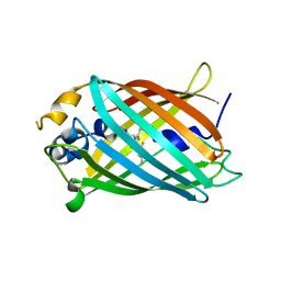
6L27
 | | X-ray crystal structure of the mutant green fluorescent protein | | 分子名称: | Green fluorescent protein | | 著者 | Adachi, M, Shimizu, R, Shibazaki, C, Kagotani, Y, Ostermann, A, Schrader, T.E. | | 登録日 | 2019-10-02 | | 公開日 | 2020-04-01 | | 最終更新日 | 2023-11-15 | | 実験手法 | X-RAY DIFFRACTION (0.77 Å) | | 主引用文献 | Direct Observation of the Protonation States in the Mutant Green Fluorescent Protein.
J Phys Chem Lett, 11, 2020
|
|
6UHN
 | | Crystal Structure of C148 mGFP-cDNA-1 | | 分子名称: | C148 mGFP-cDNA-1, UNKNOWN LIGAND | | 著者 | Winegar, P.W, Hayes, O.G, McMillan, J.R, Figg, C.A, Focia, P.J, Mirkin, C.A. | | 登録日 | 2019-09-27 | | 公開日 | 2020-03-18 | | 最終更新日 | 2023-11-15 | | 実験手法 | X-RAY DIFFRACTION (1.92 Å) | | 主引用文献 | DNA-Directed Protein Packing within Single Crystals.
Chem, 6, 2020
|
|
6UHQ
 | | Crystal Structure of C148 mGFP-cDNA-3 | | 分子名称: | C148 mGFP-cDNA-3, UNKNOWN LIGAND | | 著者 | Winegar, P.W, Hayes, O.G, McMillan, J.R, Figg, C.A, Focia, P.J, Mirkin, C.A. | | 登録日 | 2019-09-27 | | 公開日 | 2020-03-18 | | 最終更新日 | 2023-11-15 | | 実験手法 | X-RAY DIFFRACTION (2.85 Å) | | 主引用文献 | DNA-Directed Protein Packing within Single Crystals.
Chem, 6, 2020
|
|
6UHK
 | | Crystal Structure of C176 mGFP | | 分子名称: | C176 mGFP | | 著者 | Winegar, P.W, Hayes, O.G, McMillan, J.R, Figg, C.A, Focia, P.J, Mirkin, C.A. | | 登録日 | 2019-09-27 | | 公開日 | 2020-03-18 | | 最終更新日 | 2023-11-15 | | 実験手法 | X-RAY DIFFRACTION (1.9 Å) | | 主引用文献 | DNA-Directed Protein Packing within Single Crystals.
Chem, 6, 2020
|
|
6UHR
 | | Crystal Structure of C148 mGFP-scDNA-2 | | 分子名称: | C148 mGFP-scDNA-2 | | 著者 | Winegar, P.W, Hayes, O.G, McMillan, J.R, Figg, C.A, Focia, P.J, Mirkin, C.A. | | 登録日 | 2019-09-27 | | 公開日 | 2020-03-18 | | 最終更新日 | 2023-11-15 | | 実験手法 | X-RAY DIFFRACTION (3 Å) | | 主引用文献 | DNA-Directed Protein Packing within Single Crystals.
Chem, 6, 2020
|
|
6UHJ
 | | X-ray Structure of C148 mGFP | | 分子名称: | C148 mGFP | | 著者 | Winegar, P.W, Hayes, O.G, McMillan, J.R, Figg, C.A, Focia, P.J, Mirkin, C.A. | | 登録日 | 2019-09-27 | | 公開日 | 2020-03-18 | | 最終更新日 | 2023-11-15 | | 実験手法 | X-RAY DIFFRACTION (1.5 Å) | | 主引用文献 | DNA-Directed Protein Packing within Single Crystals.
Chem, 6, 2020
|
|
6UHP
 | | Crystal Structure of C148 mGFP-ncDNA-1 | | 分子名称: | C148 mGFP-ncDNA-1 | | 著者 | Winegar, P.W, Hayes, O.G, McMillan, J.R, Figg, C.A, Focia, P.J, Mirkin, C.A. | | 登録日 | 2019-09-27 | | 公開日 | 2020-03-18 | | 最終更新日 | 2023-11-15 | | 実験手法 | X-RAY DIFFRACTION (2.9 Å) | | 主引用文献 | DNA-Directed Protein Packing within Single Crystals.
Chem, 6, 2020
|
|
6UHL
 | | Crystal Structure of C148 mGFP-scDNA-1 | | 分子名称: | C148 mGFP-scDNA-1, UNKNOWN LIGAND | | 著者 | Winegar, P.W, Hayes, O.G, McMillan, J.R, Figg, C.A, Focia, P.J, Mirkin, C.A. | | 登録日 | 2019-09-27 | | 公開日 | 2020-03-18 | | 最終更新日 | 2023-11-15 | | 実験手法 | X-RAY DIFFRACTION (1.91 Å) | | 主引用文献 | DNA-Directed Protein Packing within Single Crystals.
Chem, 6, 2020
|
|
6UHM
 | | Crystal Structure of a Physical Mixture of C148 mGFP and scDNA-1 | | 分子名称: | C148 mGFP | | 著者 | Winegar, P.W, Hayes, O.G, McMillan, J.R, Figg, C.A, Focia, P.J, Mirkin, C.A. | | 登録日 | 2019-09-27 | | 公開日 | 2020-03-18 | | 最終更新日 | 2023-11-15 | | 実験手法 | X-RAY DIFFRACTION (2.1 Å) | | 主引用文献 | DNA-Directed Protein Packing within Single Crystals.
Chem, 6, 2020
|
|
6UHO
 | | Crystal Structure of C148 mGFP-cDNA-2 | | 分子名称: | C148 mGFP-cDNA-2, UNKNOWN LIGAND | | 著者 | Winegar, P.W, Hayes, O.G, McMillan, J.R, Figg, C.A, Focia, P.J, Mirkin, C.A. | | 登録日 | 2019-09-27 | | 公開日 | 2020-03-18 | | 最終更新日 | 2023-11-15 | | 実験手法 | X-RAY DIFFRACTION (1.95 Å) | | 主引用文献 | DNA-Directed Protein Packing within Single Crystals.
Chem, 6, 2020
|
|
6T39
 | | Crystal structure of rsEGFP2 in its off-state determined by SFX | | 分子名称: | Green fluorescent protein | | 著者 | Woodhouse, J, Coquelle, N, Adam, V, Barends, T.R.M, De La Mora, E, Bourgeois, D, Colletier, J.P, Schlichting, I, Weik, M. | | 登録日 | 2019-10-10 | | 公開日 | 2020-02-19 | | 最終更新日 | 2024-01-24 | | 実験手法 | X-RAY DIFFRACTION (1.6 Å) | | 主引用文献 | Photoswitching mechanism of a fluorescent protein revealed by time-resolved crystallography and transient absorption spectroscopy.
Nat Commun, 11, 2020
|
|


