5W2K
 
 | |
5W2R
 | |
5W2X
 | |
5W3B
 | |
5W37
 | |
6LVP
 | |
1TVR
 | | HIV-1 RT/9-CL TIBO | | 分子名称: | 4-CHLORO-8-METHYL-7-(3-METHYL-BUT-2-ENYL)-6,7,8,9-TETRAHYDRO-2H-2,7,9A-TRIAZA-BENZO[CD]AZULENE-1-THIONE, REVERSE TRANSCRIPTASE | | 著者 | Das, K, Ding, J, Hsiou, Y, Arnold, E. | | 登録日 | 1996-04-16 | | 公開日 | 1997-03-12 | | 最終更新日 | 2024-02-14 | | 実験手法 | X-RAY DIFFRACTION (3 Å) | | 主引用文献 | Crystal structures of 8-Cl and 9-Cl TIBO complexed with wild-type HIV-1 RT and 8-Cl TIBO complexed with the Tyr181Cys HIV-1 RT drug-resistant mutant.
J.Mol.Biol., 264, 1996
|
|
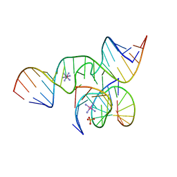
3IVK
 | | Crystal Structure of the Catalytic Core of an RNA Polymerase Ribozyme Complexed with an Antigen Binding Antibody Fragment | | 分子名称: | CADMIUM ION, CHLORIDE ION, Fab heavy chain, ... | | 著者 | Koldobskaya, Y, Duguid, E.M, Shechner, D.M, Koide, S, Kossiakoff, A.A, Bartel, D.P, Piccirilli, J.A. | | 登録日 | 2009-09-01 | | 公開日 | 2010-03-02 | | 最終更新日 | 2017-11-01 | | 実験手法 | X-RAY DIFFRACTION (3.1 Å) | | 主引用文献 | Crystal structure of the catalytic core of an RNA-polymerase ribozyme.
Science, 326, 2009
|
|
5W2V
 | |
5W1G
 | | CR1-07 unliganded Fab | | 分子名称: | CR1-07 Fab heavy chain, CR1-07 Fab light chain | | 著者 | Raymond, D.D, Clark, L.E, Abraham, J. | | 登録日 | 2017-06-03 | | 公開日 | 2018-05-30 | | 最終更新日 | 2020-01-01 | | 実験手法 | X-RAY DIFFRACTION (2 Å) | | 主引用文献 | Vaccine-elicited receptor-binding site antibodies neutralize two New World hemorrhagic fever arenaviruses.
Nat Commun, 9, 2018
|
|
5W30
 | | Crystal structure of mutant CJ YCEI protein (CJ-N48C) with monobromobimane guest structure | | 分子名称: | 3-(bromomethyl)-2,5,6-trimethyl-1H,7H-pyrazolo[1,2-a]pyrazole-1,7-dione, Putative periplasmic protein, SULFATE ION, ... | | 著者 | Huber, T.R, Snow, C.D. | | 登録日 | 2017-06-07 | | 公開日 | 2018-01-03 | | 最終更新日 | 2023-10-04 | | 実験手法 | X-RAY DIFFRACTION (2.75 Å) | | 主引用文献 | Installing Guest Molecules at Specific Sites within Scaffold Protein Crystals.
Bioconjug. Chem., 29, 2018
|
|
1VF6
 | | 2.1 Angstrom crystal structure of the PALS-1-L27N and PATJ L27 heterodimer complex | | 分子名称: | MAGUK p55 subfamily member 5, PALS1-associated tight junction protein | | 著者 | Li, Y, Lavie, A, Margolis, B, Karnak, D. | | 登録日 | 2004-04-09 | | 公開日 | 2004-04-20 | | 最終更新日 | 2023-12-27 | | 実験手法 | X-RAY DIFFRACTION (2.1 Å) | | 主引用文献 | Structural basis for L27 domain-mediated assembly of signaling and cell polarity complexes.
Embo J., 23, 2004
|
|
1SJS
 | |
1T2M
 | | Solution Structure Of The Pdz Domain Of AF-6 | | 分子名称: | AF-6 protein | | 著者 | Zhou, H, Wu, J.H, Xu, Y.Q, Huang, A.D, Shi, Y.Y. | | 登録日 | 2004-04-22 | | 公開日 | 2005-02-08 | | 最終更新日 | 2024-05-29 | | 実験手法 | SOLUTION NMR | | 主引用文献 | Solution Structure of AF-6 PDZ Domain and Its Interaction with the C-terminal Peptides from Neurexin and Bcr
J.Biol.Chem., 280, 2005
|
|
5DWX
 | |
7ZNK
 | | Structure of an endogenous human TREX complex bound to mRNA | | 分子名称: | RNA, Spliceosome RNA helicase DDX39B, THO complex subunit 1, ... | | 著者 | Pacheco-Fiallos, F.B, Vorlaender, M.K, Plaschka, C. | | 登録日 | 2022-04-21 | | 公開日 | 2023-05-03 | | 最終更新日 | 2023-05-24 | | 実験手法 | ELECTRON MICROSCOPY (3.9 Å) | | 主引用文献 | mRNA recognition and packaging by the human transcription-export complex.
Nature, 616, 2023
|
|
5EST
 | | Crystallographic analysis of the inhibition of porcine pancreatic elastase by a peptidyl boronic acid: structure of a reaction intermediate | | 分子名称: | CALCIUM ION, ELASTASE, N~2~-[(benzyloxy)carbonyl]-N-[(1R,2S)-1-(dihydroxyboranyl)-2-methylbutyl]-L-alaninamide, ... | | 著者 | Takahashi, L.H, Radhakrishnan, R, Rosenfieldjunior, R.E, Meyerjunior, E.F. | | 登録日 | 1989-05-15 | | 公開日 | 1992-04-15 | | 最終更新日 | 2024-06-05 | | 実験手法 | X-RAY DIFFRACTION (2.09 Å) | | 主引用文献 | Crystallographic analysis of the inhibition of porcine pancreatic elastase by a peptidyl boronic acid: structure of a reaction intermediate.
Biochemistry, 28, 1989
|
|
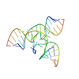
1X9C
 | | An all-RNA Hairpin Ribozyme with mutation U39C | | 分子名称: | 5'-R(*CP*GP*GP*UP*GP*AP*GP*AP*AP*GP*GP*G)-3', 5'-R(*GP*GP*CP*AP*GP*AP*GP*AP*AP*AP*CP*AP*CP*AP*CP*GP*A)-3', 5'-R(*UP*CP*CP*CP*(A2M)P*GP*UP*CP*CP*AP*CP*CP*G)-3', ... | | 著者 | Alam, S, Grum-Tokars, V, Krucinska, J, Kundracik, M.L, Wedekind, J.E. | | 登録日 | 2004-08-20 | | 公開日 | 2005-11-22 | | 最終更新日 | 2023-08-23 | | 実験手法 | X-RAY DIFFRACTION (2.19 Å) | | 主引用文献 | Conformational Heterogeneity at Position U37 of an All-RNA Hairpin Ribozyme with Implications for Metal Binding and the Catalytic Structure of the S-Turn.
Biochemistry, 44, 2005
|
|
1Q55
 | | W-shaped trans interactions of cadherins model based on fitting C-cadherin (1L3W) to 3D map of desmosomes obtained by electron tomography | | 分子名称: | 2-acetamido-2-deoxy-alpha-D-glucopyranose, 2-acetamido-2-deoxy-beta-D-glucopyranose, CALCIUM ION, ... | | 著者 | He, W, Cowin, P, Stokes, D.L. | | 登録日 | 2003-08-06 | | 公開日 | 2003-10-07 | | 最終更新日 | 2020-07-29 | | 実験手法 | ELECTRON MICROSCOPY (30 Å) | | 主引用文献 | Untangling Desmosomal Knots with Electron Tomography
Science, 302, 2003
|
|
1X9K
 | | An all-RNA Hairpin Ribozyme with mutation U39C | | 分子名称: | 5'-R(*AP*AP*UP*AP*GP*AP*GP*AP*AP*GP*CP*GP*A)-3', 5'-R(*GP*GP*CP*AP*GP*AP*GP*AP*AP*AP*CP*AP*CP*AP*CP*GP*A)-3', 5'-R(*UP*CP*GP*CP*AP*GP*UP*CP*CP*UP*AP*UP*U)-3', ... | | 著者 | Alam, S, Grum-Tokars, V, Krucinska, J, Kundracik, M.L, Wedekind, J.E. | | 登録日 | 2004-08-21 | | 公開日 | 2005-11-22 | | 最終更新日 | 2023-08-23 | | 実験手法 | X-RAY DIFFRACTION (3.17 Å) | | 主引用文献 | Conformational Heterogeneity at Position U37 of an All-RNA Hairpin Ribozyme with Implications for Metal Binding and the Catalytic Structure of the S-Turn.
Biochemistry, 44, 2005
|
|
1YMG
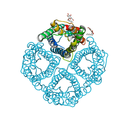 | | The Channel Architecture of Aquaporin O at 2.2 Angstrom Resolution | | 分子名称: | Lens fiber major intrinsic protein, nonyl beta-D-glucopyranoside | | 著者 | Harries, W.E.C, Akhavan, D, Miercke, L.J.W, Khademi, S, Stroud, R.M. | | 登録日 | 2005-01-20 | | 公開日 | 2005-02-08 | | 最終更新日 | 2023-08-23 | | 実験手法 | X-RAY DIFFRACTION (2.24 Å) | | 主引用文献 | The Channel Architecture of Aquaporin 0 at a 2.2-A Resolution
Proc.Natl.Acad.Sci.USA, 101, 2004
|
|
4R0Z
 | | A conserved phosphorylation switch controls the interaction between cadherin and beta-catenin in vitro and in vivo | | 分子名称: | FORMIC ACID, Protein humpback-2 | | 著者 | Choi, H.-J, Loveless, T, Lynch, A, Bang, I, Hardin, J, Weis, W.I. | | 登録日 | 2014-08-03 | | 公開日 | 2015-04-29 | | 最終更新日 | 2023-11-08 | | 実験手法 | X-RAY DIFFRACTION (2.005 Å) | | 主引用文献 | A Conserved Phosphorylation Switch Controls the Interaction between Cadherin and beta-Catenin In Vitro and In Vivo
Dev.Cell, 33, 2015
|
|
1Q5B
 | | lambda-shaped TRANS and CIS interactions of cadherins model based on fitting C-cadherin (1L3W) to 3D map of desmosomes obtained by electron tomography | | 分子名称: | 2-acetamido-2-deoxy-alpha-D-glucopyranose, 2-acetamido-2-deoxy-beta-D-glucopyranose, CALCIUM ION, ... | | 著者 | He, W, Cowin, P, Stokes, D.L. | | 登録日 | 2003-08-06 | | 公開日 | 2003-10-07 | | 最終更新日 | 2020-07-29 | | 実験手法 | ELECTRON MICROSCOPY (30 Å) | | 主引用文献 | Untangling Desmosomal Knots with Electron Tomography
Science, 302, 2003
|
|
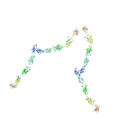
7UUW
 | | Cryogenic electron microscopy 3D map of F-actin bound by the Actin Binding Domain of alpha-catenin ortholog, HMP1 | | 分子名称: | ADENOSINE-5'-DIPHOSPHATE, Actin, alpha skeletal muscle, ... | | 著者 | Rangarajan, E.S, Smith, E.W, Izard, T. | | 登録日 | 2022-04-29 | | 公開日 | 2023-01-18 | | 最終更新日 | 2023-01-25 | | 実験手法 | ELECTRON MICROSCOPY (3.36 Å) | | 主引用文献 | The nematode alpha-catenin ortholog, HMP1, has an extended alpha-helix when bound to actin filaments.
J.Biol.Chem., 299, 2022
|
|
1Q5C
 | | S-S-lambda-shaped TRANS and CIS interactions of cadherins model based on fitting C-cadherin (1L3W) to 3D map of desmosomes obtained by electron tomography | | 分子名称: | 2-acetamido-2-deoxy-alpha-D-glucopyranose, 2-acetamido-2-deoxy-beta-D-glucopyranose, CALCIUM ION, ... | | 著者 | He, W, Cowin, P, Stokes, D.L. | | 登録日 | 2003-08-06 | | 公開日 | 2003-10-07 | | 最終更新日 | 2020-07-29 | | 実験手法 | ELECTRON MICROSCOPY (30 Å) | | 主引用文献 | Untangling Desmosomal Knots with Electron Tomography
Science, 302, 2003
|
|


