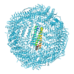
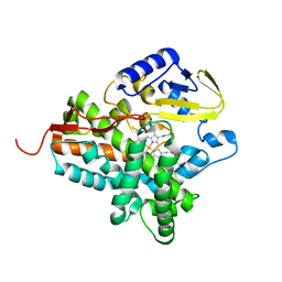
4LPJ
 
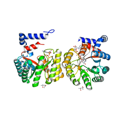
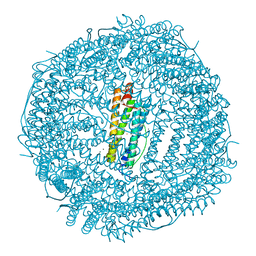 | | One minute iron loaded frog M ferritin | | 分子名称: | CHLORIDE ION, FE (II) ION, Ferritin, ... | | 著者 | Mangani, S, Di Pisa, F, Pozzi, C, Turano, P, Lalli, D. | | 登録日 | 2013-07-16 | | 公開日 | 2014-08-06 | | 最終更新日 | 2023-09-20 | | 実験手法 | X-RAY DIFFRACTION (1.27 Å) | | 主引用文献 | Time-lapse anomalous X-ray diffraction shows how Fe(2+) substrate ions move through ferritin protein nanocages to oxidoreductase sites.
Acta Crystallogr.,Sect.D, 71, 2015
|
|
2WJU
 | |
7RM8
 | |
3C50
 | |
4LPN
 | |
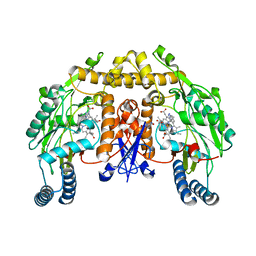
5G6L
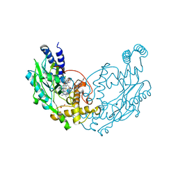 | | Structure of Bacillus subtilis Nitric Oxide Synthase in complex with 7-((4-Chloro-3-((methylamino)methyl)phenoxy)methyl) quinolin-2-amine | | 分子名称: | 5,6,7,8-TETRAHYDROBIOPTERIN, 7-[[4-chloranyl-3-(methylaminomethyl)phenoxy]methyl]quinolin-2-amine, CHLORIDE ION, ... | | 著者 | Holden, J.K, Poulos, T.L. | | 登録日 | 2016-06-18 | | 公開日 | 2016-09-21 | | 最終更新日 | 2024-01-10 | | 実験手法 | X-RAY DIFFRACTION (2.034 Å) | | 主引用文献 | Targeting Bacterial Nitric Oxide Synthase with Aminoquinoline-Based Inhibitors.
Biochemistry, 55, 2016
|
|
7R8J
 | |
7LA8
 | |
3DQ2
 | |
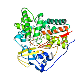
2WIO
 | | Structure of the histidine tagged, open cytochrome P450 Eryk from S. erythraea | | 分子名称: | ERYTHROMYCIN B/D C-12 HYDROXYLASE, PROTOPORPHYRIN IX CONTAINING FE | | 著者 | Savino, C, Montemiglio, L.C, Sciara, G, Miele, A.E, Kedrew, S.G, Gianni, S, Vallone, B. | | 登録日 | 2009-05-14 | | 公開日 | 2009-07-21 | | 最終更新日 | 2023-12-13 | | 実験手法 | X-RAY DIFFRACTION (2 Å) | | 主引用文献 | Investigating the Structural Plasticity of a Cytochrome P450: Three-Dimensional Structures of P450 Eryk and Binding to its Physiological Substrate.
J.Biol.Chem., 284, 2009
|
|
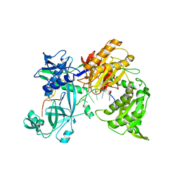
7QZO
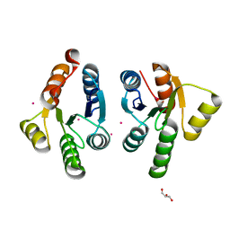 | | Crystal structure of GacS D1 domain | | 分子名称: | CADMIUM ION, GLYCEROL, Histidine kinase | | 著者 | Fadel, F, Bassim, V, Botzanowski, T, Francis, V.I, Legrand, P, Porter, S.L, Bourne, Y, Cianferani, S, Vincent, F. | | 登録日 | 2022-01-31 | | 公開日 | 2022-07-06 | | 最終更新日 | 2024-01-31 | | 実験手法 | X-RAY DIFFRACTION (1.45 Å) | | 主引用文献 | Insights into the atypical autokinase activity of the Pseudomonas aeruginosa GacS histidine kinase and its interaction with RetS.
Structure, 30, 2022
|
|
3DQC
 | |
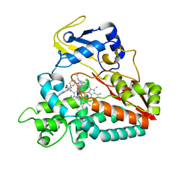
2WM4
 | | X-ray structure of Mycobacterium tuberculosis cytochrome P450 CYP124 in complex with phytanic acid | | 分子名称: | (3R,7S,11S)-3,7,11,15-tetramethylhexadecanoic acid, CALCIUM ION, PROTOPORPHYRIN IX CONTAINING FE, ... | | 著者 | Johnston, J.B, Kells, P.M, Podust, L.M, Ortiz de Montellano, P.R. | | 登録日 | 2009-06-30 | | 公開日 | 2009-10-06 | | 最終更新日 | 2023-12-13 | | 実験手法 | X-RAY DIFFRACTION (2.11 Å) | | 主引用文献 | Biochemical and structural characterization of CYP124: a methyl-branched lipid omega-hydroxylase from Mycobacterium tuberculosis.
Proc. Natl. Acad. Sci. U.S.A., 106, 2009
|
|
8WMV
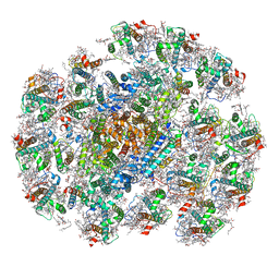 | | The structure of PSI-14CAC complex at stationary growth phase | | 分子名称: | (1~{R})-3,5,5-trimethyl-4-[(3~{E},5~{E},7~{E},9~{E},11~{E},13~{E},15~{E})-3,7,12,16-tetramethyl-18-[(4~{R})-2,6,6-trimethyl-4-oxidanyl-cyclohexen-1-yl]octadeca-3,5,7,9,11,13,15-heptaen-1,17-diynyl]cyclohex-3-en-1-ol, (1~{R})-3,5,5-trimethyl-4-[(3~{E},5~{E},7~{E},9~{E},11~{E},13~{E},15~{E},17~{E})-3,7,12,16-tetramethyl-18-(2,6,6-trimethylcyclohexen-1-yl)octadeca-3,5,7,9,11,13,15,17-octaen-1-ynyl]cyclohex-3-en-1-ol, 1,2-DIPALMITOYL-PHOSPHATIDYL-GLYCEROLE, ... | | 著者 | Zhang, S.M, Si, L, Li, M. | | 登録日 | 2023-10-04 | | 公開日 | 2024-05-29 | | 実験手法 | ELECTRON MICROSCOPY (2.94 Å) | | 主引用文献 | Growth phase-dependent reorganization of cryptophyte photosystem I antennae.
Commun Biol, 7, 2024
|
|
2EEB
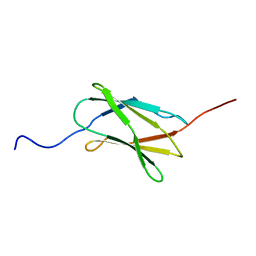 | | Solution structure of the 22th filamin domain from human Filamin-B | | 分子名称: | Filamin-B | | 著者 | Tomizawa, T, Koshiba, S, Watanabe, S, Harada, T, Kigawa, T, Yokoyama, S, RIKEN Structural Genomics/Proteomics Initiative (RSGI) | | 登録日 | 2007-02-15 | | 公開日 | 2007-08-21 | | 最終更新日 | 2024-05-29 | | 実験手法 | SOLUTION NMR | | 主引用文献 | Solution structure of the 22th filamin domain from human Filamin-B
To be Published
|
|
3DQR
 | | Structure of neuronal NOS D597N/M336V mutant heme domain in complex with a inhibitor (+-)-N1-{cis-4'-[(6"-aminopyridin-2"-yl)methyl]pyrrolidin-3'-yl}ethane-1,2-diamine | | 分子名称: | 5,6,7,8-TETRAHYDROBIOPTERIN, ACETATE ION, N-{(3S,4S)-4-[(6-aminopyridin-2-yl)methyl]pyrrolidin-3-yl}ethane-1,2-diamine, ... | | 著者 | Igarashi, J, Li, H, Poulos, T.L. | | 登録日 | 2008-07-09 | | 公開日 | 2009-03-31 | | 最終更新日 | 2024-02-21 | | 実験手法 | X-RAY DIFFRACTION (2.4 Å) | | 主引用文献 | Crystal structures of constitutive nitric oxide synthases in complex with de novo designed inhibitors.
J.Med.Chem., 52, 2009
|
|
5D7K
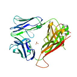 | | Structure of MR1-reactive MAV36 TCR | | 分子名称: | MAV36 TCR Alpha Chain, MAV36 TCR Beta Chain, SULFATE ION | | 著者 | Keller, A.N, Rossjohn, J. | | 登録日 | 2015-08-14 | | 公開日 | 2016-01-27 | | 最終更新日 | 2023-09-27 | | 実験手法 | X-RAY DIFFRACTION (1.9 Å) | | 主引用文献 | Diversity of T Cells Restricted by the MHC Class I-Related Molecule MR1 Facilitates Differential Antigen Recognition.
Immunity, 44, 2016
|
|
4IPP
 | |
7LMW
 | | Receptor for Advanced Glycation End Products VC1 domain in complex with 3-(3-((4-(4-carboxyphenoxy)benzyl)oxy)phenyl)-1H-indole-2-carboxylic acid | | 分子名称: | 7-methyl-3-(1~{H}-pyrazol-4-yl)-1~{H}-indole-2-carboxylic acid, ACETATE ION, Advanced glycosylation end product-specific receptor, ... | | 著者 | Salay, L.E, Kozlyuk, N, Gilston, B.A, Gogliotti, R.D, Christov, P.P, Kim, K, Ovee, M, Waterson, A.G, Chazin, W.J. | | 登録日 | 2021-02-06 | | 公開日 | 2021-07-28 | | 最終更新日 | 2023-10-18 | | 実験手法 | X-RAY DIFFRACTION (2.5 Å) | | 主引用文献 | A fragment-based approach to discovery of Receptor for Advanced Glycation End products inhibitors.
Proteins, 89, 2021
|
|
2EKG
 | |
2WHF
 | | Interaction of Mycobacterium tuberculosis CYP130 with heterocyclic arylamines | | 分子名称: | 1-(3-METHYLPHENYL)-1H-BENZIMIDAZOL-5-AMINE, PROTOPORPHYRIN IX CONTAINING FE, PUTATIVE CYTOCHROME P450 130 | | 著者 | Podust, L.M, Ouellet, H, von Kries, J.P, Ortiz de Montellano, P.R. | | 登録日 | 2009-05-04 | | 公開日 | 2009-07-14 | | 最終更新日 | 2023-12-13 | | 実験手法 | X-RAY DIFFRACTION (1.58 Å) | | 主引用文献 | Interaction of Mycobacterium tuberculosis CYP130 with heterocyclic arylamines.
J. Biol. Chem., 284, 2009
|
|
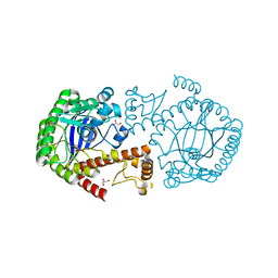
3C7M
 | | Crystal structure of reduced DsbL | | 分子名称: | CADMIUM ION, CHLORIDE ION, DI(HYDROXYETHYL)ETHER, ... | | 著者 | Stirnimann, C.U, Grimshaw, J.P.A, Glockshuber, R, Grutter, M.G, Capitani, G. | | 登録日 | 2008-02-07 | | 公開日 | 2008-07-15 | | 最終更新日 | 2024-04-03 | | 実験手法 | X-RAY DIFFRACTION (1.55 Å) | | 主引用文献 | DsbL and DsbI form a specific dithiol oxidase system for periplasmic arylsulfate sulfotransferase in uropathogenic Escherichia coli.
J.Mol.Biol., 380, 2008
|
|
7R94
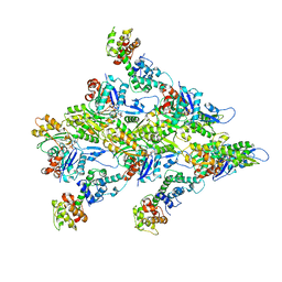 | | T-Plastin-F-actin complex | | 分子名称: | ADENOSINE-5'-DIPHOSPHATE, Actin, alpha skeletal muscle, ... | | 著者 | Mei, L, Alushin, G.M. | | 登録日 | 2021-06-28 | | 公開日 | 2022-07-06 | | 最終更新日 | 2022-09-21 | | 実験手法 | ELECTRON MICROSCOPY (2.6 Å) | | 主引用文献 | Structural mechanism for bidirectional actin cross-linking by T-plastin.
Proc.Natl.Acad.Sci.USA, 119, 2022
|
|
3DNC
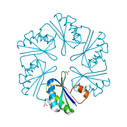 | | Carboxysome shell protein, CcmK2 C-terminal deletion mutant, with a closer spacing between hexamers | | 分子名称: | Carbon dioxide-concentrating mechanism protein ccmK homolog 2, GLYCEROL, SULFATE ION | | 著者 | Tanaka, S, Sawaya, M.R, Yeates, T.O. | | 登録日 | 2008-07-01 | | 公開日 | 2009-01-20 | | 最終更新日 | 2023-08-30 | | 実験手法 | X-RAY DIFFRACTION (2.05 Å) | | 主引用文献 | Insights from multiple structures of the shell proteins from the beta-carboxysome.
Protein Sci., 18, 2009
|
|
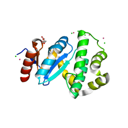
5D6X
 | | Crystal structure of double tudor domain of human lysine demethylase KDM4A | | 分子名称: | Lysine-specific demethylase 4A, SULFATE ION | | 著者 | Wang, F, Su, Z, Denu, J.M, Phillips Jr, G.N, Enzyme Discovery for Natural Product Biosynthesis (NatPro) | | 登録日 | 2015-08-13 | | 公開日 | 2015-11-25 | | 最終更新日 | 2024-03-06 | | 実験手法 | X-RAY DIFFRACTION (2.153 Å) | | 主引用文献 | Reader domain specificity and lysine demethylase-4 family function.
Nat Commun, 7, 2016
|
|


