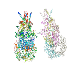
8P2Z
 
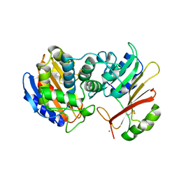
 | | Structure of human SIT1 bound to L-pipecolate (focussed map / refinement) | | 分子名称: | (2S)-piperidine-2-carboxylic acid, 2-acetamido-2-deoxy-beta-D-glucopyranose, CHLORIDE ION, ... | | 著者 | Li, H.Z, Pike, A.C.W, Chi, G, Hansen, J.S, Lee, S.G, Rodstrom, K.E.J, Bushell, S.R, Speedman, D, Evans, A, Wang, D, He, D, Shrestha, L, Nasrallah, C, Chalk, R, Moreira, T, MacLean, E.M, Marsden, B, Bountra, C, Burgess-Brown, N.A, Dafforn, T.R, Carpenter, E.P, Sauer, D.B. | | 登録日 | 2023-05-16 | | 公開日 | 2024-06-12 | | 最終更新日 | 2024-07-10 | | 実験手法 | ELECTRON MICROSCOPY (3.5 Å) | | 主引用文献 | Structure and function of the SIT1 proline transporter in complex with the COVID-19 receptor ACE2.
Nat Commun, 15, 2024
|
|
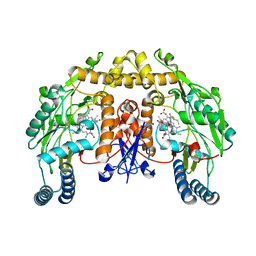
1IRK
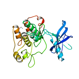 | | CRYSTAL STRUCTURE OF THE TYROSINE KINASE DOMAIN OF THE HUMAN INSULIN RECEPTOR | | 分子名称: | ETHYL MERCURY ION, INSULIN RECEPTOR TYROSINE KINASE DOMAIN | | 著者 | Hubbard, S.R, Wei, L, Ellis, L, Hendrickson, W.A. | | 登録日 | 1995-01-02 | | 公開日 | 1995-02-27 | | 最終更新日 | 2024-02-07 | | 実験手法 | X-RAY DIFFRACTION (2.1 Å) | | 主引用文献 | Crystal structure of the tyrosine kinase domain of the human insulin receptor.
Nature, 372, 1994
|
|
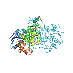
1ISW
 | | Crystal structure of xylanase from Streptomyces olivaceoviridis E-86 complexed with xylobiose | | 分子名称: | beta-D-xylopyranose, beta-D-xylopyranose-(1-4)-beta-D-xylopyranose, endo-1,4-beta-D-xylanase | | 著者 | Fujimoto, Z, Kuno, A, Kaneko, S, Kobayashi, H, Kusakabe, I, Mizuno, H. | | 登録日 | 2001-12-27 | | 公開日 | 2002-02-20 | | 最終更新日 | 2023-10-25 | | 実験手法 | X-RAY DIFFRACTION (2.1 Å) | | 主引用文献 | Crystal structures of the sugar complexes of Streptomyces olivaceoviridis E-86 xylanase: sugar binding structure of the family 13 carbohydrate binding module.
J.Mol.Biol., 316, 2002
|
|
1ITB
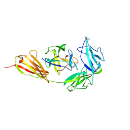 | | TYPE-1 INTERLEUKIN-1 RECEPTOR COMPLEXED WITH INTERLEUKIN-1 BETA | | 分子名称: | INTERLEUKIN-1 BETA, TYPE 1 INTERLEUKIN-1 RECEPTOR | | 著者 | Vigers, G.P.A, Anderson, L.J, Caffes, P, Brandhuber, B.J. | | 登録日 | 1997-01-15 | | 公開日 | 1998-02-04 | | 最終更新日 | 2011-07-13 | | 実験手法 | X-RAY DIFFRACTION (2.5 Å) | | 主引用文献 | Crystal structure of the type-I interleukin-1 receptor complexed with interleukin-1beta.
Nature, 386, 1997
|
|
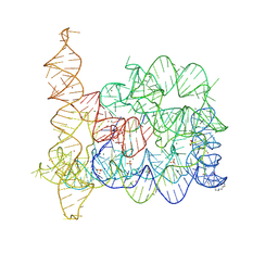
1J5A
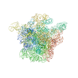 | | STRUCTURAL BASIS FOR THE INTERACTION OF ANTIBIOTICS WITH THE PEPTIDYL TRANSFERASE CENTER IN EUBACTERIA | | 分子名称: | 23S RRNA, CLARITHROMYCIN, MAGNESIUM ION, ... | | 著者 | Schluenzen, F, Zarivach, R, Harms, J, Bashan, A, Tocilj, A, Albrecht, R, Yonath, A, Franceschi, F. | | 登録日 | 2002-03-06 | | 公開日 | 2002-03-08 | | 最終更新日 | 2023-12-27 | | 実験手法 | X-RAY DIFFRACTION (3.5 Å) | | 主引用文献 | Structural basis for the interaction of antibiotics with the peptidyl transferase centre in eubacteria.
Nature, 413, 2001
|
|
1ILK
 | |
1IDE
 | | ISOCITRATE DEHYDROGENASE Y160F MUTANT STEADY-STATE INTERMEDIATE COMPLEX (LAUE DETERMINATION) | | 分子名称: | ISOCITRATE DEHYDROGENASE, ISOCITRIC ACID, MAGNESIUM ION, ... | | 著者 | Bolduc, J.M, Dyer, D.H, Scott, W.G, Singer, P, Sweet, R.M, Koshland Junior, D.E, Stoddard, B.L. | | 登録日 | 1995-01-18 | | 公開日 | 1996-03-08 | | 最終更新日 | 2024-02-07 | | 実験手法 | X-RAY DIFFRACTION (2.5 Å) | | 主引用文献 | Mutagenesis and Laue structures of enzyme intermediates: isocitrate dehydrogenase.
Science, 268, 1995
|
|
1IXM
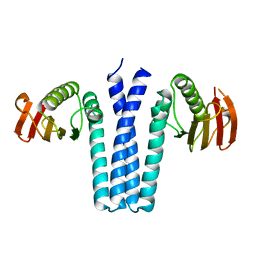 | | CRYSTAL STRUCTURE OF SPOOB FROM BACILLUS SUBTILIS | | 分子名称: | PROTEIN (SPORULATION RESPONSE REGULATORY PROTEIN) | | 著者 | Varughese, K.I. | | 登録日 | 1998-11-06 | | 公開日 | 1998-11-25 | | 最終更新日 | 2023-12-27 | | 実験手法 | X-RAY DIFFRACTION (2.6 Å) | | 主引用文献 | Formation of a novel four-helix bundle and molecular recognition sites by dimerization of a response regulator phosphotransferase.
Mol.Cell, 2, 1998
|
|
8OLY
 | |
1IXX
 | | CRYSTAL STRUCTURE OF COAGULATION FACTORS IX/X-BINDING PROTEIN (IX/X-BP) FROM VENOM OF HABU SNAKE WITH A HETERODIMER OF C-TYPE LECTIN DOMAINS | | 分子名称: | CALCIUM ION, COAGULATION FACTORS IX/X-BINDING PROTEIN | | 著者 | Mizuno, H, Fujimoto, Z, Koizumi, M, Kano, H. | | 登録日 | 1997-05-01 | | 公開日 | 1998-05-06 | | 最終更新日 | 2011-07-13 | | 実験手法 | X-RAY DIFFRACTION (2.5 Å) | | 主引用文献 | Structure of coagulation factors IX/X-binding protein, a heterodimer of C-type lectin domains.
Nat.Struct.Biol., 4, 1997
|
|
1JVV
 | |
1JTG
 | | CRYSTAL STRUCTURE OF TEM-1 BETA-LACTAMASE / BETA-LACTAMASE INHIBITOR PROTEIN COMPLEX | | 分子名称: | BETA-LACTAMASE INHIBITORY PROTEIN, BETA-LACTAMASE TEM, CALCIUM ION | | 著者 | Strynadka, N.C.J, Jensen, S.E, Alzari, P.M, James, M.N. | | 登録日 | 2001-08-20 | | 公開日 | 2001-10-17 | | 最終更新日 | 2024-04-03 | | 実験手法 | X-RAY DIFFRACTION (1.73 Å) | | 主引用文献 | Crystal structure and kinetic analysis of beta-lactamase inhibitor protein-II in complex with TEM-1 beta-lactamase.
Nat.Struct.Biol., 8, 2001
|
|
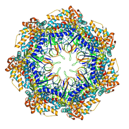
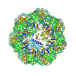
8P4M
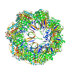 | | CryoEM structure of a C7-symmetrical GroEL7-GroES7 cage in presence of ADP-BeFx | | 分子名称: | ADENOSINE-5'-DIPHOSPHATE, BERYLLIUM TRIFLUORIDE ION, Chaperonin GroEL, ... | | 著者 | Wagner, J, Beck, F, Bracher, A, Caravajal, A.I, Wan, W, Bohn, S, Koerner, R, Baumeister, W, Fernandez-Busnadiego, R, Hartl, F.U. | | 登録日 | 2023-05-23 | | 公開日 | 2024-07-03 | | 最終更新日 | 2024-08-21 | | 実験手法 | ELECTRON MICROSCOPY (2.5 Å) | | 主引用文献 | Visualizing chaperonin function in situ by cryo-electron tomography
Nature, 2024
|
|
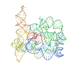
8OLZ
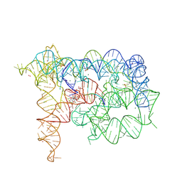 | | Structure of Oceanobacillus iheyensis group II intron in the presence of K+, Mg2+, 5'-exon, and intronistat B after 2h30 soaking | | 分子名称: | 4-(2-HYDROXYETHYL)-1-PIPERAZINE ETHANESULFONIC ACID, 5'-exon, DOMAINS 1-5, ... | | 著者 | Silvestri, I, Marcia, M. | | 登録日 | 2023-03-30 | | 公開日 | 2024-06-19 | | 最終更新日 | 2024-07-03 | | 実験手法 | X-RAY DIFFRACTION (3.31 Å) | | 主引用文献 | Targeting the conserved active site of splicing machines with specific and selective small molecule modulators.
Nat Commun, 15, 2024
|
|
8P4R
 | | In situ structure average of GroEL14-GroES14 complexes in Escherichia coli cytosol obtained by cryo electron tomography | | 分子名称: | ADENOSINE-5'-TRIPHOSPHATE, Chaperonin GroEL, Co-chaperonin GroES, ... | | 著者 | Wagner, J, Caravajal, A.I, Beck, F, Bracher, A, Wan, W, Bohn, S, Koerner, R, Baumeister, W, Fernandez-Busnadiego, R, Hartl, F.U. | | 登録日 | 2023-05-23 | | 公開日 | 2024-07-03 | | 最終更新日 | 2024-08-21 | | 実験手法 | ELECTRON MICROSCOPY (11.9 Å) | | 主引用文献 | Visualizing chaperonin function in situ by cryo-electron tomography
Nature, 2024
|
|
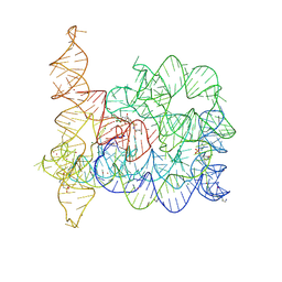
8OLV
 | | Structure of Oceanobacillus iheyensis group II intron in the presence of K+, Mg2+ and ARN25850 | | 分子名称: | 2-[2,6-bis(bromanyl)-3,4,5-tris(oxidanyl)phenyl]carbonyl-~{N}-(2-pyrrolidin-1-ylethyl)-1-benzofuran-5-carboxamide, 4-(2-HYDROXYETHYL)-1-PIPERAZINE ETHANESULFONIC ACID, Group IIC intron, ... | | 著者 | Silvestri, I, Marcia, M. | | 登録日 | 2023-03-30 | | 公開日 | 2024-06-19 | | 最終更新日 | 2024-07-03 | | 実験手法 | X-RAY DIFFRACTION (2.81 Å) | | 主引用文献 | Targeting the conserved active site of splicing machines with specific and selective small molecule modulators.
Nat Commun, 15, 2024
|
|
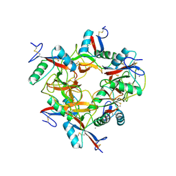
1K2E
 | | crystal structure of a nudix protein from Pyrobaculum aerophilum | | 分子名称: | ACETIC ACID, GLYCEROL, NICKEL (II) ION, ... | | 著者 | Wang, S, Mura, C, Sawaya, M.R, Cascio, D, Eisenberg, D. | | 登録日 | 2001-09-26 | | 公開日 | 2002-04-03 | | 最終更新日 | 2023-08-16 | | 実験手法 | X-RAY DIFFRACTION (1.8 Å) | | 主引用文献 | Structure of a Nudix protein from Pyrobaculum aerophilum reveals a dimer with two intersubunit beta-sheets.
Acta Crystallogr.,Sect.D, 58, 2002
|
|
8P4N
 | | CryoEM structure of a GroEL7-GroES7 cage with encapsulated disordered substrate MetK in the presence of ADP-BeFx | | 分子名称: | ADENOSINE-5'-DIPHOSPHATE, BERYLLIUM TRIFLUORIDE ION, Chaperonin GroEL, ... | | 著者 | Wagner, J, Beck, F, Bracher, A, Caravajal, A.I, Wan, W, Bohn, S, Koerner, R, Baumeister, W, Fernandez-Busnadiego, R, Hartl, F.U. | | 登録日 | 2023-05-23 | | 公開日 | 2024-07-03 | | 最終更新日 | 2024-08-21 | | 実験手法 | ELECTRON MICROSCOPY (2.9 Å) | | 主引用文献 | Visualizing chaperonin function in situ by cryo-electron tomography
Nature, 2024
|
|
1K2T
 | | Structure of rat brain nNOS heme domain complexed with S-ethyl-N-phenyl-isothiourea | | 分子名称: | 2-ETHYL-1-PHENYL-ISOTHIOUREA, 5,6,7,8-TETRAHYDROBIOPTERIN, ACETATE ION, ... | | 著者 | Li, H, Martasek, P, Masters, B.S.S, Poulos, T.L, Raman, C.S. | | 登録日 | 2001-09-28 | | 公開日 | 2003-03-04 | | 最終更新日 | 2024-02-07 | | 実験手法 | X-RAY DIFFRACTION (2.2 Å) | | 主引用文献 | Structure of rat brain nNOS heme domain
To be Published
|
|
1JAW
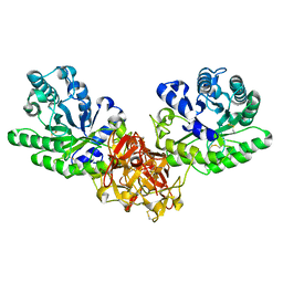
 | | AMINOPEPTIDASE P FROM E. COLI LOW PH FORM | | 分子名称: | ACETATE ION, AMINOPEPTIDASE P, MANGANESE (II) ION | | 著者 | Wilce, M.C.J, Bond, C.S, Lilley, P.E, Dixon, N.E, Freeman, H.C, Guss, J.M. | | 登録日 | 1997-12-22 | | 公開日 | 1999-04-06 | | 最終更新日 | 2024-02-07 | | 実験手法 | X-RAY DIFFRACTION (2.7 Å) | | 主引用文献 | Structure and mechanism of a proline-specific aminopeptidase from Escherichia coli.
Proc.Natl.Acad.Sci.USA, 95, 1998
|
|
8P4P
 | | Structure average of GroEL14 complexes found in the cytosol of Escherichia coli overexpressing GroEL obtained by cryo electron tomography | | 分子名称: | ADENOSINE-5'-DIPHOSPHATE, ADENOSINE-5'-TRIPHOSPHATE, Chaperonin GroEL, ... | | 著者 | Wagner, J, Caravajal, A.I, Beck, F, Bracher, A, Wan, W, Bohn, S, Koerner, R, Baumeister, W, Fernandez-Busnadiego, R, Hartl, F.U. | | 登録日 | 2023-05-23 | | 公開日 | 2024-07-03 | | 最終更新日 | 2024-08-21 | | 実験手法 | ELECTRON MICROSCOPY (9.6 Å) | | 主引用文献 | Visualizing chaperonin function in situ by cryo-electron tomography
Nature, 2024
|
|
8OLS
 | |
8P4O
 | | CryoEM structure of a GroEL7-GroES7 cage with encapsulated ordered substrate MetK in the presence of ADP-BeFx | | 分子名称: | ADENOSINE-5'-DIPHOSPHATE, BERYLLIUM TRIFLUORIDE ION, Chaperonin GroEL, ... | | 著者 | Wagner, J, Beck, F, Bracher, A, Caravajal, A.I, Wan, W, Bohn, S, Koerner, R, Baumeister, W, Fernandez-Busnadiego, R, Hartl, F.U. | | 登録日 | 2023-05-23 | | 公開日 | 2024-07-03 | | 最終更新日 | 2024-08-21 | | 実験手法 | ELECTRON MICROSCOPY (3.04 Å) | | 主引用文献 | Visualizing chaperonin function in situ by cryo-electron tomography
Nature, 2024
|
|
8OZQ
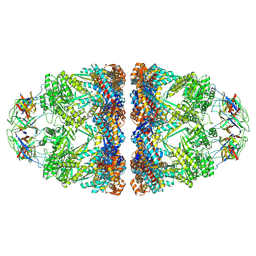
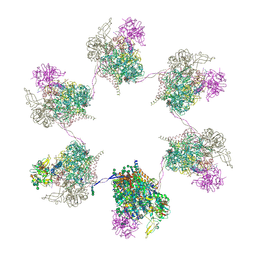 | | In situ subtomogram average of Prototype Foamy Virus Env hexamer of trimers | | 分子名称: | 2-acetamido-2-deoxy-beta-D-glucopyranose, 2-acetamido-2-deoxy-beta-D-glucopyranose-(1-2)-alpha-D-mannopyranose-(1-3)-[alpha-D-mannopyranose-(1-6)]beta-D-mannopyranose-(1-4)-2-acetamido-2-deoxy-beta-D-glucopyranose-(1-4)-2-acetamido-2-deoxy-beta-D-glucopyranose, 2-acetamido-2-deoxy-beta-D-glucopyranose-(1-4)-2-acetamido-2-deoxy-beta-D-glucopyranose, ... | | 著者 | Calcraft, T, Nans, A, Rosenthal, P.B. | | 登録日 | 2023-05-09 | | 公開日 | 2024-07-10 | | 最終更新日 | 2024-08-21 | | 実験手法 | ELECTRON MICROSCOPY (10 Å) | | 主引用文献 | Integrated cryoEM structure of a spumaretrovirus reveals cross-kingdom evolutionary relationships and the molecular basis for assembly and virus entry.
Cell, 187, 2024
|
|
8OZJ
 | | In situ cryoEM structure of Prototype Foamy Virus Env dimer of trimers | | 分子名称: | 2-acetamido-2-deoxy-beta-D-glucopyranose, 2-acetamido-2-deoxy-beta-D-glucopyranose-(1-2)-alpha-D-mannopyranose-(1-3)-[alpha-D-mannopyranose-(1-6)]beta-D-mannopyranose-(1-4)-2-acetamido-2-deoxy-beta-D-glucopyranose-(1-4)-2-acetamido-2-deoxy-beta-D-glucopyranose, 2-acetamido-2-deoxy-beta-D-glucopyranose-(1-4)-2-acetamido-2-deoxy-beta-D-glucopyranose, ... | | 著者 | Calcraft, T, Nans, A, Rosenthal, P.B. | | 登録日 | 2023-05-09 | | 公開日 | 2024-07-10 | | 最終更新日 | 2024-08-21 | | 実験手法 | ELECTRON MICROSCOPY (3.7 Å) | | 主引用文献 | Integrated cryoEM structure of a spumaretrovirus reveals cross-kingdom evolutionary relationships and the molecular basis for assembly and virus entry.
Cell, 187, 2024
|
|


