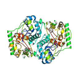
Loading
PDBj
メニューPDBj@FacebookPDBj@TwitterPDBj@YouTubewwPDB FoundationwwPDB
RCSB PDBPDBeBMRBAdv. SearchSearch help
PDB: 3 件

1AFW
DownloadVisualize
BU of 1afw by Molmil
THE 1.8 ANGSTROM CRYSTAL STRUCTURE OF THE DIMERIC PEROXISOMAL THIOLASE OF SACCHAROMYCES CEREVISIAE
分子名称: (4R)-2-METHYLPENTANE-2,4-DIOL, 3-KETOACETYL-COA THIOLASE
著者Mathieu, M, Wierenga, R.K.
登録日1997-03-15
公開日1997-06-16
最終更新日2024-02-07
実験手法X-RAY DIFFRACTION (1.8 Å)
主引用文献The 1.8 A crystal structure of the dimeric peroxisomal 3-ketoacyl-CoA thiolase of Saccharomyces cerevisiae: implications for substrate binding and reaction mechanism.
J.Mol.Biol., 273, 1997
3W15
DownloadVisualize
BU of 3w15 by Molmil
Structure of peroxisomal targeting signal 2 (PTS2) of Saccharomyces cerevisiae 3-ketoacyl-CoA thiolase in complex with Pex7p and Pex21p
分子名称: 3-ketoacyl-CoA thiolase, peroxisomal, Maltose-binding periplasmic protein, ...
著者Pan, D, Nakatsu, T, Kato, H.
登録日2012-11-06
公開日2013-07-03
最終更新日2017-08-16
実験手法X-RAY DIFFRACTION (1.8 Å)
主引用文献Crystal structure of peroxisomal targeting signal-2 bound to its receptor complex Pex7p-Pex21p
Nat.Struct.Mol.Biol., 20, 2013
1PXT
DownloadVisualize
BU of 1pxt by Molmil
THE 2.8 ANGSTROMS STRUCTURE OF PEROXISOMAL 3-KETOACYL-COA THIOLASE OF SACCHAROMYCES CEREVISIAE: A FIVE LAYERED A-B-A-B-A STRUCTURE, CONSTRUCTED FROM TWO CORE DOMAINS OF IDENTICAL TOPOLOGY
分子名称: PEROXISOMAL 3-KETOACYL-COA THIOLASE
著者Mathieu, M, Wierenga, R.K.
登録日1994-07-04
公開日1994-08-31
最終更新日2024-02-14
実験手法X-RAY DIFFRACTION (2.8 Å)
主引用文献The 2.8 A crystal structure of peroxisomal 3-ketoacyl-CoA thiolase of Saccharomyces cerevisiae: a five-layered alpha beta alpha beta alpha structure constructed from two core domains of identical topology.
Structure, 2, 1994

222415

件を2024-07-10に公開中

PDB statisticsPDBj update infoContact PDBjnumon