5NG2
 
 | |
5NG0
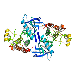
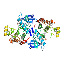 | | Structure of RIP2K(L294F) with bound AMPPCP | | 分子名称: | COBALT (II) ION, MAGNESIUM ION, PHOSPHOMETHYLPHOSPHONIC ACID ADENYLATE ESTER, ... | | 著者 | Pellegrini, E, Cusack, S. | | 登録日 | 2017-03-16 | | 公開日 | 2017-06-07 | | 最終更新日 | 2024-01-17 | | 実験手法 | X-RAY DIFFRACTION (2 Å) | | 主引用文献 | Structures of the inactive and active states of RIP2 kinase inform on the mechanism of activation.
PLoS ONE, 12, 2017
|
|
5NG3
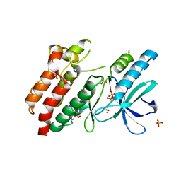 | | Structure of inactive kinase RIP2K(K47R) | | 分子名称: | Receptor-interacting serine/threonine-protein kinase 2, SULFATE ION | | 著者 | Pellegrini, E, Cusack, S. | | 登録日 | 2017-03-16 | | 公開日 | 2017-06-28 | | 最終更新日 | 2024-01-17 | | 実験手法 | X-RAY DIFFRACTION (2.6 Å) | | 主引用文献 | Structures of the inactive and active states of RIP2 kinase inform on the mechanism of activation.
PLoS ONE, 12, 2017
|
|


