Loading
PDBj
メニューPDBj@FacebookPDBj@TwitterPDBj@YouTubewwPDB FoundationwwPDB
RCSB PDBPDBeBMRBAdv. SearchSearch help
PDB: 4 件

7V8G
DownloadVisualize
BU of 7v8g by Molmil
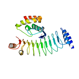
Crystal structure of HOIP RING1 domain bound to IpaH1.4 LRR domain
分子名称: E3 ubiquitin-protein ligase RNF31, RING-type E3 ubiquitin transferase, ZINC ION
著者Liu, J, Wang, Y, Pan, L.
登録日2021-08-23
公開日2022-03-30
最終更新日2023-11-29
実験手法X-RAY DIFFRACTION (2.75 Å)
主引用文献Mechanistic insights into the subversion of the linear ubiquitin chain assembly complex by the E3 ligase IpaH1.4 of Shigella flexneri.
Proc.Natl.Acad.Sci.USA, 119, 2022
7V8E
DownloadVisualize
BU of 7v8e by Molmil
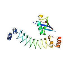
Crystal structure of IpaH1.4 LRR domain bound to HOIL-1L UBL domain.
分子名称: RING-type E3 ubiquitin transferase, RanBP-type and C3HC4-type zinc finger-containing protein 1
著者Liu, J, Wang, Y, Pan, L.
登録日2021-08-22
公開日2022-03-30
最終更新日2023-11-29
実験手法X-RAY DIFFRACTION (1.9 Å)
主引用文献Mechanistic insights into the subversion of the linear ubiquitin chain assembly complex by the E3 ligase IpaH1.4 of Shigella flexneri.
Proc.Natl.Acad.Sci.USA, 119, 2022
7V8F
DownloadVisualize
BU of 7v8f by Molmil
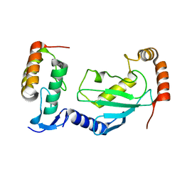
Crystal structure of UBE2L3 bound to HOIP RING1 domain.
分子名称: E3 ubiquitin-protein ligase RNF31, Ubiquitin-conjugating enzyme E2 L3, ZINC ION
著者Liu, J, Wang, Y, Pan, L.
登録日2021-08-22
公開日2022-03-30
最終更新日2023-11-29
実験手法X-RAY DIFFRACTION (1.66 Å)
主引用文献Mechanistic insights into the subversion of the linear ubiquitin chain assembly complex by the E3 ligase IpaH1.4 of Shigella flexneri.
Proc.Natl.Acad.Sci.USA, 119, 2022
7V8H
DownloadVisualize
BU of 7v8h by Molmil
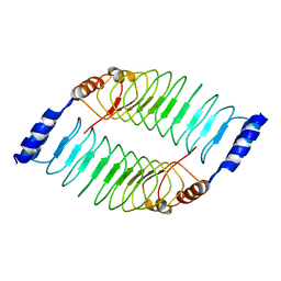
Crystal structure of LRR domain from Shigella flexneri IpaH1.4
分子名称: RING-type E3 ubiquitin transferase
著者Liu, J, Wang, Y, Pan, L.
登録日2021-08-23
公開日2022-03-30
最終更新日2023-11-29
実験手法X-RAY DIFFRACTION (2.46 Å)
主引用文献Mechanistic insights into the subversion of the linear ubiquitin chain assembly complex by the E3 ligase IpaH1.4 of Shigella flexneri.
Proc.Natl.Acad.Sci.USA, 119, 2022

222624

件を2024-07-17に公開中

PDB statisticsPDBj update infoContact PDBjnumon